Abstract
Purpose
To analyze the clinical features and determine the factors that affect glaucomatous change of patients with Posner-Schlossman syndrome (PSS).
Methods
A retrospective analysis of 51 eyes of 51 patients diagnosed with PSS was performed. We analyzed the factors including age of first attack, highest intraocular pressure (IOP), duration of the disease, number of the attacks and interval between attacks among the patients who developed glaucoma and those who did not and compared the 2 groups.
Results
The age of first attack was 34.73 ± 10.77 years, and highest IOP was 47.75 ± 9.43 mm Hg. Duration of the disease was 62.06 ± 69.84 months, number of the attacks was 6.20 ± 7.73 times, and interval between attacks was 12.65 ± 8.95 months. Of 51 eyes of 51 patients, 12 eyes (23.5%) of 12 patients showed significant glaucomatous change. In the glaucoma group, highest IOP was 52.81 ± 7.87 mm Hg, number of attacks was 11.91 ± 10.63 times, and interval between attacks was 8.07 ± 3.97 months. In the non-glaucomatous group highest IOP was 46.19 ± 9.14 mm Hg, number of attacks was 4.59 ± 5.94 times, and interval between attacks was 14.59 ± 9.79 months, respectively. Highest IOP was significantly greater, number of attacks was higher, and interval was shorter with statistical significance in the glaucoma group (p = 0.025, p = 0.001, p = 0.028).
Go to : 
References
1. Posner A, Schlossman A. Syndrome of unilateral recurrent attacks of glaucoma with cyclitic symptoms. Arch Ophthal. 1948; 39:517–35.

2. Posner A, Schlossman A. Further observations on the syndrome of glaucomatocyclitic crises. Trans Am Acad Ophthalmol Otolaryngol. 1953; 57:531–6.
3. Masuda K, Izawa Y, Mishima S. Prostaglandins and glaucomatocyclitic crisis. Jpn J Ophthalmol. 1975; 19:368–75.
5. Yamamoto S, Pavan-Langston D, Tada R, et al. Possible role of herpes simplex virus in the origin of Posner-Schlossman syndrome. Am J Ophthalmol. 1995; 119:796–8.

6. Bloch-Michel E, Dussaix E, Cerqueti P, Patarin D. Possible role of cytomegalovirus infection in the etiology of the Posner-Schlossmann syndrome. Int Ophthalmol. 1987; 11:95–6.

7. Teoh SB, Thean L, Koay E. Cytomegalovirus in aetiology of Posner-Schlossman syndrome: evidence from quantitative polymerase chain reaction. Eye (Lond). 2005; 19:1338–40.

8. Spivey BE, Armaly MF. Tonographic findings in glaucomatocyclitic crises. Am J Ophthalmol. 1963; 55:47–51.

10. Kass MA, Becker B, Kolker AE. Glaucomatocyclitic crisis and primary open-angle glaucoma. Am J Ophthalmol. 1973; 75:668–73.

11. Jap A, Sivakumar M, Chee SP. Is Posner Schlossman syndrome be-nign? Ophthalmology. 2001; 108:913–8.

12. Park WH, Jung YS, Han KS, Sohn YH. Clinical factors of glaucomatous change in patients with Posner-Schlossman syndrome. J Korean Ophthalmol Soc. 2005; 46:671–5.
13. Caprioli J, Miller JM. Correlation of structure and function in glaucoma. Quantitative measurements of disc and field. Ophthalmology. 1988; 95:723–7.
14. Sommer A, Pollack I, Maumenee AE. Optic disc parameters and onset of glaucomatous field loss. II. Static screening criteria. Arch Ophthalmol. 1979; 97:1449–54.
15. Pederson JE, Anderson DR. The mode of progressive disc cupping in ocular hypertension and glaucoma. Arch Ophthalmol. 1980; 98:490–5.

16. Zeyen TG, Caprioli J. Progression of disc and field damage in early glaucoma. Arch Ophthalmol. 1993; 111:62–5.

17. Yamada N, Mills RP, Leen MM, et al. Probability maps of sequential glaucoma-scope images help identify significant change. J Glaucoma. 1997; 6:279–87.

18. Darchuk V, Sampaolesi J, Mato L, et al. Optic nerve head behavior in Posner-Schlossman syndrome. Int Ophthalmol. 2001; 23:373–9.

19. Kim TH, Kim JL, Kee C. Optic disc atrophy in patient with Posner-Schlossman syndrome. Korean J Ophthalmol. 2012; 26:473–7.

20. Mitchell P, Smith W, Attebo K, Healey PR. Prevalence of open-angle glaucoma in Australia. The Blue Mountains Eye Study. Ophthalmology. 1996; 103:1661–9.

21. Dielemans I, Vingerling JR, Wolfs RC, et al. The prevalence of primary open-angle glaucoma in a population-based study in The Netherlands. The Rotterdam Study. Ophthalmology. 1994; 101:1851–5.

22. Coleman AL, Miglior S. Risk factors for glaucoma onset and progression. Surv Ophthalmol. 2008; 53(Suppl 1):S3–10.

23. Suzuki Y, Iwase A, Araie M, et al. Risk factors for open-angle glaucoma in a Japanese population: the Tajimi Study. Ophthalmology. 2006; 113:1613–7.
24. Leske MC, Wu SY, Hennis A, et al. Risk factors for incident open-angle glaucoma: the Barbados Eye Studies. Ophthalmology. 2008; 115:85–93.
Go to : 
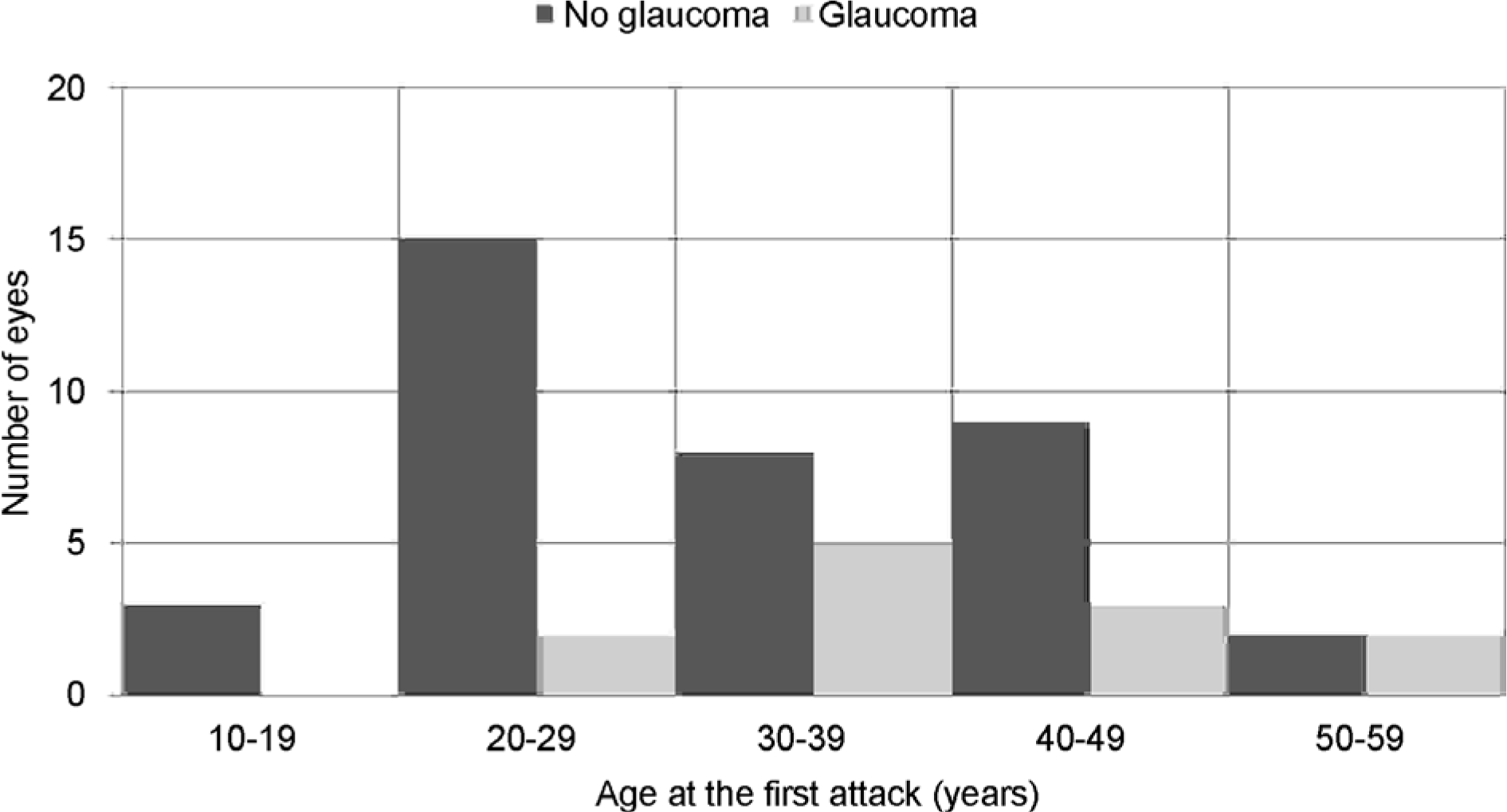
 | Figure 1.Distribution of age at the first attack in patients with Posner-Schlossman syndrome. In patients with no glaucomatous change, the age at first attack is distributed mostly from twenties to thirties and in patients with glaucomatous change, the age at first attack showed tendency toward a slightly older age. No glaucoma= patients without glaucoma development; Glaucoma= patients with glaucoma development. |
Table 1.
Patients clinical characteristics
| Clinical characteristics | Total (n=51) | No glaucomatous change (n=39) | Glaucomatous change (n=12) | p-value |
|---|---|---|---|---|
| Age of first attack (years, range) | 34.73 ± 10.77 (13-57) | 33.26 ± 11.21 (13-57) | 39.5 ± 7.76 (29-52) | 0.050* |
| Sex (male/female) | 37/14 | 27/12 | 10/2 | 0.34† |
| Highest IOP (mm Hg, range) | 47.75 ± 9.43 (31-70) | 46.19 ± 9.14 (31-63) | 52.81 ± 7.87 (42-70) | 0.025* |
| Duration (month, range) | 62.06 ± 69.84 (1-312) | 59.24 ± 70.51 (1-312) | 74.00 ± 69.64 (4-198) | 0.480* |
| Number of attack (times, range) | 6.20 ± 7.73 (1-35) | 4.59 ± 5.94 (1-30) | 11.91 ± 10.63 (3-35) | 0.001* |
| Interval between attacks (month, range) | 12.65 ± 8.95 (1.33-38.70) | 14.59 ± 9.79 (3.0-38.7) | 8.07 ± 3.97 (1.33-13.67) | 0.028* |
| Baseline MD (dB, range) | -2.57 ± 4.87 (-20.66-2.08) | -0.61 ± 1.20 (-4.21-2.08) | -7.21 ± 6.94 (-20.66~-0.84) | 0.000* |
| CCT (μ m, range) | 547.0 ± 39.85 (482-635) | 553.00 ± 36.02 (492-635) | 535.50 ± 47.73 (482-623) | 0.350* |
Table 2.
Logistic regression analysis with the dependent variable being the presence of a glaucomatous change
| Factors |
Univariate analysis |
Multivariable analysis* |
Multivariable analysis* |
|||
|---|---|---|---|---|---|---|
| OR (95% CI) | p-value | OR (95% CI) | p-value | OR (95% CI) | p-value | |
| Age of first attack (years) | 1.057 (0.990-1.129) | 0.097 | 1.177 (1.020-1.358)† | 0.025 | 0.112 (0.989-1.250) | 0.076 |
| Highest IOP (mm Hg) | 1.102 (1.011-1.202)† | 0.028 | 1.206 (1.017-1.431)† | 0.031 | 1.146 (0.990-1.327) | 0.067 |
| Number of attack (times) | 1.113 (1.017-1.207)† | 0.020 | 1.321 (1.037-1.683)† | 0.024 | - | - |
| Interval between attacks (month) | 0.862 (0.738-1.006) | 0.060 | - | - | 0.816 (0.648-1.028) | 0.084 |
Table 3.
Characteristics of patients with Posner-Schlossman syndrome who developed glaucoma
| Patient | Sex | Age at first attack (years) | Duration of the disease (month) | Highest IOP (mm Hg) | Total number of attacks (times) | Interval between attacks (month) | Baseline MD (dB) | Last MD (dB) | CCT (μ m) | Glaucoma surgery |
|---|---|---|---|---|---|---|---|---|---|---|
| 1 | M | 29 | -* | 55 | 8 | - | -10.69 | -20.65 | 543 | No |
| 2 | F | 38 | 111 | 47 | 15 | 7.93 | -5.67 | -4.09 | 489 | No |
| 3 | M | 32 | 174 | 60 | 29 | 6.21 | -0.96 | -20.24 | - | No |
| 4 | M | 42 | 40 | 50 | 4 | 13.33 | -2.72 | -2.49 | - | No |
| 5 | M | 46 | 52 | 54 | 6 | 10.40 | -6.95 | -13.61 | 578 | No |
| 6 | M | 44 | 22 | 52 | 6 | 4.40 | -2.81 | -6.39 | - | No |
| 7 | M | 30 | 198 | 70 | 11 | 9.78 | -0.84 | -5.65 | - | No |
| 8 | M | 51 | 41 | 42 | 4 | 13.67 | -11.37 | -15.25 | 623 | No |
| 9 | M | 40 | 145 | 47 | 35 | 3.88 | -0.99 | - | 535 | Yes |
| 10 | F | 33 | -† | 58 | 9 | - | -8.17 | -10.33 | 498 | Yes |
| 11 | M | 37 | 22 | 43 | 3 | 11.00 | -20.42 | -21.24 | - | Yes |
| 12 | M | 52 | 12 | 46 | 10 | 1.33 | -20.66 | -17.71 | 482 | Yes |


 PDF
PDF ePub
ePub Citation
 Print
Print


 XML Download