Abstract
Purpose
To compare the clinical outcomes between refractive-type multifocal intraocular lenses (IOL) (Lentis Mplus® LS 313, Oculentis GmbH., Berlin, Germany) and diffractive-type multifocal IOL (Acrysof Restor®; SN6AD1, Alcon Lab., Fort Worth, TX, USA) with same near added.
Methods
We evaluated 30 eyes implanted with Lentis Mplus® IOL and 33 eyes implanted with Acrysof Restor® IOL after phacoemulsification. The distant, intermediate, and near uncorrected visual acuities of the 2 groups were evaluated at 2 weeks and 1, 3, and 6 months postoperatively. Optical quality obtained using the Optical Quality Analysis System II (OQAS II®, Visiometrics, Castelldefels, Barcelona, Spain), higher-order aberrations (HOAs), and patient satisfaction questionnaire of the 2 groups were evaluated at 3 months postoperatively.
Results
The visual acuity of intermediate 100 cm was statistically better in the Lentis Mplus® group (p < 0.05). There were no significant differences between the 2 groups with distant, intermediate 63 cm, and near vision. At the 3-month postoperative fol-low-up, objective scatter index, modulation transfer function (MTF) cutoff value, and pseudo-accommodation range measured by OQAS II® showed no differences between the 2 groups, but Strhel ratio was higher in the Acrysof Restor® group. HOAs of 5 mm and 6 mm increased significantly in the Lentis Mplus® group. No significant differences were found in the patient satisfaction questionnaire.
Go to : 
References
1. Alfonso JF, Fernández-Vega L, Baamonde MB, Montés-Micó R. Prospective visual evaluation of apodized diffractive intraocular lenses. J Cataract Refract Surg. 2007; 33:1235–43.

2. Lee HS, Park SH, Kim MS. Clinical results and some problems of multifocal apodized diffractive intraocular lens implantation. J Korean Ophthalmol Soc. 2008; 49:1235–41.

3. de Vries NE, Webers CA, Montés-Micó R, et al. Long-term fol-low-up of a multifocal apodized diffractive intraocular lens after cataract surgery. J Cataract Refract Surg. 2008; 34:1476–82.

4. Woodward MA, Randleman JB, Stulting RD. Dissatisfaction after multifocal intraocular lens implantation. J Cataract Refract Surg. 2009; 35:992–7.

5. Cheon MH, Lee JE, Kim JH, et al. One-year outcome of monocular implant of aspheric multifocal IOL. J Korean Ophthalmol Soc. 2010; 51:822–8.

6. Kim SM, Kim CH, Chung ES, Chung TY. Visual outcome and patient satisfaction after implantation of multifocal IOLs: three- month follow-up results. J Korean Ophthalmol Soc. 2012; 53:230–7.
7. de Vries NE, Nuijts RM. Multifocal intraocular lenses in cataract surgery: literature review of benefits and side effects. J Cataract Refract Surg. 2013; 39:268–78.

8. Alfonso JF, Puchades C, Fernández-Vega L, et al. Contrast sensitivity comparison between AcrySof ReSTOR and Acri.LISA aspheric intraocular lenses. J Refract Surg. 2010; 26:471–7.

9. Castillo-Gómez A, Carmona-González D, Martínez-de-la-Casa JM, et al. Evaluation of image quality after implantation of 2 diffractive multifocal intraocular lens models. J Cataract Refract Surg. 2009; 35:1244–50.

10. Alfonso JF, Fernández-Vega L, Blázquez JI, Montés-Micó R. Visual function comparison of 2 aspheric multifocal intraocular lenses. J Cataract Refract Surg. 2012; 38:242–8.

11. Blaylock JF, Si Z, Vickers C. Visual and refractive status at different focal distances after implantation of the ReSTOR multifocal intraocular lens. J Cataract Refract Surg. 2006; 32:1464–73.

12. Kohnen T, Allen D, Boureau C, et al. European multicenter study of the AcrySof ReSTOR apodized diffractive intraocular lens. Ophthalmology. 2006; 113:578–84.e1.

13. Nochez Y, Majzoub S, Pisella PJ. Effect of interaction of macro-aberrations and scattered light on objective quality of vision in pseudophakic eyes with aspheric monofocal intraocular lenses. J Cataract Refract Surg. 2012; 38:633–40.

14. McAlinden C, Moore JE. Multifocal intraocular lens with a sur-face-embedded near section: short-term clinical outcomes. J Cataract Refract Surg. 2011; 37:441–5.

15. Alió JL, Plaza-Puche AB, Piñero DP, et al. Comparative analysis of the clinical outcomes with 2 multifocal intraocular lens models with rotational asymmetry. J Cataract Refract Surg. 2011; 37:1605–14.

16. Muñoz G, Albarrán-Diego C, Ferrer-Blasco T, et al. Visual function after bilateral implantation of a new zonal refractive aspheric multifocal intraocular lens. J Cataract Refract Surg. 2011; 37:2043–52.

17. Alió JL, Plaza-Puche AB, Montalban R, Javaloy J. Visual outcomes with a single-optic accommodating intraocular lens and a low-addition-power rotational asymmetric multifocal intraocular lens. J Cataract Refract Surg. 2012; 38:978–85.

18. Alio JL, Plaza-Puche AB, Javaloy J, et al. Comparison of a new refractive multifocal intraocular lens with an inferior segmental near add and a diffractive multifocal intraocular lens. Ophthalmology. 2012; 119:555–63.

19. Chung YK, Park CW, Hwang JH, Joo CK. Clinical outcomes of M-plus intraocular lenses. J Korean Ophthalmol Soc. 2014; 55:519–26.

20. Hayashi K, Manabe S, Yoshida M, Hayashi H. Effect of astigmatism on visual acuity in eyes with a diffractive multifocal intraocular lens. J Cataract Refract Surg. 2010; 36:1323–9.

21. Nanavaty MA, Spalton DJ, Marshall J. Effect of intraocular lens asphericity on vertical coma aberration. J Cataract Refract Surg. 2010; 36:215–21.

22. Nishi T, Nawa Y, Ueda T, et al. Effect of total higher-order aberrations on accommodation in pseudophakic eyes. J Cataract Refract Surg. 2006; 32:1643–9.

23. Nochez Y, Majzoub S, Pisella PJ. Effect of interaction of macro-aberrations and scattered light on objective quality of vision in pseudophakic eyes with aspheric monofocal intraocular lenses. J Cataract Refract Surg. 2012; 38:633–40.

24. Diaz-Valle D, Arriola-Villalobos P, García-Vidal SE, et al. Effect of lubricating eyedrops on ocular light scattering as a measure of vision quality in patients with dry eye. J Cataract Refract Surg. 2012; 38:1192–7.

25. Cabot F, Saad A, McAlinden C, et al. Objective assessment of crystalline lens opacity level by measuring ocular light scattering with a double-pass system. Am J Ophthalmol. 2013; 155:629–35. 635.e1-2.

26. Lee K, Ahn JM, Kim EK, Kim TI. Comparison of optical quality parameters and ocular aberrations after wavefront-guided laser in-situ keratomileusis versus wavefront-guided laser epithelial keratomileusis for myopia. Graefes Arch Clin Exp Ophthalmol. 2013; 251:2163–9.

Go to : 
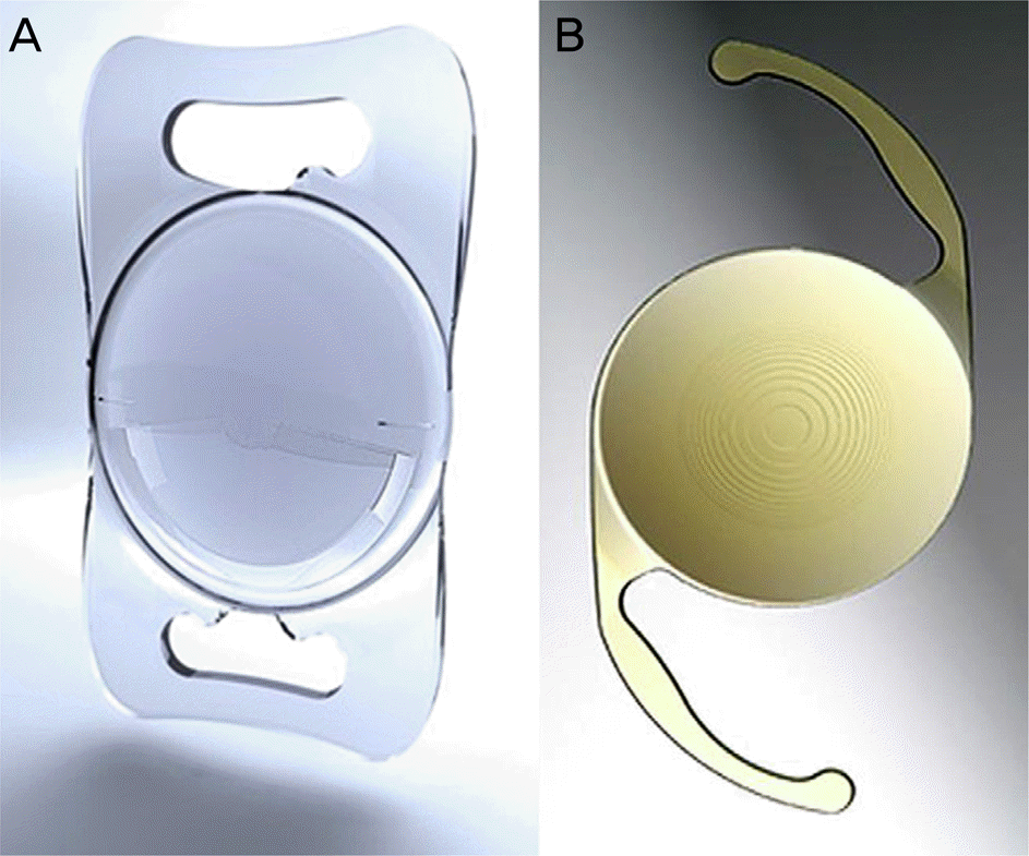
 | Figure 1.(A) Lentis Mplus LS-313 intraocular lenses (IOL) and (B) Acrysof Restor SN6AD1 IOL. |
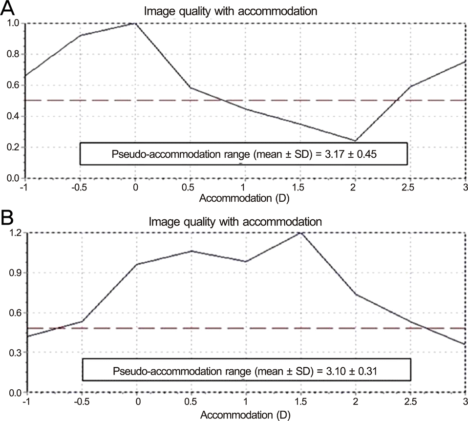
 | Figure 2.An example of a image quality with pseudo-accom-modation measured by Optical quality analysis system II® at post-operation 3 months of (A) Restor® and (B) Lentis Mplus®. (A) Graph A shows good optical quality at distant (0.0 D) and near (3.0 D). (B) Graph B shows good optical quality at distant (0.0 D) and intermediate distance (1.5 D). At the 3-month postoperative visit, the mean pseudo-accom-modation range of total enrolled patients was 3.17 ± 0.45 D for Restor® and 3.10 ± 0.31 D for Lentis Mplus®. |
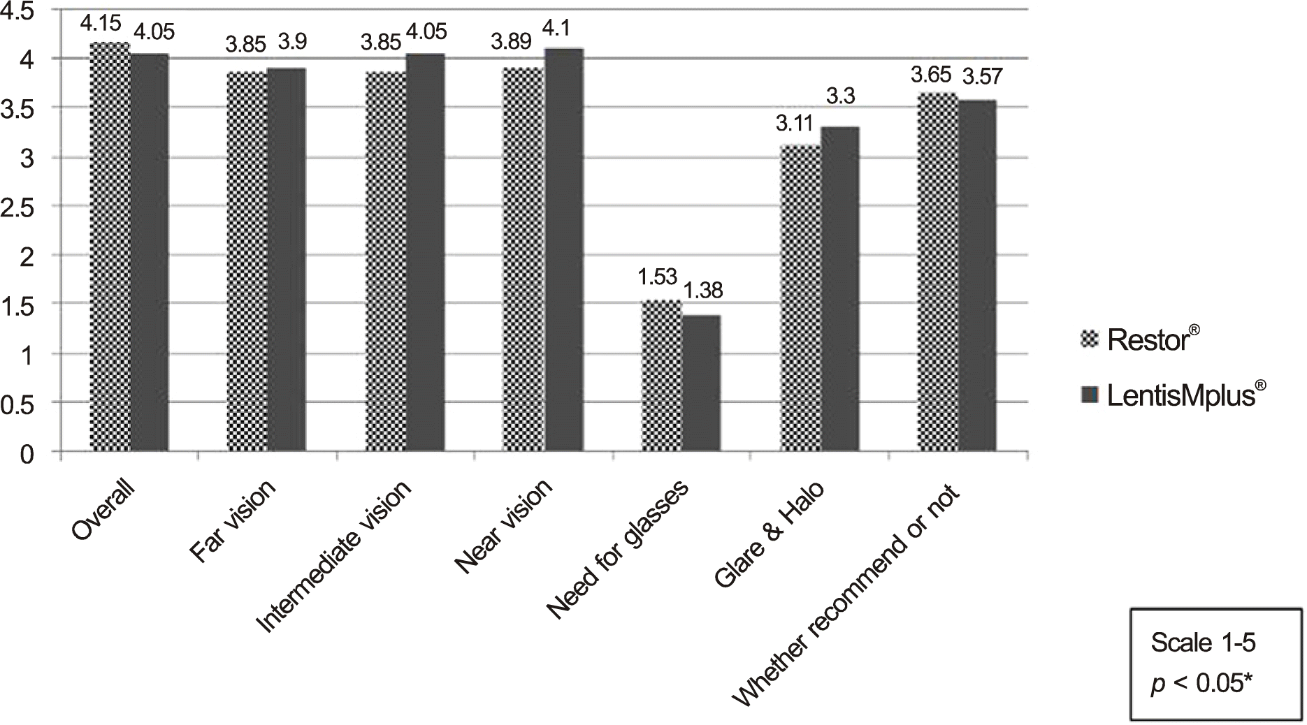
 | Figure 3.Satisfaction questionnaire of the patients at post-operative 3 months. There was no statistically significant difference in satisfaction between the two groups. *Student's t-test. |
Table 1.
Demographics of the study group
| Restor® | Lentis Mplus® | p-value* | |
|---|---|---|---|
| No. of eyes (patients) | 33 (28) | 30 (24) | |
| Male:female (eyes) | 16:17 | 18:12 | |
| Mean age (years) | 52.58 ± 5.06 | 55.50 ± 7.21 | 0.065 |
| Preoperative S.E. (D) | -0.55 ± 2.31 | 0.29 ± 1.29 | 0.179 |
| IOL power (D) (range) | 20.36 ± 1.42 (17.5-24.0) | 19.50 ± 1.64 (17.0-22.0) | 0.088 |
| Axial length (mm) | 23.64 ± 0.73 | 23.69 ± 0.72 | 0.795 |
| Preoperative UCVA (log MAR) | 0.53 ± 0.37 | 0.42 ± 0.28 | 0.383 |
Table 2.
Postoperative visual acuity outcomes over time (log MAR)
| UCDVA | UNVA | UIVA 63 cm | UIVA 100 cm* | ||
|---|---|---|---|---|---|
| Restor® | Postoperative 2 weeks | 0.03 ± 0.06 | 0.05 ± 0.07 | 0.16 ± 0.13 | 0.30 ± 0.16 |
| Lentis Mplus® | 0.04 ± 0.06 | 0.08 ± 0.07 | 0.13 ± 0.12 | 0.10 ± 0.12 | |
| Restor® | Postoperative 1 month | 0.03 ± 0.04 | 0.05 ± 0.07 | 0.17 ± 0.14 | 0.28 ± 0.16 |
| Lentis Mplus® | 0.04 ± 0.05 | 0.08 ± 0.08 | 0.16 ± 0.27 | 0.10 ± 0.10 | |
| Restor® | Postoperative 3 months | 0.03 ± 0.05 | 0.07 ± 0.09 | 0.15 ± 0.15 | 0.27 ± 0.17 |
| Lentis Mplus® | 0.04 ± 0.05 | 0.08 ± 0.09 | 0.12 ± 0.07 | 0.08 ± 0.10 | |
| Restor® | Postoperative 6 months | 0.03 ± 0.05 | 0.04 ± 0.06 | 0.16 ± 0.15 | 0.28 ± 0.17 |
| Lentis Mplus® | 0.04 ± 0.05 | 0.08 ± 0.10 | 0.16 ± 0.09 | 0.12 ± 0.11 |
Table 3.
Postoperative refractive outcomes over time
| Postoperative 2 weeks | Postoperative 1 month | Postoperative 3 months | Postoperative 6 months | p-value* | ||
|---|---|---|---|---|---|---|
| S.E. (MR) | Restor® | -0.13 ± 0.31 | -0.12 ± 0.33 | -0.10 ± 0.29 | -0.13 ± 0.27 | 0.035 |
| Lentis Mplus® | -0.08 ± 0.27 | -0.05 ± 0.23 | -0.04 ± 0.29 | 0.02 ± 0.28 | ||
| S.E. (AR) | Restor® | -0.16 ± 0.27 | -0.16 ± 0.28 | -0.08 ± 0.29 | -0.14 ± 0.25 | <0.001 |
| Lentis Mplus® | -1.19 ± 0.32 | -1.16 ± 0.40 | -1.03 ± 0.46 | -1.01 ± 0.42 | ||
| S.E. (MR) and target | Restor® | 0.29 ± 0.25 | 0.32 ± 0.20 | 0.27 ± 0.21 | 0.30 ± 0.20 | 0.440 |
| ref. difference | Lentis Mplus® | 0.23 ± 0.19 | 0.20 ± 0.16 | 0.26 ± 0.21 | 0.25 ± 0.21 |
Table 4.
Optical quality parameters measured by OQASⅡ® at postoperative 3 months
| Restor® | Lentis Mplus® | p-value | |
|---|---|---|---|
| OSI | 1.07 ± 0.40 | 0.88 ± 0.39 | 0.065 |
| MTF-cut off | 40.98 ± 8.07 | 37.44 ± 8.66 | 0.098 |
| Strhel ratio | 0.25 ± 0.07 | 0.18 ± 0.05 | 0.000* |
Table 5.
Ocular aberrometry analysis (μ m) measured by Zywave II® at postoperative 3 months
| Restor® | Lentis Mplus® | p-value | |
|---|---|---|---|
| HOA 5 mm | 0.29 ± 0.08 | 0.59 ± 0.17 | 0.000* |
| HOA 6 mm | 0.50 ± 0.13 | 1.07 ± 0.22 | 0.000* |
| Vertical trefoil | 0.21 ± 0.14 | 0.31 ± 0.31 | 0.203 |
| Horizontal trefoil | 0.16 ± 0.11 | 0.20 ± 0.22 | 0.473 |
| Vertical coma | 0.15 ± 0.10 | 0.47 ± 0.32 | 0.000* |
| Horizontal coma | 0.11 ± 0.10 | 0.22 ± 0.22 | 0.046* |
| 4th order spherical aberration | 0.28 ± 0.09 | 0.56 ± 0.17 | 0.000* |
| Quadrafoil | 0.07 ± 0.07 | 0.07 ± 0.10 | 0.857 |
| 2nd astigmatism | 0.05 ± 0.03 | 0.06 ± 0.07 | 0.413 |
| Pentafoil | 0.04 ± 0.04 | 0.06 ± 0.05 | 0.111 |
| 2nd vertical trefoil | 0.02 ± 0.02 | 0.03 ± 0.03 | 0.192 |
| 2nd horizontal trefoil | 0.03 ± 0.03 | 0.03 ± 0.03 | 0.843 |
| 2nd vertical coma | 0.03 ± 0.03 | 0.05 ± 0.04 | 0.087 |
| 2nd horizontal coma | 0.02 ± 0.03 | 0.03 ± 0.03 | 0.564 |


 PDF
PDF ePub
ePub Citation
 Print
Print


 XML Download