Journal List > J Korean Ophthalmol Soc > v.56(6) > 1010291

Lee, Kim, Choi, Park, and Lee: Comparison of Ocular Adnexal Mantle Cell Lymphoma and MALT Lymphoma

Abstract

Purpose

Mantle cell lymphoma (MCL) is known to have systemic dissemination with poor prognosis, but very few cases have been reported in Korea. Therefore, we analyzed clinical features and prognosis of MCL by comparing 3 cases of ocular adnexal MCL and mucosa-associated lymphoid tissue (MALT) lymphoma.

Methods

The medical records of patients with ocular adnexal MALT lymphoma and MCL from Chungnam National University Hospital and Pusan National University Hospital from January 1999 to April 2014 were retrospectively reviewed. Immunohistochemical examination was selectively used to diagnose lymphoma subtypes. Systemic dissemination, treatment response, and recurrence were identified using radiological testing and bone marrow aspiration.

Results

This study included 39 patients with ocular and ocular adnexal MALT lymphoma and 1 patient with MCL from Chungnam National University Hospital and 53 patients with MALT lymphoma and 2 patients with MCL from Pusan National University Hospital. All 3 (100%) patients diagnosed with ocular adnexal MCL were over 60 years of age. However, 28 of 92 (30.4%) patients diagnosed with MALT lymphoma were over 60 years of age. In MALT lymphoma, 3 of 92 patients presented with systemic dissemination and most patients recovered with radiotherapy. Conversely, all 3 patients with MCL showed systemic dissemination and recurrence after radiotherapy or chemotherapy, therefore, additional chemotherapy was required.

Conclusions

Contrary to MALT lymphoma, ocular and ocular adnexal MCL usually presented with systemic dissemination and complete remission was difficult even though many regimens of chemotherapy were attempted. Because MCL can be misdiagnosed as MALT lymphoma, careful evaluation should be performed.

References

1. Cahill M, Barnes C, Moriarty P, et al. Ocular adnexal lymphoma-comparison of MALT lymphoma with other histological types. Br J Ophthalmol. 1999; 83:742–7.
crossref
2. Swerdlow SH, Williams ME. From centrocytic to mantle cell lymphoma: a clinicopathologic and molecular review of 3 decades. Hum Pathol. 2002; 33:7–20.
crossref
3. Andersen NS, Jensen MK, de Nully Brown P, Geisler CH. A Danish population-based analysis of 105 mantle cell lymphoma patients: incidences, clinical features, response, survival and prognostic factors. Eur J Cancer. 2002; 38:401–8.
4. Looi A, Gascoyne RD, Chhanabhai M, et al. Mantle cell lymphoma in the ocular adnexal region. Ophthalmology. 2005; 112:114–9.
crossref
5. Jenkins C, Rose GE, Bunce C, et al. Histological features of ocular adnexal lymphoma (REAL classification) and their association with patient morbidity and survival. Br J Ophthalmol. 2000; 84:907–13.
crossref
6. Lee JS, Song SW, Kim MS. A case of primary mantle cell lymphoma on the conjunctiva. J Korean Ophthalmol Soc. 1999; 40:2010–4.
7. Freeman C, Berg JW, Cutler SJ. Occurrence and prognosis of ex-tranodal lymphomas. Cancer. 1972; 29:252–60.
crossref
8. Bairey O, Kremer I, Rakowsky E, et al. Orbital and adnexal involvement in systemic non-Hodgkins lymphoma. Cancer. 1994; 73:2395–9.
crossref
9. Coupland SE, Krause L, Delecluse HJ, et al. Lymphoproliferative lesions of the ocular adnexa. Analysis of 112 cases. Ophthalmology. 1998; 105:1430–41.
10. Shields JA, Shields CL, Scartozzi R. Survey of 1264 patients with orbital tumors and simulating lesions: The 2002 Montgomery Lecture, Part 1. Ophthalmology. 2004; 111:997–1008.
11. Nola M, Lukenda A, Bollmann M, et al. Outcome and prognostic factors in ocular adnexal lymphoma. Croat Med J. 2004; 45:328–32.
12. Oh DE, Kim YD. Lymphoprolfierative diseases of the ocular ad-nexa in Korea. Arch Ophthalmol. 2007; 125:1668–73.
13. Looi A, Gascoyne RD, Chhanabhai M, et al. Mantle cell lymphoma in the ocular adnexal region. Ophthalmology. 2005; 112:114–9.
crossref
14. Romaguera JE, Medeiros LJ, Hagemeister FB, et al. Frequency of gastrointestinal involvement and its clinical significance in mantle cell lymphoma. Cancer. 2003; 97:586–91.
crossref
15. Rasmussen P, Sjö LD, Prause JU, et al. Mantle cell lymphoma in the orbital and adnexal region. Br J Ophthalmol. 2009; 93:1047–51.
crossref
16. Weisenburger DD, Armitage JO. Mantle cell lymphoma-an entity comes of age. Blood. 1996; 87:4483–94.
17. A clinical evaluation of the International Lymphoma Study Group classification of non-Hodgkin's lymphoma. The Non-Hodgkin's Lymphoma Classification Project. Blood. 1997; 89:3909–18.
18. Liu Z, Dong HY, Gorczyca W, et al. CD5- mantle cell lymphoma. Am J Clin Pathol. 2002; 118:216–24.
crossref
19. Witzig TE. Current treatment approaches for mantle-cell lymphoma. J Clin Oncol. 2005; 23:6409–14.
crossref
20. Schulz H, Bohlius J, Skoetz N, et al. Chemotherapy plus rituximab versus chemotherapy alone for B-cell non-Hodgkin's lymphoma. Cochrane Database Syst Rev. 2007; (4):CD003805.
crossref

Figure 1.
The photograph shows the both lower palpebral conjunctival mass.
jkos-56-815f1.tif
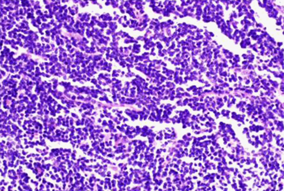
Figure 2.
High-power photomicrograph shows a monotonous proliferation of small to medium sized lymphocyte with irreg-ular, hyperchromatic nuclei, and scant cytoplasm. There are hyalinized vessels in the background (H&E; ×400).
jkos-56-815f2.tif
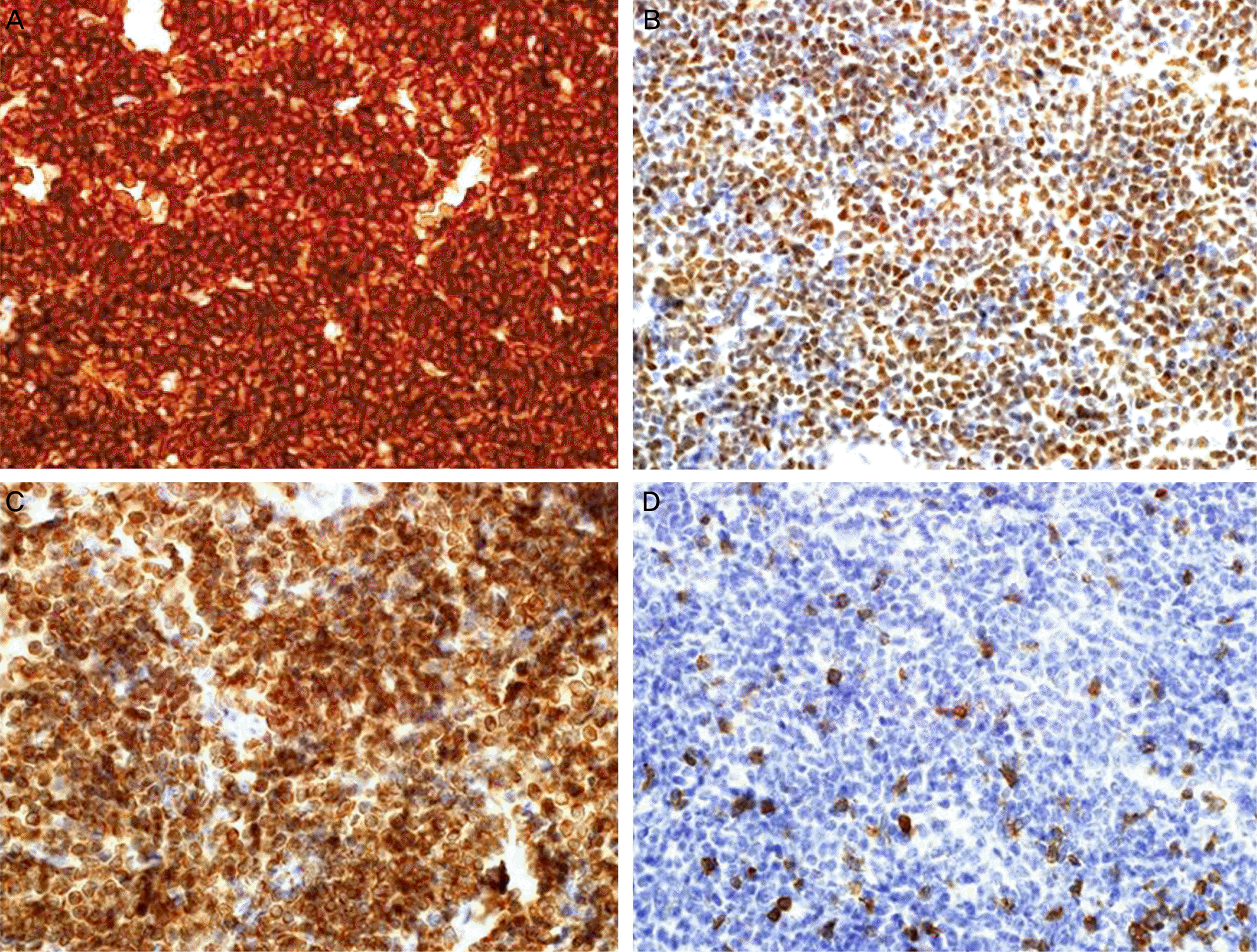
Figure 3.
Immunohistochemistry showing positive nuclear staining for CD20 (A), cyclin D1 (B), bcl-2 (C), and negative nuclear staining for CD5 (D), respectively.
jkos-56-815f3.tif
Table 1.
Characteristics of 95 patients with MCL and MALT lymphoma
Characteristics MCL (n = 3) MALT lymphoma (n = 92) All patients (n = 95)
Age      
  Mean age 67.7 50.4  
  ≥60 3 (100.0) 28 (30.4) 31 (32.6)
  <60 0 (0.0) 64 (69.6) 64 (67.4)
Gender (n, %)      
  Male 2 (66.7) 35 (38.0) 37 (38.9)
  Female 1 (33.3) 57 (62.0) 58 (61.1)
Laterality (n, %)      
  Unilateral 1 (33.3) 74 (80.4) 75 (78.9)
  Bilateral 2 (66.7) 18 (19.6) 20 (21.1)
Location (n, %)      
  Orbit 1 (33.3) 12 (13.0) 13 (13.7)
  Conjunctiva 1 (33.3) 53 (57.6) 54 (56.8)
  Lacrimal gland 0 (0.0) 8 (8.7) 8 (8.4)
  Eyelid 1 (33.3) 17 (18.5) 18 (19.0)
  Sclera 0 (0.0) 2 (2.2) 2 (2.1)
Ann Arbor stage (n, %)      
  Ⅰ 0 (0.0) 84 (91.3) 84 (88.4)
  Ⅱ 0 (0.0) 2 (2.1) 2 (2.1)
  Ⅲ 0 (0.0) 3 (3.3) 3 (3.2)
  Ⅳ 3 (100.0) 3 (3.3) 6 (6.3)
Bone marrow involvement (n, %)      
  Involvement 3 (100.0) 2 (2.2) 5 (5.3)
  No involvement 0 (0.0) 90 (97.8) 90 (34.7)
Metastasis (n, %) 3 (100.0) 3 (3.3) 6 (6.3)
Overall treatment (n, %)      
  Radiotherapy only 0 (0.0) 49 (53.3) 49 (51.6)
  Chemotherapy only 2 (66.7) 12 (13.0) 14 (14.7)
  Radiotherapy + chemotherapy 1 (33.3) 11 (12.0) 12 (12.6)
  Observation 0 (0.0) 14 (15.2) 14 (14.7)
Recurrence (n, %) 3 (100.0) 5 (5.4) 8 (8.4)
Follow-up period (months) 76.0 37.6 38.8

Values are presented as n (%).

MCL = mantle cell lymphoma; MALT = mucosa-associated lymphoid tissue.

Table 2.
Clinical features of 3 patients with mantle cell lymphoma
Case no. 1 2 3
Sex/age (years) M/79 M/60 F/64
Initial symptom Both conjunctival mass Inguinal mass Both conjunctival mass
  Both foggy vision    
Initial diagnosis Low grade marginal zone lymphoma of MALT Mantle cell lymphoma Diffuse mixed small and large cell type B cell lymphoma
Initial treatment CVP #8 R-CHOP #8 CHOP #6
Primary orbital site Conjunctiva Orbit Eyelid
Immunohistochemistry CD20, cyclin D1, bcl-2(+) CD20, CD5, cyclin D1(+) CD20, cyclin D1, Bcl-2(+)
  CD3, CD5(-) CD3, bcl-6, CD10(-) CD3, CD5(+)
Final diagnosis Mantle cell lymphoma Mantle cell lymphoma Mantle cell lymphoma
Site of metastasis Both parotid gland left thyroid gland cervical, thoracic, abdominal lymph node bone marrow Cecum, bone marrow, orbit marrow Nasopharynx, endometrium, bone
Additional treatment FC #4 IEOX #6 HyperCVAD + MA #6
  Bortezomib #6 IVIG + rituximab #4 ESHAP #5
    ESHAP #4 MINE #1
Follow-up period (months) 60 132 36

MALT = mucosa-associated lymphoid tissue; CVP = cyclophosphamide, vincristine, prednisolone; R-CHOP = rituximab, cyclo-phosphamide, hydroxydaunorubicin, oncovin, prednisolone; CHOP = cyclophosphamide, hydroxydaunorubicin, oncovin, prednisolone; FC = fludarabine, cyclophosphamide; IEOX = ifosfamide, etoposide, oxaliplatin, mesna; IVIG = intravenous immunoglobulin; ESHAP = eto-poside, cisplatin, high dose cytarabine, methylprednisolone; HyperCVAD = hyperfractionated cyclophosphamide, vincristine, doxorubicin, dexamethasone; MA = methotrexate/cytarabine; MINE = mesna, ifosfamide, mitoxantrone, etoposide.

TOOLS
Similar articles