

 PDF
PDF ePub
ePub Citation
 Print
Print


Abstract
Purpose
To investigate the characteristics of non-glaucomatous eyes with peripapillary retinoschisis.
Methods
Six non-glaucomatous eyes with peripapillary retinoschisis were enrolled. Age, sex, refractive error, intraocular pressure, location and changes of peripapillary retinoschisis, and the presence of accompanied abnormalities were assessed. To determine possible abnormalities of the optic nerve head and macula, fundus photographs and cross-sectional images of the optic nerve head and macula obtained by optical coherence tomography were inspected.
Results
Three males and 3 females were enrolled. Age of the subjects ranged from 11 to 59 years (median, 45 years). Refractive error ranged from -6.25 to +1.00 diopter (median, -0.50 diopter). Peripapillary retinoschisis was located in the superior quadrant in four eyes, in the nasal quadrant in one eye, and in the inferior quadrant in one eye, respectively. No additional abnor-malities were found in fundus photographs or in the cross-sectional images of the optic nerve head and macula that were obtained by optical coherence tomography. Longitudinal follow-up was available for two eyes and spontaneous resolution of peri-papillary retinoschisis was observed in these eyes 6 and 9 months later, respectively.
Go to : 
References
1. Yu H, Li T, Luo Y. . Long-term outcomes of vitrectomy for progressive X-linked retinoschisis. Am J Ophthalmol. 2012; 154:394–402.e2.

2. George ND, Yates JR, Moore AT. Clinical features in affected males with X-linked retinoschisis. Arch Ophthalmol. 1996; 114:274–80.
3. Brockhurst RJ. Photocoagulation in congenital retinoschisis. Arch Ophthalmol. 1970; 84:158–65.
4. Hirakata A, Hida T, Ogasawara A, Iizuka N. Multilayered retinoschisis associated with optic disc pit. Jpn J Ophthalmol. 2005; 49:414–6.
5. Scott IU, Moshfeghi AA, Flynn HW Jr. Surgical management of macular retinoschisis associated with high myopia. Arch Ophthalmol. 2006; 124:1197–9.
6. Kahook MY, Noecker RJ, Ishikawa H. . Peripapillary schisis in glaucoma patients with narrow angles and increased intraocular pressure. Am J Ophthalmol. 2007; 143:697–9.
7. Farjad H, Besada E, Frauens BJ. Peripapillary schisis with serous detachment in advanced glaucoma. Optom Vis Sci. 2010; 87:E205–17.
8. Hubschman JP, Reddy S, Kaines A, Law S. Nasal Retinoschisis Associated with Glaucoma. Ophthalmic Surg Lasers Imaging. 2010 Mar; 9:1–4. [Epub ahead of print].
9. Hwang YH, Kim YY, Kim HK, Sohn YH. Effect of peripapillary retinoschisis on retinal nerve fibre layer thickness measurement in glaucomatous eyes. Br J Ophthalmol. 2014; 98:669–74.
10. Lee EJ, Kim TW, Kim M, Choi YJ. Peripapillary retinoschisis in glaucomatous eyes. PLoS One. 2014; 9:e90129.
11. Hwang YH, Kim YY, Jin S. . Errors in neuroretinal rim measurement by Cirrus highdefinition optical coherence tomography in myopic eyes. Br J Ophthalmol. 2012; 96:1386–90.
12. Shimada N, Ohno-Matsui K, Nishimuta A. . Peripapillary changes detected by optical coherence tomography in eyes with high myopia. Ophthalmology. 2007; 114:2070–6.
13. Rutledge BK, Puliafito CA, Duker JS. . Optical coherence tomography of macular lesions associated with optic nerve head pits. Ophthalmology. 1996; 103:1047–53.
14. Song IS, Shin JW, Shin YW, Uhm KB. Optic disc pit with peripapillary retinoschisis presenting as a localized retinal nerve fiber layer defect. Korean J Ophthalmol. 2011; 25:455–8.
15. Brown GC, Shields JA, Patty BE, Goldberg RE. Congenital pits of the optic nerve head. I. Experimental studies in collie dogs. Arch Ophthalmol. 1979; 97:1341–4.
16. Georgalas I, Ladas I, Georgopoulos G, Petrou P. Optic disc pit: a review. Graefes Arch Clin Exp Ophthalmol. 2011; 249:1113–22.
17. Sun CB, Liu Z, Xue AQ, Yao K. Natural evolution from macular retinoschisis to fullthickness macular hole in highly myopic eyes. Eye (Lond). 2010; 24:1787–91.
18. Imamura Y, Zweifel SA, Fujiwara T. . High-resolution optical coherence tomography findings in optic pit maculopathy. Retina. 2010; 30:1104–12.
19. Tatham AJ, Miki A, Weinreb RN. . Defects of the lamina cribrosa in eyes with localized retinal nerve fiber layer loss. Ophthalmology. 2014; 121:110–8.
20. Zhao M, Li X. Macular retinoschisis associated with normal tension glaucoma. Graefes Arch Clin Exp Ophthalmol. 2011; 249:1255–8.
21. Hollander DA, Barricks ME, Duncan JL, Irvine AR. Macular schisis detachment associated with angle-closure glaucoma. Arch Ophthalmol. 2005; 123:270–2.
22. Zumbro DS, Jampol LM, Folk JC. . Macular schisis and detachment associated with presumed acquired enlarged optic nerve head cups. Am J Ophthalmol. 2007; 144:70–4.
Go to :
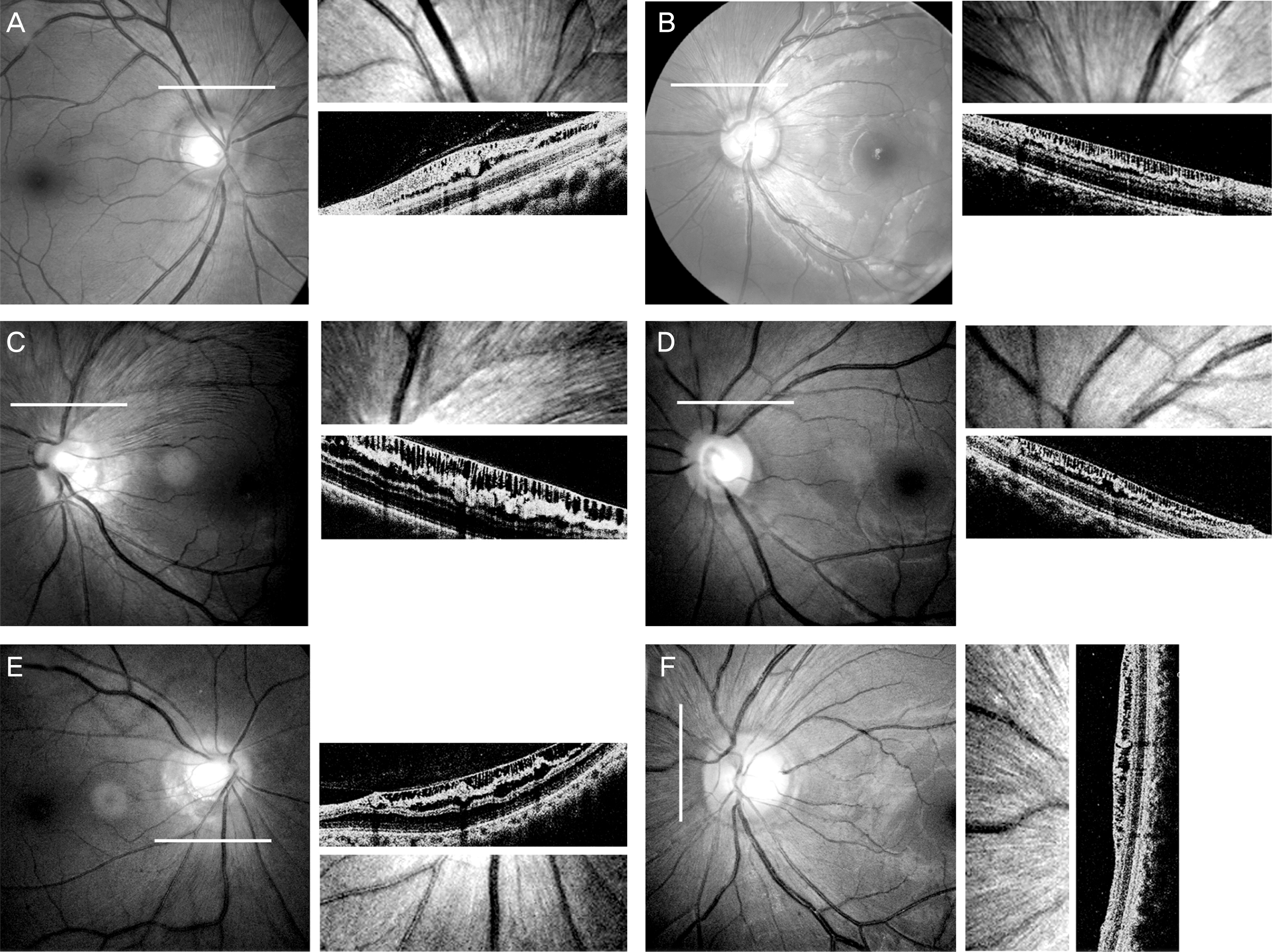 | Figure 1.Fundus photographs, magnified images and cross-sectional images of the fundus in areas with peripapillary retinoschisis (white lines) obtained by optical coherence tomography in non-glaucomatous eyes with peripapillary retinoschisis (A, case 1; B, case 2; C, case 3; D, case 4; E, case 5; F, case 6). |
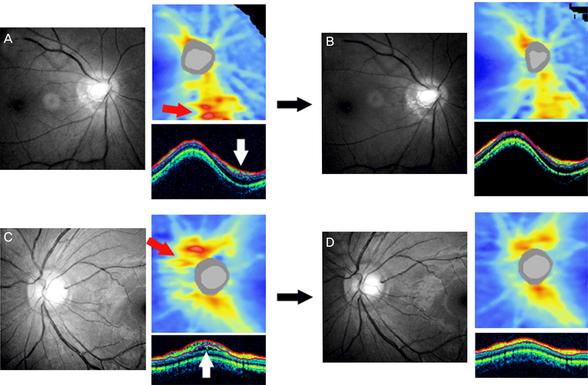 | Figure 2.Fundus photographs, optical coherence tomography (OCT) images of eyes with peripapillary retinoschisis (A & B, case 5; C & D, case 6) at the time of retinoschisis formation (A, C) and after the spontaneous decrease in retinoschisis (B, D). Retinoschisis is observed in cross-sectional circumpapillary retinal nerve fiber layer (RNFL) images obtained by OCT (white ar-rows). OCT RNFL thickness maps show transient increase in RNFL thickness (red arrows). |
Table 1.
Clinical characteristics of subjects with peripapillary retinoschisis
 XML Download