초록
Purpose:
To report a case of persistent shallow anterior chamber after silicone tube intubation, recovered by fibrin glue in glaucoma drainage device implantation (GDI).
Case summary:
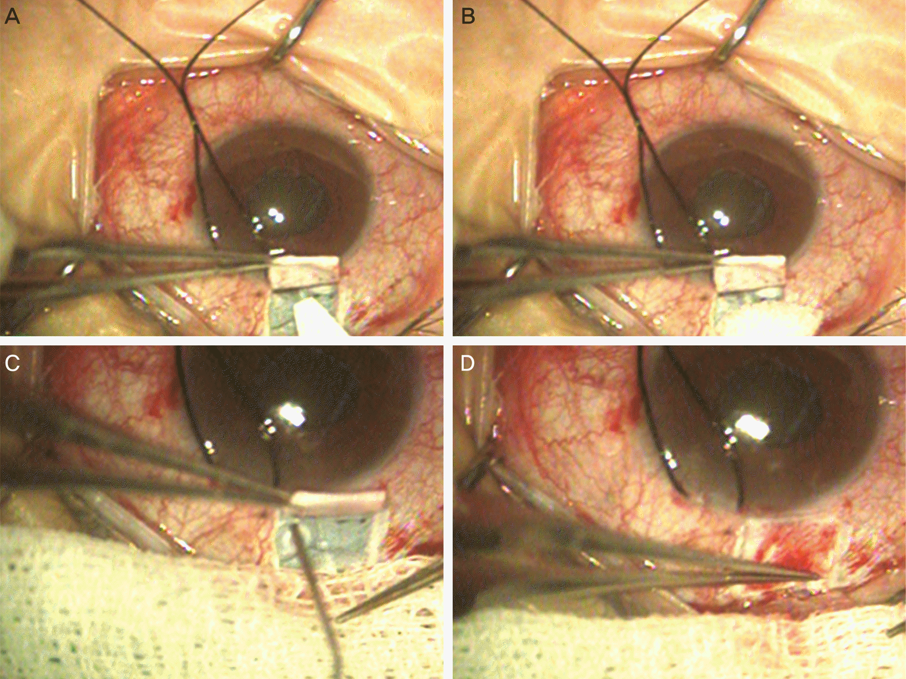
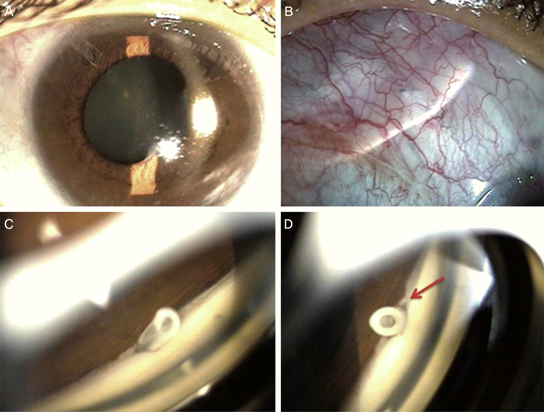
A 42-year-old female, diagnosed with neovascular glaucoma at a local clinic visited our clinic for uncontrolled intraocular pressure (IOP) in her right eye. We performed GDI on her right eye. Scleral flap and paracentesis of the anterior chamber were performed. Then, a silicone tube was inserted into the anterior chamber. Despite repetitive infusion of balanced salt solution (BSS), the anterior chamber became persistently shallow due to peritubular leakage. After dropping the fibrin glue in the peritubular space and beneath the scleral flap, attachment occurred. No additional leakage was observed near the scleral flap and after infusion of BSS, a deep anterior chamber was maintained. One day after surgery, IOP in the right eye was 3 mm Hg, deep anterior chamber was maintained, and no leakage of aqueous humor into the conjunctiva occurred. Two months after surgery, IOP was 16 mm Hg and a deep anterior chamber was maintained.
Conclusions:
In cases of persistent shallow anterior chamber after silicone tube intubation in intraoperative GDI, the best methods to maintain the anterior chamber is by suture ligation of the peritubular loosened site or infusion of viscoelastic agent to anterior chamber. In the present case, applying the fibrin glue beneath the scleral flap apparently obstructed the peritubular infiltration.
Go to : 
References
1. Kahook MY, Noecker RJ. Fibrin glue-assisted glaucoma drainage device surgery. Br J Ophthalmol. 2006; 90:1486–9.

2. Välimäki J. Fibrin glue for preventing immediate postoperative hypotony following glaucoma drainage implant surgery. Acta Ophthalmol Scand. 2006; 84:372–4.

3. Allingham RR, Damji K, Freedman SF, et al. Shields’ textbook of glaucoma. 6th ed.Wolters Kluwer: Lippincott Williams & Wilkins company;2005. p. 532.
4. Hong CH, Arosemena A, Zurakowski D, Ayyala RS. Glaucoma drainage devices: a systematic literature review and current controversies. Surv Ophthalmol. 2005; 50:48–60.

6. García-Feijoó J, Cuiña-Sardiña R, Méndez-Fernández C, et al. Peritubular filtration as cause of severe hypotony after Ahmed valve implantation for glaucoma. Am J Ophthalmol. 2001; 132:571–2.
7. Choudhari NS, Neog A, Sharma A, et al. Our experience of fibrin sealant-assisted implantation of Ahmed glaucoma valve. Indian J Ophthalmol. 2013; 61:23–7.

8. Welder JD, Pandya HK, Nassiri N, Djalilian AR. Conjunctival limbal autograft and allograft transplantation using fibrin glue. Ophthalmic Surg Lasers Imaging. 2012; 43:323–7.

9. Ganekal S, Venkataratnam S, Dorairaj S, Jhanji V. Comparative evaluation of suture-assisted and fibrin glue-assisted scleral fix-ated intraocular lens implantation. J Refract Surg. 2012; 28:249–52.

10. Nassiri N, Pandya HK, Djalilian AR. Limbal allograft transplantation using fibrin glue. Arch Ophthalmol. 2011; 129:218–22.

11. Dal Pizzol MM, Roggia MF, Kwitko S, et al. Use of fibrin glue in ocular surgery. Arq Bras Oftalmol. 2009; 72:308–12.
Go to : 
 | Figure 1.Intraoperative anterior segment photographs. (A) Surgical spear was approximated to suspicious leaking site beneath the scleral flap. (B) Surgical spear expanded due to peritubular leak. (C) Fibrin glue was applied in the peritublar space and beneath the scleral flap. (D) Scleral flap was attached to the lower sclera. |


 PDF
PDF ePub
ePub Citation
 Print
Print


 XML Download