초록
Purpose:
To report a case of decreased endothelial cell density 7 years after posterior chamber phakic intraocular lens implantation.
Case summary:
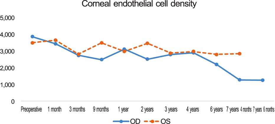
A 45-year-old man with high myopia combined with astigmatism was treated with Toric implantable Collamer Lens (ICL) implantation. The patient’s best corrected visual acuity was 0.7 in both eyes before the operation. After the treatment, his uncorrected visual acuity was 0.9 and corrected visual acuity was 1.0 in both eyes, indicating an improvement in visual function. Preoperative endothelial cell density measured 3,063 cells/mm2 in the right eye and 3,126 cells/mm2 in the left eye. At 5 years postoperatively, measurements were 2,897 cells/mm2 in the right eye and 2,974 cells/mm2 in the left, showing little change. However, a 6-year postoperative measurement of 2,198 cells/mm2 in the right eye and 2,803 cells/mm2 in the left showed a slight decrease in endothelial cell density in the right eye, and a follow-up measurement one year later displayed a rapid decline to 1,272 cells/mm2 in the right eye and 2,852 cells/mm2 in the left eye. The Toric ICL lens was removed from the right eye and phacoemulsification and posterior chamber intraocular lens implantation was performed. Two-month postoperative endothelial cell density was 1,257 cells/mm2 and endothelial cell damage from the operation itself was minimal.
Go to : 
References
1. Heitzmann J, Binder PS, Kassar BS, Nordan LT. The correction of high myopia using the excimer laser. Arch Ophthalmol. 1993; 111:1627–34.

2. Geggel HS, Talley AR. Delayed onset keratectasia following laser in situ keratomileusis. J Cataract Refract Surg. 1999; 25:582–6.

3. Holladay JT, Dudeja DR, Chang J. Functional vision and corneal changes after laser in situ keratomileusis determined by contrast sensitivity, glare testing, and corneal topography. J Cataract Refract Surg. 1999; 25:663–9.

4. Dejaco-Ruhswurm I, Scholz U, Pieh S, et al. Long-term endothelial changes in phakic eyes with posterior chamber intraocular lenses. J Cataract Refract Surg. 2002; 28:1589–93.

5. Javitt JC. Clear-lens extraction for high myopia. Is this an idea whose time has come? Arch Ophthalmol. 1994; 112:321–3.
6. Sanders DR, Doney K, Poco M; ICL in Treatment of Myopia Study Group. United States Food and Drug Administration clinical trial of the Implantable Collamer Lens (ICL) for moderate to high myopia: three-year follow-up. Ophthalmology. 2004; 111:1683–92.
7. Sarikkola AU, Sen HN, Uusitalo RJ, Laatikainen L. Traumatic cataract and other adverse events with the implantable contact lens. J Cataract Refract Surg. 2005; 31:511–24.

8. Smallman DS, Probst L, Rafuse PE. Pupillary block glaucoma secondary to posterior chamber phakic intraocular lens implantation for high myopia. J Cataract Refract Surg. 2004; 30:905–7.

9. Kodjikian L, Gain P, Donate D, et al. Malignant glaucoma induced by a phakic posterior chamber intraocular lens for myopia. J Cataract Refract Surg. 2002; 28:2217–21.

10. Bourne RR, Minassian DC, Dart JK, et al. Effect of cataract surgery on the corneal endothelium: modern phacoemulsification compared with extracapsular cataract surgery. Ophthalmology. 2004; 111:679–85.
11. Jacobs PM, Cheng H, Price NC, et al. Endothelial cell loss after cataract surgery-the problem of interpretation. Trans Ophthalmol Soc U K. 1982; 102(pt 2):291–3.
12. Han SY, Lee KH. Long term effect of ICL implantation to treat high myopia. J Korean Ophthalmol Soc. 2007; 48:465–72.
13. Chun YS, Lee JH, Lee JM, et al. Outcomes after implantable contact lens for moderate to high myopia. J Korean Ophthalmol Soc. 2004; 45:480–9.
14. Lee SY, Cheon HJ, Baek TM, Lee KH. Implantable contact lens to correct high myopia (clinical study with 24 months follow-up). J Korean Ophthalmol Soc. 2000; 41:1515–22.
15. Pesando PM, Ghiringhello MP, Di Meglio G, Fanton G. Posterior chamber phakic intraocular lens (ICL) for hyperopia: ten-year fol-low-up. J Cataract Refract Surg. 2007; 33:1579–84.

16. Sanders DR, Doney K, Poco M; ICL in Treatment of Myopia Study Group. United States Food and Drug Administration clinical trial of the Implantable Collamer Lens (ICL) for moderate to high myopia: three-year follow-up. Ophthalmology. 2004; 111:1683–92.
17. Edelhauser HF, Sanders DR, Azar R, Lamielle H; ICL in Treatment of Myopia Study Group. Corneal endothelial assessment after ICL implantation. J Cataract Refract Surg. 2004; 30:576–83.

18. Fyodorov SN, Zuev VK, Tumanyan ER, Larionov Y. Analysis of long term clinical and functional results of intraocular correction of high myopia. Ophthalmosurg (Moscow). 1990; 2:3–6.
19. Fyodorov SN, Zuyev VK, Aznabayev BM. Intraocular correction of high myopia with negative posterior chamber lens. Ophthalmosurg. 1991; 3:57–8.
20. Jiménez-Alfaro I, Gómez-Tellería G, Bueno JL, Puy P. Contrast sensitivity after posterior chamber phakic intraocular lens implantation for high myopia. J Refract Surg. 2001; 17:641–5.

21. Choi WS, Lee HY, Seo SG, Her J. Clinical outcomes of implantable contact lens and iris-fixed intraocular lens for correction of myopia. J Korean Ophthalmol Soc. 2008; 49:1406–14.

22. Han SY, Lee KH. Long term effect of ICL implantation to treat high myopia. J Korean Ophthalmol Soc. 2007; 48:465–72.
23. Igarashi A, Shimizu K, Kamiya K. Eight-year follow-up of posterior chamber phakic intraocular lens implantation for moderate to high myopia. Am J Ophthalmol. 2014; 157:532–9.e1.

24. Jiménez-Alfaro I, Benítez del Castillo JM, García-Feijoó J, et al. Safety of posterior chamber phakic intraocular lenses for the correction of high myopia: anterior segment changes after posterior chamber phakic intraocular lens implantation. Ophthalmology. 2001; 108:90–9.
Go to : 
 | Figure 1.Corneal endothelial cell density of the right eye. (A) Before ICL implantation operation. (B) Seven years after ICL implantation operation. R = right; T = thickness; N = number; SD = standard deviation; CV = coefficient of variation; CD = cell density; ICL = implantable contact lens. |
 | Figure 2.Changes in corneal endothelial cell density. OD = oculus dexter (the right eye); OS = oculus sinister (the left eye). |
Table 1.
Changes in corneal endothelial cell density (CD, cells/mm )


 PDF
PDF ePub
ePub Citation
 Print
Print


 XML Download