초록
Purpose:
To investigate the clinical significance of the internal lens signal measured using dual Scheimpflug anterior segment analyzer (Galilei TM, Ziemer, Switzerland) in patients receiving cataract surgery.
Methods:
The present study included 151 eyes of 148 patients who received surgery for senile cataracts from February 2012 to January 2013. Preoperative internal lens signals were measured preoperatively. The depth of anterior chamber and anterior angles were measured using dual Scheimpflug anterior segment analyzer preoperatively and 1 month postoperatively. Preoperative and postoperative best-corrected visual acuities (BCVAs) were measured. The relationships between preoperative internal lens signal and the changes in BCVA or anterior angles were evaluated.
Results:
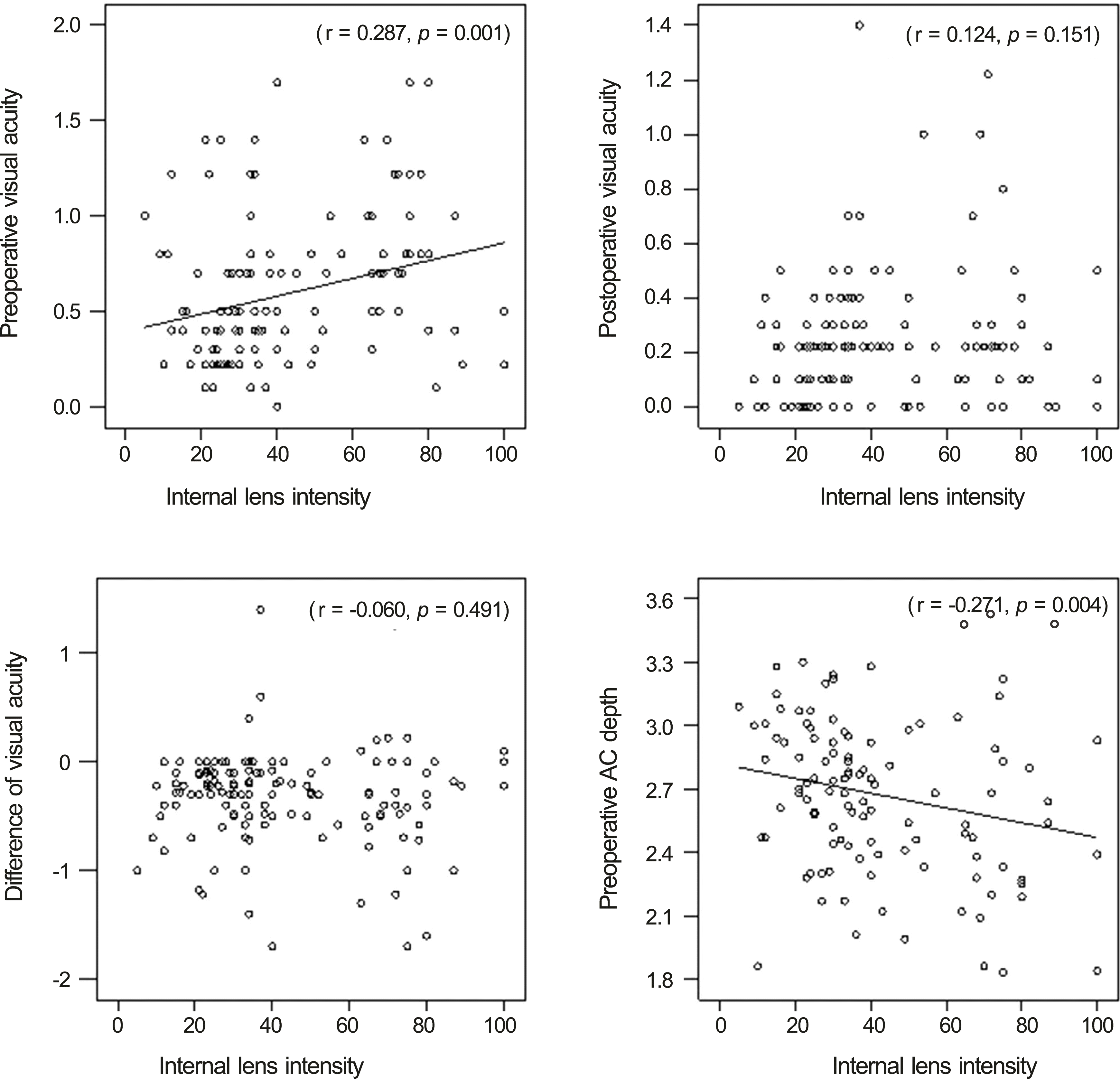
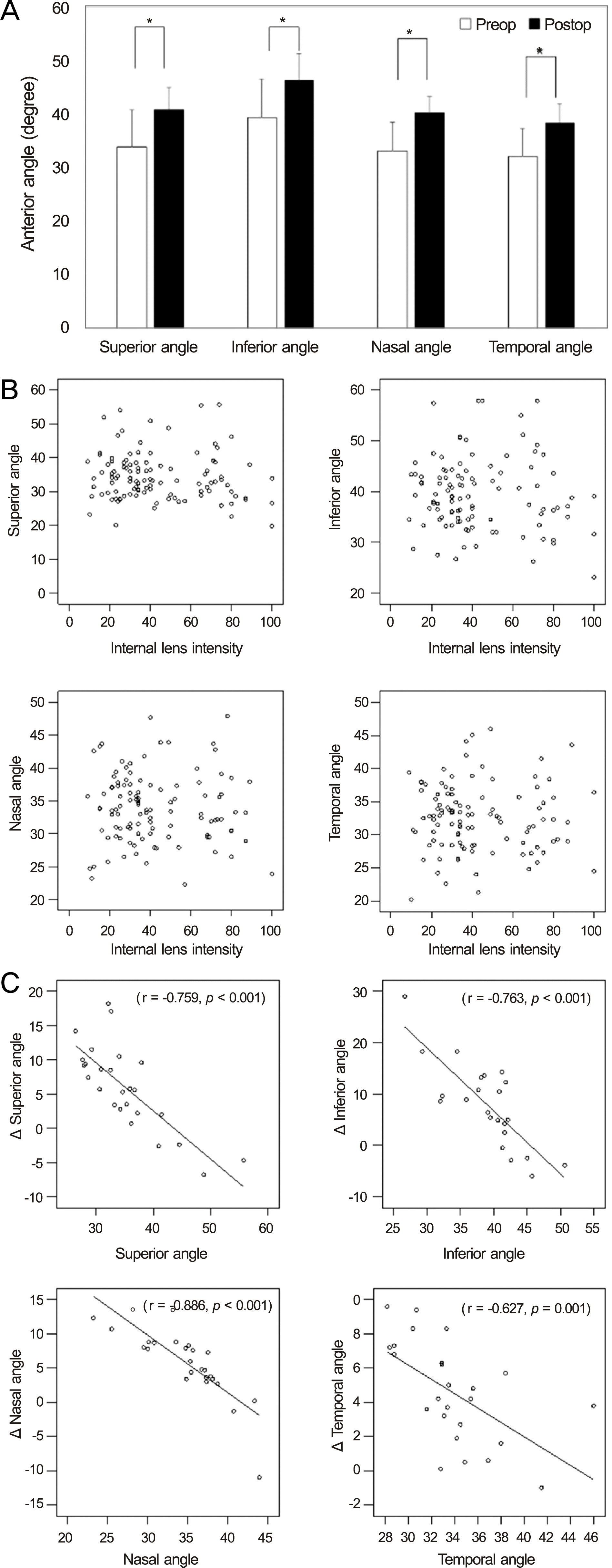
Internal lens signal and preoperative BCVA (log MAR) or preoperative anterior chamber depth were highly correlated (r = 0.287, p = 0.001 and r = -0.271, p = 0.004, respectively). Anterior angles increased 1 month after surgery compared with the preoperative values ( p < 0.001). The amount of change between preoperative and postoperative anterior angles correlated with preoperative anterior angles ( p < 0.001). However, no statistically significant correlation was observed between internal lens signal and preoperative anterior angles or postoperative BCVA. Internal lens signal correlated with changes in postoperative anterior angles ( p < 0.001).
Conclusions:
Internal lens signal correlated with preoperative visual acuity and may help evaluate the cataract severity quantita-tively and objectively. Internal lens signal may aid in understanding the structure of anterior segments by predicting the lens volume. Knowing the effect of visual impairment due to cataracts and predicting visual improvement after cataract surgery is necessary.
Go to : 
References
1. Foster PJ, Wong TY, Machin D, et al. Risk factors for nuclear, cortical and posterior subcapsular cataracts in the Chinese population of Singapore: the Tanjong Pagar Survey. Br J Ophthalmol. 2003; 87:1112–20.

2. Brown NA, Bron AJ, Ayliffe W, et al. The objective assessment of cataract. Eye (Lond). 1987; 1(Pt 2):234–46.

3. Mehra V, Minassian DC. A rapid method of grading cataract in epi-demiological studies and eye surveys. Br J Ophthalmol. 1988; 72:801–3.

4. Chylack LT Jr, Wolfe JK, Singer DM, et al. The Lens Opacities Classification System III. The Longitudinal Study of Cataract Study Group. Arch Ophthalmol. 1993; 111:831–6.
5. Klein BE, Klein R, Linton KL, et al. Assessment of cataracts from photographs in the Beaver Dam Eye Study. Ophthalmology. 1990; 97:1428–33.

6. Sparrow JM, Bron AJ, Brown NA, et al. The Oxford Clinical Cataract Classification and Grading System. Int Ophthalmol. 1986; 9:207–25.

7. Hockwin O, Dragomirescu V, Laser H. Measurements of lens transparency or its disturbances by densitometric image analysis of Scheimpflug photographs. Graefes Arch Clin Exp Ophthalmol. 1982; 219:255–62.

8. Hall NF, Lempert P, Shier RP, et al. Grading nuclear cataract: reproducibility and validity of a new method. Br J Ophthalmol. 1999; 83:1159–63.

9. Sasaki K, Shibata T, Obazawa H, et al. Classification system for cataracts. Application by the Japanese Cooperative Cataract Epidemiology Study Group. Ophthalmic Res. 1990; 22(Suppl 1):46–50.
10. Foo KP, Maclean H. Measured changes in cataract over six months: sensitivity of the Nidek EAS-1000. Ophthalmic Res. 1996; 28(Suppl 2):32–6.
11. West SK, Taylor HR. The detection and grading of cataract: an epi-demiologic perspective. Surv Ophthalmol. 1986; 31:175–84.

12. McCarty CA, Keeffe JE, Taylor HR. The need for cataract surgery: projections based on lens opacity, visual acuity, and personal concern. Br J Ophthalmol. 1999; 83:62–5.

13. Rouhiainen P, Rouhiainen H, Notkola IL, Salonen JT. Comparison of the lens opacities classification system II and Lensmeter 701. Am J Ophthalmol. 1993; 116:617–21.

14. Duncan DD, Shukla OB, West SK, Schein OD. New objective classification system for nuclear opacification. J Opt Soc Am A Opt Image Sci Vis. 1997; 14:1197–204.

15. Tan AC, Loon SC, Choi H, Thean L. Lens Opacities Classification System III: cataract grading variability between junior and senior staff at a Singapore hospital. J Cataract Refract Surg. 2008; 34:1948–52.

16. Pei X, Bao Y, Chen Y, Li X. Correlation of lens density measured using the Pentacam Scheimpflug system with the Lens Opacities Classification System III grading score and visual acuity in age-related nuclear cataract. Br J Ophthalmol. 2008; 92:1471–5.

17. Cheung CY, Li H, Lamoureux EL, et al. Validity of a new com-puter-aided diagnosis imaging program to quantify nuclear cataract from slit-lamp photographs. Invest Ophthalmol Vis Sci. 2011; 52:1314–9.

18. Kanellopoulos AJ, Asimellis G. Clear-cornea cataract surgery: pupil size and shape changes, along with anterior chamber volume and depth changes. A Scheimpflug imaging study. Clin Ophthalmol. 2014; 8:2141–50.

19. Jonas JB, Nangia V, Gupta R, et al. Lens thickness and associated factors. Clin Experiment Ophthalmol. 2012; 40:583–90.

20. Pan AP, Wang QM, Huang F, et al. Correlation among lens opacities classification system III grading, visual function index-14, pentacam nucleus staging, and objective scatter index for cataract assessment. Am J Ophthalmol. 2015; 159:241–7.e2.

22. Mehra V, Minassian DC. A rapid method of grading cataract in epi-demiological studies and eye surveys. Br J Ophthalmol. 1988; 72:801–3.

23. Sasaki H, Kawakami Y, Ono M, et al. Localization of cortical cataract in subjects of diverse races and latitude. Invest Ophthalmol Vis Sci. 2003; 44:4210–4.

24. Datiles MB 3rd, Magno BV, Freidlin V. Study of nuclear cataract progression using the National Eye Institute Scheimpflug system. Br J Ophthalmol. 1995; 79:527–34.

25. Mitchell P, Cumming RG, Attebo K, Panchapakesan J. Prevalence of cataract in Australia: the Blue Mountains eye study. Ophthalmology. 1997; 104:581–8.
26. Maraini G, Pasquini P, Sperduto RD, et al. The effect of cataract severity and morphology on the reliability of the Lens Opacities Classification System II (LOCS II). Invest Ophthalmol Vis Sci. 1991; 32:2400–3.
27. Kirwan JF, Venter L, Stulting AA, Murdoch IE. LOCS III examination at the slit lamp, do settings matter? Ophthalmic Epidemiol. 2003; 10:259–66.

28. Savini G, Barboni P, Carbonelli M, Hoffer KJ. Repeatability of au-tomatic measurements by a new Scheimpflug camera combined with Placido topography. J Cataract Refract Surg. 2011; 37:1809–16.

29. Magalhães FP, Costa EF, Cariello AJ, et al. Comparative analysis of the nuclear lens opalescence by the Lens Opacities Classification System III with nuclear density values provided by Oculus Pentacam: a cross-section study using Pentacam Nucleus Staging software. Arq Bras Oftalmol. 2011; 74:110–3.

30. Arai M, Ohzuno I, Zako M. Anterior chamber depth after posterior chamber intraocular lens implantation. Acta Ophthalmol (Copenh). 1994; 72:694–7.

31. Yoshida S, Hashiba H, Tsukuda M, Ohara Y. Significance of angle of intraocular lens haptics on anterior chamber depth. Jpn J Clin Ophthalmol. 1989; 43:173–6.
Go to : 
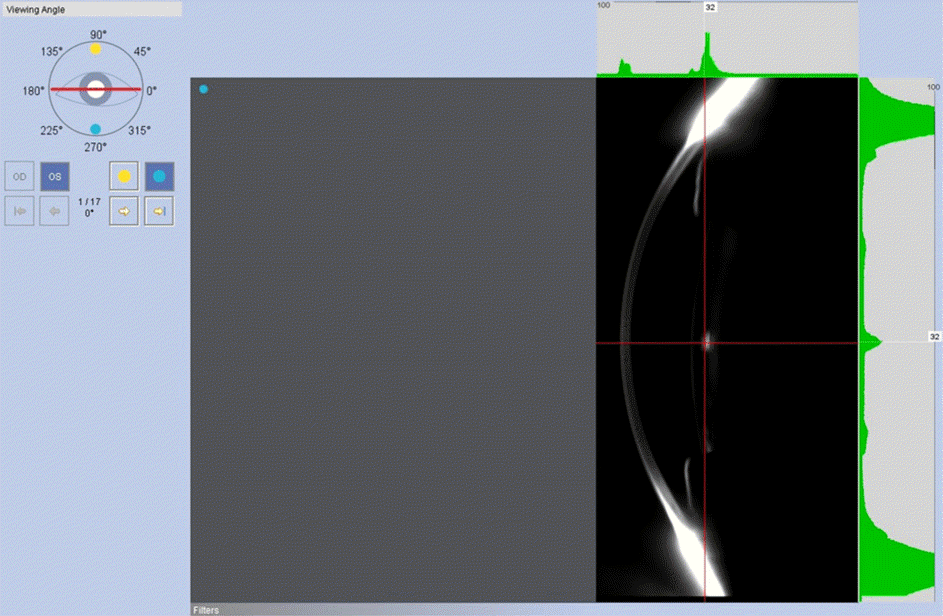
 | Figure 1.Internal intensity of the lens using dual Scheimpflug camera. Internal intensity was decided as the value at the high-est intensity. |
 | Figure 2.Internal lens signal and preoperative visual acuity, postoperative visual acuity, difference between preoperative visual acuity and postoperative visual acuity, or anterior chamber depth. Visual acuity was expressed as log MAR. AC = anterior chamber. |
 | Figure 3.(A) Difference between preoperative and postoperative anterior chamber angle. (B) The relationship between preoperative anterior chamber angle and internal lens intensity. (C) The relationship between anterior chamber angles and the difference of anterior chamber angles. Preop = preoperative; Postop = postoperative. * Statistically significant by Wilcoxon rank test. |
Table 1.
Preoperative and postoperative data
| Preoperative | One month postoperative | |
|---|---|---|
| Internal lens intensity | 42.7 ± 23.0 | |
| Corrective visual acuity (log MAR) | 0.58 ± 0.37 | 0.23 ± 0.24 |
| Anterior angle (°) | ||
| Superior | 34.0 ± 6.9 | 40.9 ± 4.3* |
| Inferior | 39.5 ± 7.2 | 46.4 ± 5.0* |
| Nasal | 33.3 ± 5.3 | 40.3 ± 3.1* |
| Temporal | 32.3 ± 5.2 | 38.4 ± 3.6* |
| Intraocular pressure (mm Hg) | 15.3 ± 3.0 | 14.2 ± 3.3 |
| Anterior chamber depth (mm) | 2.64 ± 0.37 | 3.29 ± 0.31* |


 PDF
PDF ePub
ePub Citation
 Print
Print


 XML Download