

 PDF
PDF ePub
ePub Citation
 Print
Print


Abstract
Purpose
To investigate the effects of a delay in finger temperature recovery rate on the hand cold provocation test (HCPT) and a nocturnal dip greater than 10% (dipper) on the progression of glaucomatous visual field (VF) defects in open-angle glaucoma patients when the intraocular pressure (IOP) was well controlled lower than the target pressure.
Methods
30 patients (58 eyes) with normal tension glaucoma (NTG) and 13 patients (24 eyes) with primary open angle glauco-ma, and 12 normal controls (24 eyes) were retrospectively enrolled in this study. We performed HCPT, 24 hour ambulatory blood pressure monitoring (24-hr ABPM), Goldmann applanation tonometer measurements, and VF tests on all subjects. The delay in finger temperature recovery rate was defined as a delay longer than 15% of the mean finger temperature of normal controls over 2 intervals among 5, 10, 15, and 20 minutes after the immersion of cold water. We examined the relationships among the delay in finger temperature recovery rate, dipper, and the progression of glaucomatous VF defects.
Results
The finger temperature recovery rate in NTG patients was significantly delayed more than that of normal controls at 5, 10, and 15 minutes after the immersion. The delay in finger temperature recovery rate significantly correlated with dipper in NTG patients. Glaucomatous VF defects were significantly progressed in the presence of dipper in NTG patients. Delay in finger tem-perature recovery rate was significantly related to the progression of glaucomatous VF defects in NTG patients. In the binary logistic regression test, delay in finger temperature recovery rate was the only factor that was strongly related to the progression of glaucomatous visual field in NTG patients.
Go to : 
References
1. Kass MA, Heuer DK, Higginbotham EJ. . The Ocular Hypertension Treatment Study: a randomized trial determines that topical ocular hypotensive medication delays or prevents the onset of primary open-angle glaucoma. Arch Ophthalmol. 2002; 120:701–13.
2. Heijl A, Leske MC, Bengtsson B. . Reduction of intraocular pressure and glaucoma progression: results from the Early Manifest Glaucoma Trial. Arch Ophthalmol. 2002; 120:1268–79.
3. Collaborative Normal-Tension Glaucoma Study Group. Comparison of glaucomatous progression between untreated patients with nor-mal-tension glaucoma and patients with therapeutically reduced intraocular pressures. Am J Ophthalmol. 1998; 126:487–97.
4. Levene RZ. Low tension glaucoma: a critical review and new material. Surv Ophthalmol. 1980; 24:621–64.

5. Leske MC, Heijl A, Hussein M. . Factors for glaucoma pro-gression and the effect of treatment: the early manifest glaucoma trial. Arch Ophthalmol. 2003; 121:48–56.
6. The Advanced Glaucoma Intervention Study (AGIS). 7. The relationship between control of intraocular pressure and visual field deterioration. The AGIS Investigators. Am J Ophthalmol. 2000; 130:429–40.
7. Becker B. Diabetes mellitus and primary open-angle glaucoma. The XXVII Edward Jackson Memorial Lecture. Am J Ophthalmol. 1971; 71(1 Pt 1):1–16.
8. McLeod SD, West SK, Quigley HA, Fozard JL. A longitudinal study of the relationship between intraocular and blood pressures. Invest Ophthalmol Vis Sci. 1990; 31:2361–6.
9. Bonomi L, Marchini G, Marraffa M. . Vascular risk factors for primary open angle glaucoma: the Egna-Neumarkt Study. Ophthalmology. 2000; 107:1287–93.
10. Pradalier A, Hamard P, Sellem E, Bringer L. Migraine and glauco-ma: an epidemiologic survey of French ophthalmologists. Cephalalgia. 1998; 18:74–6.
11. Broadway DC, Drance SM. Glaucoma and vasospasm. Br J Ophthalmol. 1998; 82:862–70.
12. Leske MC. Ocular perfusion pressure and glaucoma: clinical trial and epidemiologic findings. Curr Opin Ophthalmol. 2009; 20:73–8.
13. Hayreh SS. Progress in the understanding of the vascular etiology of glaucoma. Curr Opin Ophthalmol. 1994; 5:26–35.
14. Araie M, Sekine M, Suzuki Y, Koseki N. Factors contributing to the progression of visual field damage in eyes with normal-tension glaucoma. Ophthalmology. 1994; 101:1440–4.
15. Ishida K, Yamamoto T, Sugiyama K, Kitazawa Y. Disk hemorrhage is a significantly negative prognostic factor in normal-tension glaucoma. Am J Ophthalmol. 2000; 129:707–14.
16. Gherghel D, Orgül S, Gugleta K, Flammer J. Retrobulbar blood flow in glaucoma patients with nocturnal over-dipping in systemic blood pressure. Am J Ophthalmol. 2001; 132:641–7.
17. Gherghel D, Orgül S, Gugleta K. . Relationship between ocular perfusion pressure and retrobulbar blood flow in patients with glaucoma with progressive damage. Am J Ophthalmol. 2000; 130:597–605.
18. Osusky R, Rohr P, Schötzau A, Flammer J. Nocturnal dip in the optic nerve head perfusion. Jpn J Ophthalmol. 2000; 44:128–31.
19. Emre M, Orgül S, Gugleta K, Flammer J. Ocular blood flow alter-ation in glaucoma is related to systemic vascular dysregulation. Br J Ophthalmol. 2004; 88:662–6.
20. Choi J, Jeong J, Cho HS, Kook MS. Effect of nocturnal blood pressure reduction on circadian fluctuation of mean ocular perfusion pressure: a risk factor for normal tension glaucoma. Invest Ophthalmol Vis Sci. 2006; 47:831–6.
21. Choi J, Kim KH, Jeong J. . Circadian fluctuation of mean ocular perfusion pressure is a consistent risk factor for normal-tension glaucoma. Invest Ophthalmol Vis Sci. 2007; 48:104–11.
22. Flammer J, Konieczka K, Flammer AJ. The primary vascular dys-regulation syndrome: implications for eye diseases. EPMA J. 2013; 4:14.
23. SARNOFF SJ, HARDENBERGH E, WHITTENBERGER JL. Mechanism of the arterial pressure response to the Valsalva test; the basis for its use as an indicator of the intactness of the sym-pathetic outflow. Am J Physiol. 1948; 154:316–27.
24. Goldberger JJ. Sympathovagal balance: how should we measure it? Am J Physiol. 1999; 276(4 Pt 2):H1273–80.
25. Jaradeh SS, Prieto TE. Evaluation of the autonomic nervous system. Phys Med Rehabil Clin N Am. 2003; 14:287–305.
26. Nicolela MT, Ferrier SN, Morrison CA. . Effects of cold-in-duced vasospasm in glaucoma: the role of endothelin-1. Invest Ophthalmol Vis Sci. 2003; 44:2565–72.
27. Collaborative Normal-Tension Glaucoma Study Group. The effectiveness of intraocular pressure reduction in the treatment of nor-mal-tension glaucoma. Am J Ophthalmol. 1998; 126:498–505.
28. Advanced Glaucoma Intervention Study. 2. Visual field test scoring and reliability. Ophthalmology. 1994; 101:1445–55.
29. Plange N, Kaup M, Daneljan L. . 24-h blood pressure monitoring in normal tension glaucoma: night-time blood pressure variability. J Hum Hypertens. 2006; 20:137–42.
30. Graham SL, Drance SM, Wijsman K. . Ambulatory blood pressure monitoring in glaucoma. The nocturnal dip. Ophthalmology. 1995; 102:61–9.
31. Collignon N, Dewe W, Guillaume S, Collignon-Brach J. Ambulatory blood pressure monitoring in glaucoma patients. The nocturnal systolic dip and its relationship with disease progression. Int Ophthalmol. 1998; 22:19–25.
32. Seo HR, Ryu WY, Rho SH. Correlation between nocturnal dip and progression of glaucoma. J Korean Ophthalmol Soc. 2010; 51:1471–8.
33. Riva CE, Grunwald JE, Petrig BL. Autoregulation of human retinal blood flow. An investigation with laser Doppler velocimetry. Invest Ophthalmol Vis Sci. 1986; 27:1706–12.
34. Hayreh SS. The blood supply of the optic nerve head and the evaluation of it - myth and reality. Prog Retin Eye Res. 2001; 20:563–93.
35. Harris A, Rechtman E, Siesky B. . The role of optic nerve blood flow in the pathogenesis of glaucoma. Ophthalmol Clin North Am. 2005; 18:345–53.
36. Riva CE, Loebl M. Autoregulation of blood flow in the capillaries of the human macula. Invest Ophthalmol Vis Sci. 1977; 16:568–71.
37. Riva CE, Geiser M, Petrig BL; Beijing 100193, PR China Ocular Blood Flow Research Association. Ocular blood flow assessment using continuous laser Doppler flowmetry. Acta Ophthalmol. 2010; 88:622–9.
38. Pournaras CJ, Riva CE. Retinal blood flow evaluation. Ophthal- mologica. 2013; 229:61–74.
39. Hayreh SS, Zimmerman MB, Podhajsky P, Alward WL. Nocturnal arterial hypotension and its role in optic nerve head and ocular ischemic disorders. Am J Ophthalmol. 1994; 117:603–24.
40. Kaiser HJ, Flammer J. Systemic hypotension: a risk factor for glaucomatous damage? Ophthalmologica. 1991; 203:105–8.
41. Kaiser HJ, Flammer J, Graf T, Stümpfig D. Systemic blood pressure in glaucoma patients. Graefe's Arch Cli Exp Ophthalmol. 1993; 231:677–80.
42. Leske MC, Wu SY, Nemesure B, Hennis A. Incident open-angle glaucoma and blood pressure. Arch Ophthalmol. 2002; 120:954–9.
43. Gherghel D, Hosking SL, Orgül S. Autonomic nervous system, circadian rhythms, and primary openangle glaucoma. Surv Ophthalmol. 2004; 49:491–508.
44. Riccadonna M, Covi G, Pancera P. . Autonomic system activity and 24-hour blood pressure variations in subjects with normal- and hightension glaucoma. J Glaucoma. 2003; 12:156–63.
45. Kashiwagi K, Tsumura T, Ishii H. . Circadian rhythm of autonomic nervous function in patients with normaltension glaucoma compared with normal subjects using ambulatory electrocardiography. J Glaucoma. 2000; 9:239–46.
46. Flammer J, Mozaffarieh M. Autoregulation, a balancing act between supply and demand. Can J Ophthalmol. 2008; 43:317–21.
47. Alm A, Bill A. Ocular and optic nerve blood flow at normal and increased intraocular pressures in monkeys (Macaca irus): a study with radioactively labelled microspheres including flow determinations in brain and some other tissues. Exp Eye Res. 1973; 15:15–29.
48. Schmidl D, Boltz A, Kaya S. . Comparison of choroidal and optic nerve head blood flow regulation during changes in ocular perfusion pressure. Invest Ophthalmol Vis Sci. 2012; 53:4337–46.
49. Grieshaber MC, Mozaffarieh M, Flammer J. What is the link between vascular dysregulation and glaucoma? Surv Ophthalmol. 2007; 52(Suppl 2):S144–54.
50. Na KS, Lee NY, Park SH, Park CK. Autonomic dysfunction in normal tension glaucoma: the shortterm heart rate variability analysis. J Glaucoma. 2010; 19:377–81.
51. Park HY, Jung KI, Na KS. . Visual field characteristics in nor-mal-tension glaucoma patients with autonomic dysfunction and abnormal peripheral microcirculation. Am J Ophthalmol. 2012; 154:466–75.e1.
52. Kóthy P, Süveges I, Vargha P, Holló G. Cold pressor test and retinal capillary perfusion in vasospastic subjects with and without capsular glaucoma (a preliminary study). Acta Physiol Hung. 1999; 86:245–52.
Go to :
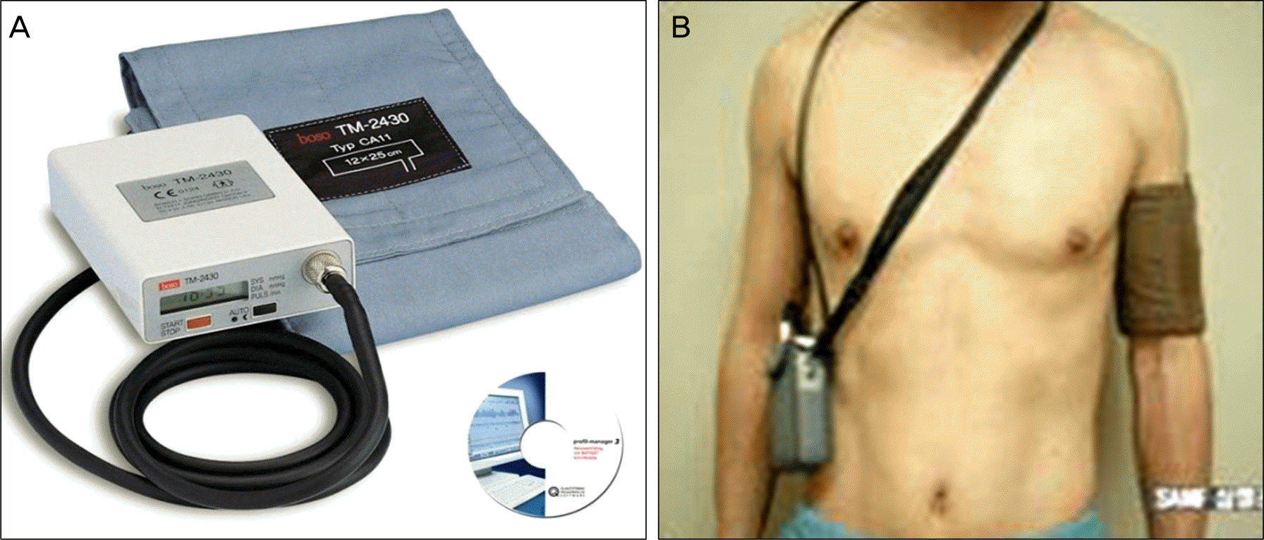
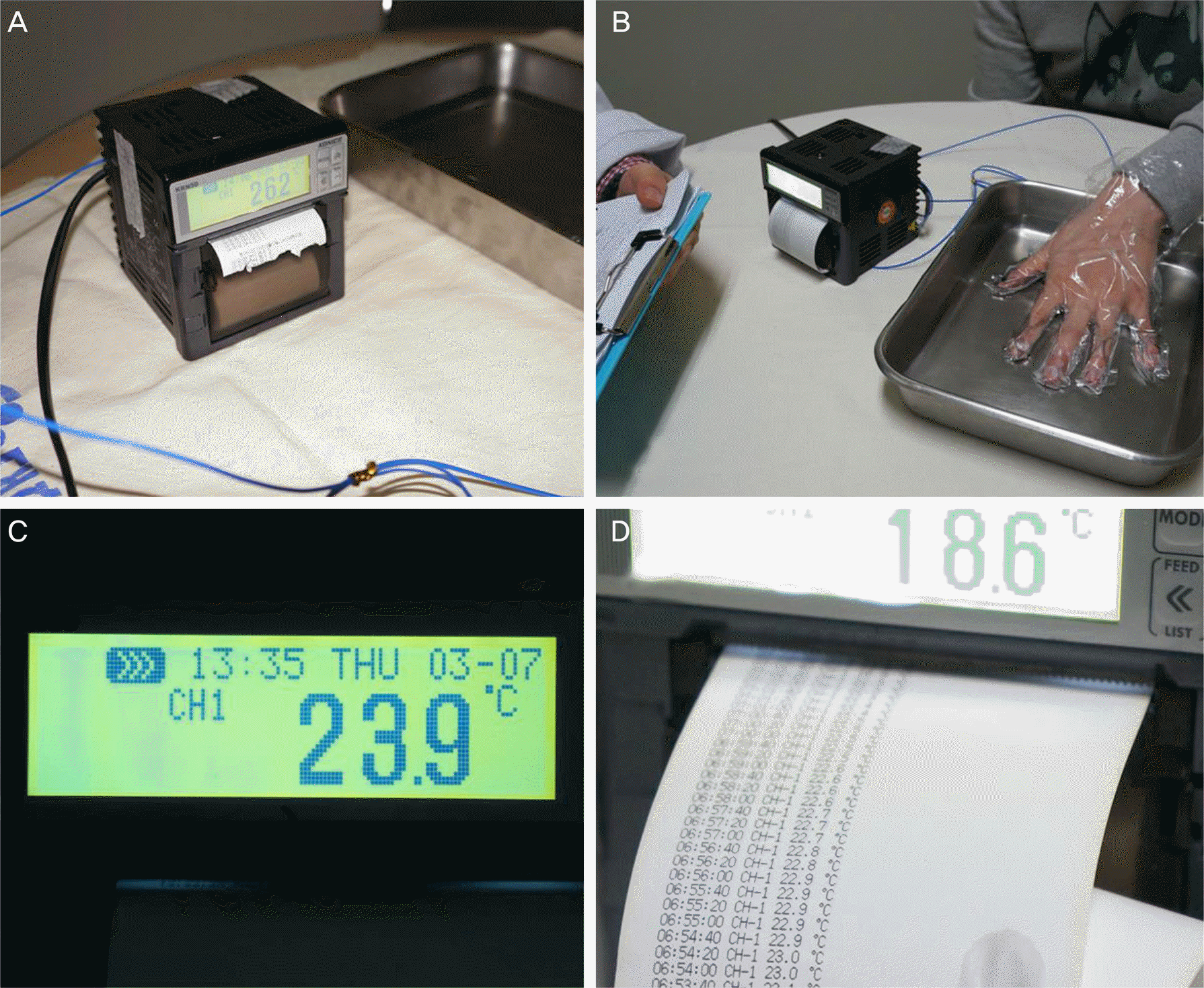 | Figure 2.Hand Cold Provocation Tester (Rose HCPT™). (A) Rose HCPT™, (B) hand cold provocation test performed, (C) information about date, time and temperature is displayed on screen, (D) temperature date printed every 20 second (preset time). |
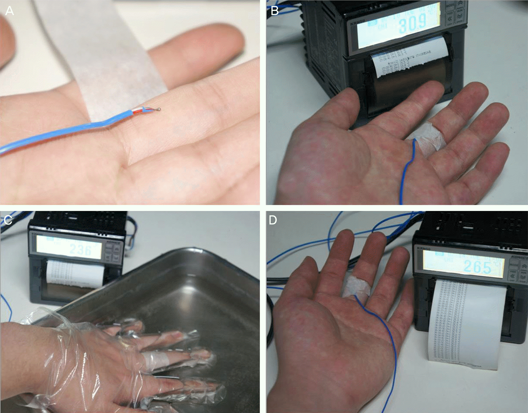 | Figure 4.The measurement process of hand cold provocation test. (A) Sensor of finger thermometer placed at proximal portion of middle finger, (B) baseline finger temperature is measured for about 5 minutes, (C) immersion process for 5 minutes in 10℃ cold water, (D) after immersion process, finger temperature recovery for 20 minutes. |
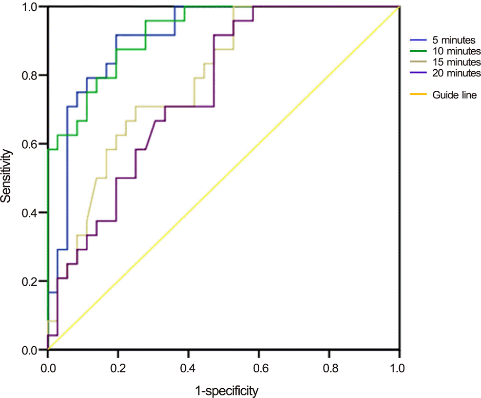 | Figure 5.ROC curve of finger temperature recovery rate in normal controls. Cut off value of delayed recovery rate of finger temperature: over 15%, sensitivity: 0.667, AUC: 0.732. ROC = receiver operating characteristic; AUC = area under the ROC curve. |
Table 1.
Demographics of patients in NTG & POAG
Table 2.
Result of hand cold provocation test in all subjects
|
Baseline (℃) |
Immersion (℃) |
Recovery (℃) |
|||||
|---|---|---|---|---|---|---|---|
| Ear | Hand | 5 min | 10 min | 15 min | 20 min | ||
| NTG | 36.24 ± 0.43 | 34.52 ± 2.16 | 19.37 ± 4.10 | 26.24 ± 5.33 | 30.51 ± 5.32 | 32.14 ± 5.08 | 33.24 ± 4.22 |
| (45.55 ± 27.11%) | (71.99 ± 31.34%) | (82.58 ± 29.02%) | (88.78 ± 23.43%) | ||||
| (p < 0.000)∗ | (p < 0.000)∗ | (p = 0.013)∗ | (p = 0.057)∗ | ||||
| POAG | 36.18 ± 0.44 | 34.63 ± 2.19 | 20.42 ± 4.10 | 27.78 ± 4.87 | 32.02 ± 3.94 | 33.33 ± 3.20 | 34.44 ± 2.29 |
| (56.59 ± 27.65%) | (79.14 ± 26.28%) | (91.63 ± 26.06%) | (93.91 ± 14.63%) | ||||
| (p = 0.285)∗ | (p = 0.552)∗ | (p = 0.968)∗ | (p = 0.812)∗ | ||||
| Control | 35.90 ± 0.67 | 35.83 ± 0.52 | 20.99 ± 4.98 | 31.87 ± 2.75 | 35.03 ± 0.73 | 35.49 ± 0.73 | 35.89 ± 0.41 |
| (75.18%) | (94.25%) | (97.28%) | (99.83%) | ||||
Table 3.
Relationship of HCPT and nocturnal dip in NTG & POAG
|
Dipper |
Chi-square test |
||||
|---|---|---|---|---|---|
| Yes | No | p-value∗ | Odds ratio | CI† | |
| NTG | |||||
| Abnormal HCPT | 30 | 9 | 0.020 | 7.000 | 2.070-32.748 |
| Normal HCPT | 5 | 14 | |||
| POAG | |||||
| Abnormal HCPT | 7 | 2 | 0.063 | 2.571 | 1.293-37.909 |
| Normal HCPT | 4 | 11 | |||
Table 4.
Relationship of nocturnal dip and VF progression in NTG & POAG
|
VF progression |
Chi-square test |
||||
|---|---|---|---|---|---|
| Yes | No | p-value∗ | Odds ratio | CI† | |
| NTG | |||||
| Dipper | 23 | 12 | 0.028 | 6.000 | 1.172-30.725 |
| Non-dipper | 5 | 18 | |||
| POAG | |||||
| Dipper | 5 | 6 | 0.805 | 0.750 | 0.084-6.710 |
| Non-dipper | 8 | 5 | |||
Table 5.
Relationship of HCPT and VF progression in NTG & POAG
|
VF progression |
Chi-square test |
||||
|---|---|---|---|---|---|
| Yes | No | p-value∗ | Odds ratio | CI† | |
| NTG | |||||
| Abnormal HCPT | 25 | 14 | 0.022 | 7.429 | 1.226-45.005 |
| Normal HCPT | 3 | 16 | |||
| POAG | |||||
| Abnormal HCPT | 7 | 2 | 0.151 | 6.667 | 0.487-91.331 |
| Normal HCPT | 6 | 9 | |||
Table 6.
Risk factors associated with glaucomatous VF progression in NTG & POAG
| p-value∗ | Odds ratio | CI† | |
|---|---|---|---|
| NTG | |||
| Sex | 0.395 | 0.527 | 0.012-2.304 |
| Age over 60 years | 0.428 | 0.544 | 0.121-2.450 |
| Dipper | 0.135 | 3.237 | 0.694-15.101 |
| Abnormal HCPT | 0.002 | 11.845 | 2.428-57.774 |
| POAG | |||
| Sex | 0.871 | 1.226 | 0.105-14.369 |
| Age over 60 years | 0.414 | 2.649 | 0.255-27.486 |
| Dipper | 0.563 | 2.435 | 0.120-49.617 |
| Abnormal HCPT | 0.632 | 0.486 | 0.025-9.312 |
 XML Download