

 PDF
PDF ePub
ePub Citation
 Print
Print


초록
Purpose:
To compare measurements of central corneal thickness (CCT) and anterior chamber depth (ACD) obtained using Galilei™, Pentacam® (Oculus, Wetzlar, Germany) and Lenstar® (Haag-Streit, Koeniz, Switzerland) and analyze the measurement agreements.
Methods:
CCT and ACD were measured using Galilei™, Pentacam® and Lenstar® in 47 eyes of 25 healthy subjects. The measurements were compared among the 3 devices.
Results:
The average CCT measurements using Galilei™, Pentacam® and Lenstar® were 552.6 ± 29.41 μ m, 543.9 ± 30.50 μ m and 537.5 ± 30.26 μ m, respectively. The measurements significantly correlated with each other (r > 0.9, p < 0.001), but were statistically significantly different ( p < 0.001). The average ACD measurements using Galilei™, Pentacam® and Lenstar® were 3.23 ± 0.360 mm, 3.22 ± 0.403 mm and 3.19 ± 0.367 mm, respectively. The measurements significantly correlated with each other (r > 0.9, p < 0.001), but were statistically significantly different ( p = 0.034). The CCT 95% limits of agreement (LoA) between Galilei™ and Pentacam®, Pentacam® and Lenstar® and Lenstar® and Galilei™ were 31.95 μ m, 44.76 μ m and 46.57 μ m, respectively and 95% ACD LoA were 0.46 mm, 0.32 mm and 0.28 mm, respectively.
Go to : 
References
1. Reddy AR, Pande MV, Finn P, El-Gogary H. Comparative estimation of anterior chamber depth by ultrasonography, Orbscan II, and IOLMaster. J Cataract Refract Surg. 2004; 30:1268–71.

2. Vetrugno M, Cardascia N, Cardia L. Anterior chamber depth measured by two methods in myopic and hyperopic phakic IOL implant. Br J Ophthalmol. 2000; 84:1113–6.
3. Olsen T. Sources of error in intraocular lens power calculation. J Cataract Refract Surg. 1992; 18:125–9.
4. Savini G, Carbonelli M, Barboni P, Hoffer KJ. Repeatability of au-tomatic measurements performed by a dual Scheimpflug analyzer in unoperated and post-refractive surgery eyes. J Cataract Refract Surg. 2011; 37:302–9.
5. Jasvinder S, Khang TF, Sarinder KK, et al. Agreement analysis of LENSTAR with other techniques of biometry. Eye (Lond). 2011; 25:717–24.
6. Anayol MA, Güler E, Yağc R, et al. Comparison of central corneal thickness, thinnest corneal thickness, anterior chamber depth, and simulated keratometry using galilei, Pentacam, and Sirius devices. Cornea. 2014; 33:582–6.
7. Borrego-Sanz L, Sáenz-Francés F, Bermudez-Vallecilla M, et al. Agreement between central corneal thickness measured using Pentacam, ultrasound pachymetry, specular microscopy and optic biometer Lenstar LS 900 and the influence of intraocular pressure. Ophthalmologica. 2014; 231:226–35.
8. Huang J, Ding X, Savini G, et al. Central and midperipheral corneal thickness measured with Scheimpflug imaging and optical coherence tomography. PLoS One. 2014; 9:e98316.
9. Huang J, Ding X, Savini G, et al. A Comparison between Scheimpflug imaging and optical coherence tomography in measuring corneal thickness. Ophthalmology. 2013; 120:1951–8.
10. Huang J, Pesudovs K, Wen D, et al. Comparison of anterior segment measurements with rotating Scheimpflug photography and partial coherence reflectometry. J Cataract Refract Surg. 2011; 37:341–8.
11. Huerva V, Ascaso FJ, Soldevila J, Lavilla L. Comparison of anterior segment measurements with optical low-coherence reflectometry and rotating dual Scheimpflug analysis. J Cataract Refract Surg. 2014; 40:1170–6.
12. OʼDonnell C, Hartwig A, Radhakrishnan H. Comparison of central corneal thickness and anterior chamber depth measured using LenStar LS900, Pentacam, and Visante AS-OCT. Cornea. 2012; 31:983–8.
13. Salouti R, Nowroozzadeh MH, Zamani M, et al. Comparison of anterior chamber depth measurements using Galilei, HR Pentacam, and Orbscan II. Optometry. 2010; 81:35–9.
14. Tai LY, Khaw KW, Ng CM, Subrayan V. Central corneal thickness measurements with different imaging devices and ultrasound pachymetry. Cornea. 2013; 32:766–71.
15. Uçakhan OÖ, Akbel V, Bıyıklı Z, Kanpolat A. Comparison of corneal curvature and anterior chamber depth measurements using the manual keratometer, Lenstar LS 900 and the Pentacam. Middle East Afr J Ophthalmol. 2013; 20:201–6.
16. Hernández-Camarena JC, Chirinos-Saldaña P, Navas A, et al. Repeatability, reproducibility, and agreement between three different Scheimpflug systems in measuring corneal and anterior segment biometry. J Refract Surg. 2014; 30:616–21.
17. Lopez de la Fuente C, Sanchez-Cano A, Segura F, et al. Repeatability of ocular measurements with a dual-Scheimpflug analyzer in healthy eyes. Biomed Res Int. 2014; 2014:808646.
18. Wang Q, Ding X, Savini G, et al. Anterior chamber depth measurements using Scheimpflug imaging and optical coherence tomography: repeatability, reproducibility, and agreement. J Cataract Refract Surg. 2015; 41:178–85.
19. Shammas HJ, Hoffer KJ. Repeatability and reproducibility of biometry and keratometry measurements using a noncontact optical low-coherence reflectometer and keratometer. Am J Ophthalmol. 2012; 153:55–61.e2.
20. Bayhan HA, Aslan Bayhan S, Can I. Comparison of central corneal thickness measurements with three new optical devices and a standard ultrasonic pachymeter. Int J Ophthalmol. 2014; 7:302–8.
21. Bland JM, Altman DG. Statistical methods for assessing agreement between two methods of clinical measurement. Lancet. 1986; 1:307–10.
22. Bland JM, Altman DG. Measurement error. BMJ. 1996; 313:744.
23. Doughty MJ, Zaman ML. Human corneal thickness and its impact on intraocular pressure measures: a review and meta-analysis approach. Surv Ophthalmol. 2000; 44:367–408.
24. Holladay JT. Standardizing constants for ultrasonic biometry, keratometry, and intraocular lens power calculations. J Cataract Refract Surg. 1997; 23:1356–70.
25. Kim YY, Jung HR. Clarifying the nomenclature for primary angle-closure glaucoma. Surv Ophthalmol. 1997; 42:125–36.
26. Crawford AZ, Patel DV, McGhee CN. Comparison and repeatability of keratometric and corneal power measurements obtained by Orbscan II, Pentacam, and Galilei corneal tomography systems. Am J Ophthalmol. 2013; 156:53–60.
27. Aramberri J, Araiz L, Garcia A, et al. Dual versus single Scheimpflug camera for anterior segment analysis: precision and agreement. J Cataract Refract Surg. 2012; 38:1934–49.
Go to :
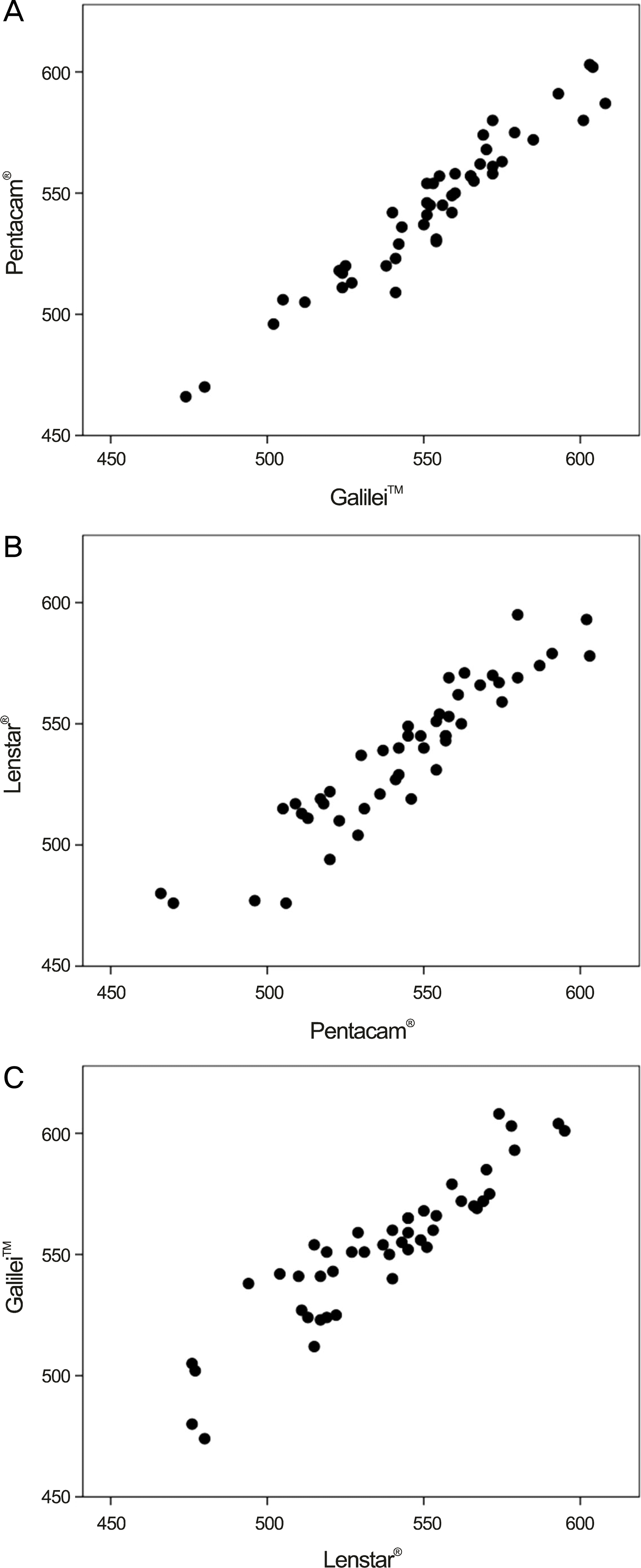 | Figure 1.Scatter plots of central corneal thickness (μ m) between (A) Galilei™ and Pentacam®, (B) Pentacam® and Lenstar®, (C) Lenstar® and Galilei™. |
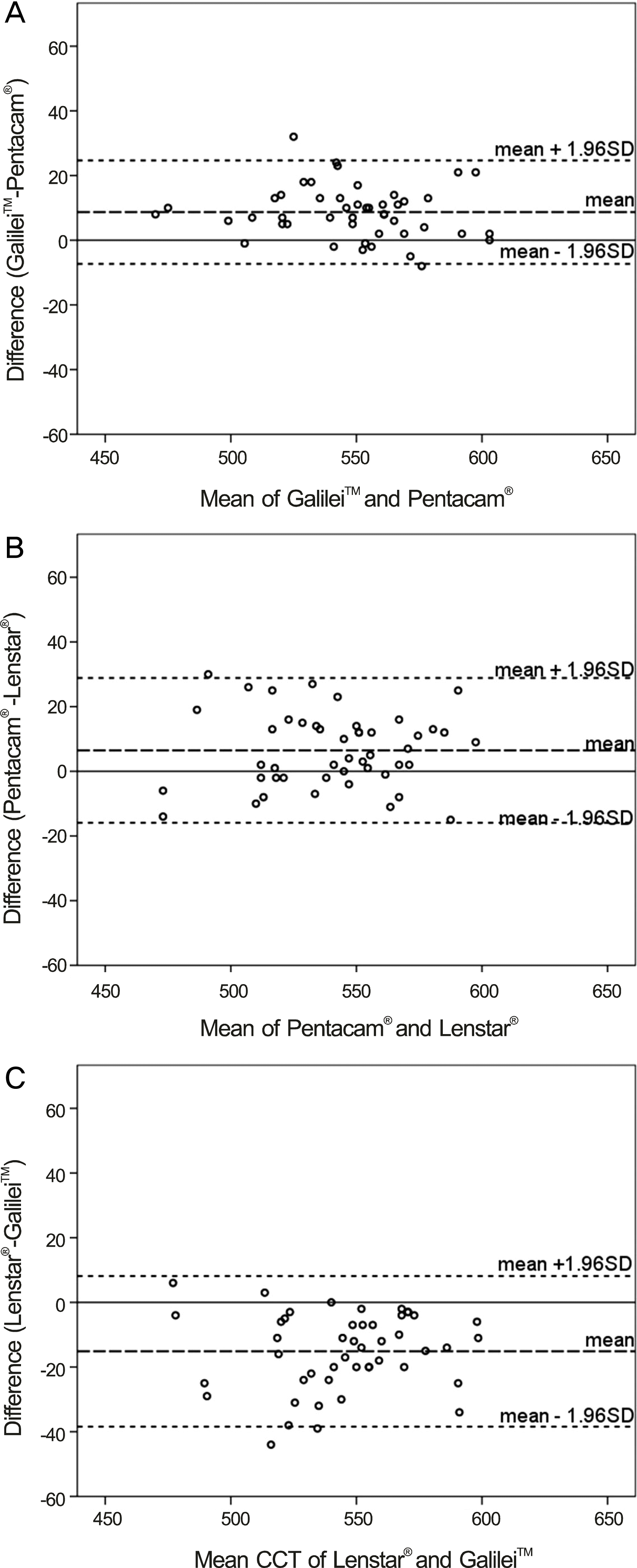 | Figure 2.Bland-Altman plots of CCT (μ m) between (A) Galilei™ and Pentacam®, (B) Pentacam® and Lenstar®, (C) Lenstar® and Galilei™. CCT = central corneal thickness. |
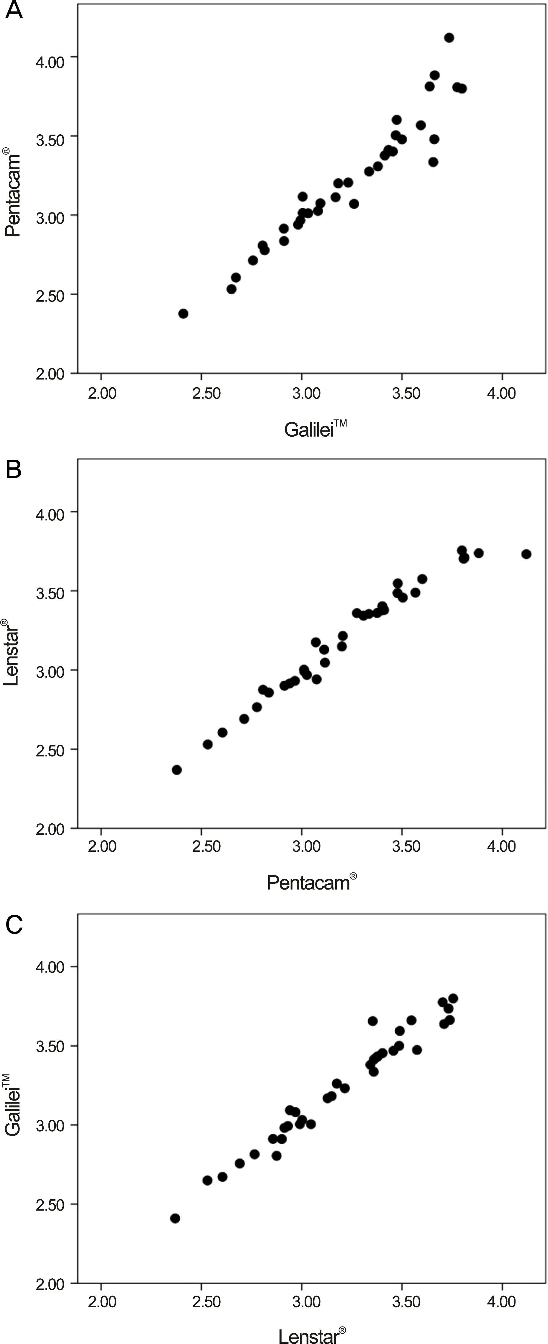 | Figure 3.Scatter plots of anterior chamber depth (mm) between (A) Galilei™ and Pentacam®, (B) Pentacam® and Lenstar®,(C) Lenstar® and Galilei™. |
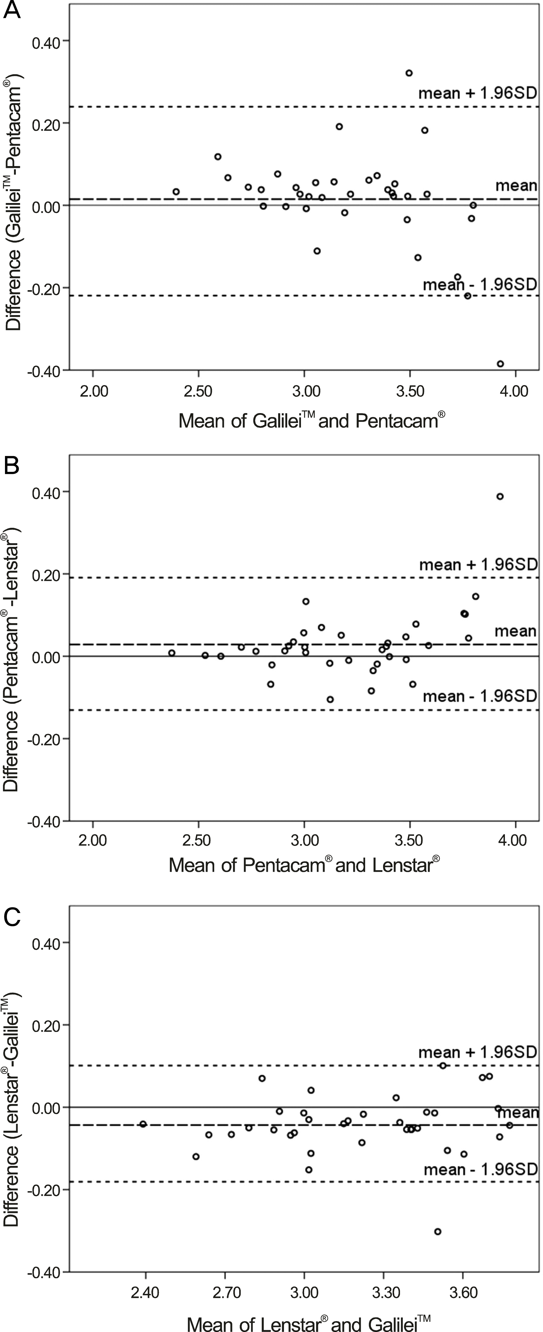 | Figure 4.Bland-Altman plots of ACD (mm) between (A) Galilei™ and Pentacam®, (B) Pentacam® and Lenstar®, (C) Lenstar® and Galilei™. ACD = anterior chamber depth. |
Table 1.
The central corneal thickness (μ m) and anterior chamber depth (mm) measured by Galilei™, Pentacam® and Lenstar®
| Galilei™ | Pentacam® | Lenstar® |
Difference |
||||
|---|---|---|---|---|---|---|---|
| Galilei™- Pentacam® | Pentacam®- Lenstar® | Lenstar®- Galilei™ | |||||
| CCT (μ m) | Mean ± SD | 552.6 ± 29.42 | 543.9 ± 30.50 | 537.5 ± 30.26 | 8.68 ± 8.15 | 6.47 ± 11.42 | -15.15 ± 11.88 |
| Min, max | 474, 608 | 466, 603 | 476, 593 | -8, 32 | -15, 30 | -44, 6 | |
| p-value* | - | - | - | <0.001 | 0.001 | <0.001 | |
| ACD (mm) | Mean ± SD | 3.23 ± 0.360 | 3.22 ± 0.403 | 3.19 ± 0.367 | 0.01 ± 0.117 | 0.03 ±0.082 | -0.04 ± 0.072 |
| Min, max | 2.41, 3.80 | 2.38, 4.12 | 2.37, 3.76 | -0.39, 0.32 | -0.11, 0.39 | -0.30, 0.10 | |
| p-value* | - | - | - | 1.000 | 0.134 | 0.003 | |
 XML Download