

 PDF
PDF ePub
ePub Citation
 Print
Print


초록
Purpose:
To evaluate the clinical efficacy and shortcomings of neodymium-doped yttrium aluminum garnet (Nd-YAG) laser membranotomy in inadvertently retained host membrane.
Methods:
Among 742 patients who underwent penetrating keratoplasty (PKP) and deep anterior lamellar keratoplasty (DALK) surgery at Seoul St. Mary’s Hospital between January 2007 and May 2013 by a single surgeon, 10 patients had a thin, opaque membrane in the anterior chamber observed under slit lamp examination and both a subjective decrease in visual acuity and decrease i best corrected visual acuity. A single surgeon performed membranotomy using the Nd-YAG laser at 4.9 months after graft surgery. In this study we compared the differences in visual acuity, endothelial cell count and correlations between distance from donor endothelium and retained host membrane and endothelial loss before and after the graft surgery.
Results:
Patients who had Nd-YAG laser membranotomy performed on the retained host membrane showed significant improvements in visual acuity ( p = 0.039). Donor endothelial cell count was significantly reduced 1 month after Nd-YAG laser. The average distance between donor endothelium and retained host membrane was 712.0 ± 217.5 µm. The distance and the decreased donor endothelial cell count were not statistically correlated (R2 = 0.39, p = 0.072). There were no significant complications after the laser membranotomy.
Go to : 
References
1. Duke-Elder S. Text-book of Ophthalmology: Injuries. Vol. 6. London: Mosby;1954.
2. Morton PL, Ormsby HL, Basu PK. Healing of endothelium and Descemet's membrane of rabbit cornea. Am J Ophthalmol. 1958; 46(1 Pt 2):62–7.

3. Chi HH, Teng CC, Katzin HM. Histopathology of primary endothelial-epithelial dystrophy of the cornea. Am J Ophthalmol. 1958; 45(4 Pt 1):518–35.
4. Stocker FW. The endothelium of the cornea and its clinical implications. Trans Am Ophthalmol Soc. 1953; 51:669–786.
5. Werb A. The postgraft membrane. Int Ophthalmol Clin. 1962; 2:771–80.
6. Hales RH, Spencer WH. Unsuccessful penetrating keratoplasties. correlation of clinical and histologic findings. Arch Ophthalmol. 1963; 70:805–10.
7. Kremer I, Dreznik A, Tessler G, Bahar I. Corneal graft failure following Nd:YAG laser membranotomy for inadvertent retained descemet's membrane after penetrating keratoplasty. Ophthalmic Surg Lasers Imaging. 2012; 43:Online. e94–8.
8. Brown SI, Dohlman CH, Boruchoff SA. Dislocation of descemet's membrane during keratoplasty. Am J Ophthalmol. 1965; 60:43–5.
9. Sinha R, Vajpayee RB, Sharma N, et al. Trypan blue assisted descemetorhexis for inadvertently retained Descemet's membranes after penetrating keratoplasty. Br J Ophthalmol. 2003; 87:654–5.
10. Henderson JW, Wolter JR. Separation of Descemet's membrane in keratoplasty. Am J Ophthalmol. 1968; 65:375–8.
11. Lazar M, Loewenstein A, Geyer O. Intentional retention of Descemet's membrane during keratoplasty. Acta Ophthalmol (Copenh). 1991; 69:111–2.
12. Loewenstein A, Geyer O, Lazar M. Intentional retention of Descemet's membrane in keratoplasty for the surgical treatment of bullous keratopathy. Acta Ophthalmol (Copenh). 1993; 71:280–2.
13. Thyagarajan S, Mearza AA, Falcon MG. Inadvertent retention of Descemet Membrane in penetrating keratoplasty. Cornea. 2006; 25:748–9.
14. Feng CS, Choi WS, Nam WH, Shin YJ. A case of retained descemet's membrane after penetrating keratoplasty. J Korean Ophthalmol Soc. 2013; 54:813–7.
15. Arenas Archila E, Ramirez Cabrera MF, Mieth Alviar A. Double Descemet's membrane in penetrating keratoplasty. Refract Corneal Surg. 1993; 9:65–6.
16. Chen YP, Lai PC, Chen PY, et al. Retained Descemet's membrane after penetrating keratoplasty. J Cataract Refract Surg. 2003; 29:1842–4.
17. Steinemann TL, Henry K, Brown MF. Nd:YAG laser treatment of retained Descemet's membrane after penetrating keratoplasty. Ophthalmic Surg. 1995; 26:80–1.
18. Patel SV, Diehl NN, Hodge DO, Bourne WM. Donor risk factors for graft failure in a 20-year study of penetrating keratoplasty. Arch Ophthalmol. 2010; 128:418–25.
19. Lass JH, Gal RL, Dontchev M, et al. Donor age and corneal endothelial cell loss 5 years after successful corneal transplantation. Specular microscopy ancillary study results. Ophthalmology. 2008; 115:627–32.e8.
20. Waring GO 3rd, Bourne WM, Edelhauser HF, Kenyon KR. The corneal endothelium. Normal and pathologic structure and function. Ophthalmology. 1982; 89:531–90.
21. Power WJ, Collum LM. Electron microscopic appearances of human corneal endothelium following Nd:YAG laser iridotomy. Ophthalmic Surg. 1992; 23:347–50.
22. Wu SC, Jeng S, Huang SC, Lin SM. Corneal endothelial damage after neodymium:YAG laser iridotomy. Ophthalmic Surg Lasers. 2000; 31:411–6.
23. Kozobolis VP, Detorakis ET, Vlachonikolis IG, Pallikaris IG. Endothelial corneal damage after neodymium:YAG laser treatment: pupillary membranectomies, iridotomies, capsulotomies. Ophthalmic Surg Lasers. 1998; 29:793–802.
24. Landers J, Craig J. Decompression retinopathy and corneal oedema following Nd:YAG laser peripheral iridotomy. Clin Experiment Ophthalmol. 2006; 34:182–4.
25. Meyer KT, Pettit TH, Straatsma BR. Corneal endothelial damage with neodymium:YAG laser. Ophthalmology. 1984; 91:1022–8.
26. Vaikoussis E, Bisogiannis Z, Margaritis L. Corneal endothelial damage after Nd:YAG laser anterior capsulotomy. An experimental study on rabbits. Doc Ophthalmol. 1993; 83:279–86.
27. Kerr Muir MG, Sherrard ES. Damage to the corneal endothelium during Nd/YAG photodisruption. Br J Ophthalmol. 1985; 69:77–85.
28. Lifshitz T, Oshry T, Rosenthal G. Retrocorneal membrane after penetrating keratoplasty. Ophthalmic Surg Lasers. 2001; 32:159–61.
29. Sherrard ES, Rycroft PV. Retrocorneal membranes. I. Their origin and structure. Br J Ophthalmol. 1967; 51:379–86.
Go to :
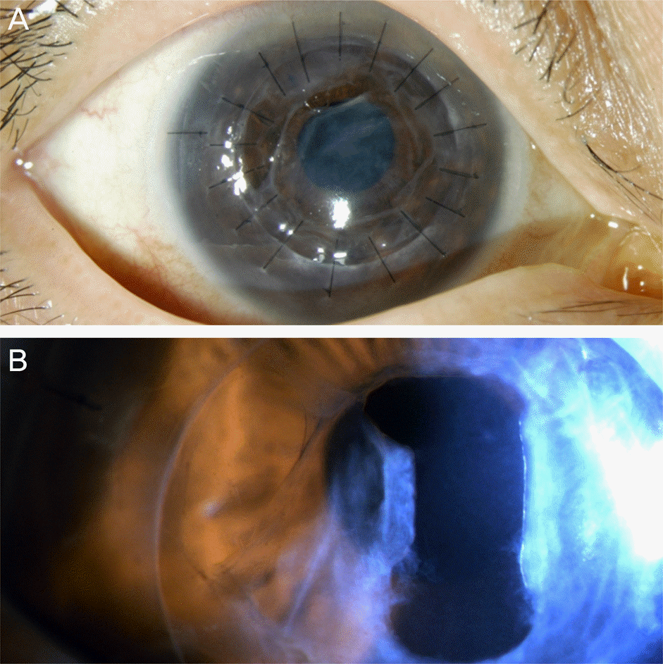 | Figure 1.Retained host membrane after keratoplasty (slit lamp photo). (A) Retained host membrane in front of the pupil is seen in slit lamp photo taken at the day after surgery. The opacity of the membrane is marked, even by naked eyes. Clear space is seen between the membrane and recipient’s peripheral cornea. (B) Retained host membrane is perforated in rec-tangular shape, 5 months after the surgery. Visual axis is well spared with 2.0 mm wide, 2.5 mm long perforation area. The cornea is transparent. |
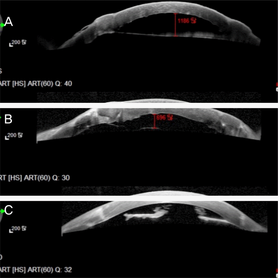 | Figure 2.Cross sectional image at the level of grafted cornea and retained host membrane. (A, B) Distance between graft endothelium and retained host membrane is measured with spectralis-domain HRA anterior OCT imaging taken at the day after surgery. Distance is variable but relatively close to the graft cornea endothelium. Retained host membrane itself is very thin. (C) Spectralis OCT taken 5 months after the surgery. Retained host membrane is perforated, graft corneal endothelium is intact. The membrane is noticeably thicker. HRA = Heidelberg retina angiograph; OCT = optical coherence tomography. |
Table 1.
Baseline ocular characteristics of the study group by primary cause and surgical procedure
| Patient | Sex | Age (years) | Primary cause of corneal decompensation | Surgical procedure | Suspected nature of the retention membrane | Membrane detection time (days)* | Laser apply time (months)† |
|---|---|---|---|---|---|---|---|
| 1 | F | 68 | Traumatic scar | DALK | Double anterior chamber | 1 | 4 |
| 2 | M | 67 | BK after surgery | PKP | Detached host DM | 1 | 7 |
| 3 | M | 48 | Chemical burn | PKP | Detached host DM | 1 | 4 |
| 4 | M | 38 | BK after surgery | PKP | Detached host DM | 1 | 5 |
| 5 | M | 64 | Lipid degeneration | DALK | Double anterior chamber | 1 | 7 |
| 6 | F | 67 | Granular dystrophy | DALK | Double anterior chamber | 1 | 4 |
| 7 | F | 73 | ICE syndrome | PKP | Detached host DM | 1 | 5 |
| 8 | F | 53 | Traumatic scar | DALK | Double anterior chamber | 1 | 4 |
| 9 | M | 69 | ICE syndrome | PKP | Detached host DM | 1 | 4 |
| 10 | F | 22 | Acanthoamoeba keratitis | DALK | Double anterior chamber | 1 | 5 |
| Average | 56.9 ± 16.5 | 1 | 4.9 ± 1.2 |
Table 2.
Pre- and post operative visual acuity (log MAR)
| Patient | BCVA 1 week before PKP (log MAR) | BCVA 1 month after PKP (log MAR)*,† | BCVA 1 month after laser apply (log MAR)* | BCVA 6 months after laser apply (log MAR)* | BCVA 12 months after laser apply (log MAR)* |
|---|---|---|---|---|---|
| 1 | 1.4 | 1.1 | 0.8 | 0.7 | 1.0 |
| 2 | 1.6 | 2.4 | 2.4 | 2.4 | 2.4 |
| 3 | 1.8 | 1.2 | 0.3 | 0.4 | 1.4 |
| 4 | 0.9 | 0.6 | 0.9 | 0.9 | 0.8 |
| 5 | 1.7 | 1.5 | 0.4 | 0.5 | 0.5 |
| 6 | 2.4 | 1.9 | 0.6 | 0.5 | 0.4 |
| 7 | 2 | 1.7 | 1.4 | 1.4 | 1.6 |
| 8 | 2 | 1.8 | 1.7 | 1.8 | 2.0 |
| 9 | 1.8 | 0.6 | 0.3 | 0.3 | 0.3 |
| 10 | 1.8 | 2.4 | 2.4 | 2.4 | 2.4 |
| Average | 1.74 ± 0.21 | 1.52 ± 0.18 | 1.12 ± 0.13 | 1.18 ± 0.78 | 1.28 ± 0.8 |
Table 3.
Pre- and postOperative endothelial cell counts by specular microscopy
| Patient | CD before PKP*,† | CD 1 month after PKP*,†,‡ | CD 1 month after laser*,† | CD 6 months after laser*,† | CD 12 months after laser*,† |
|---|---|---|---|---|---|
| 1 | Error c | 2,759 | 1,709 | 584 | 729 |
| 2 | Error c | 1,461 | Error c | Error c | Error c |
| 3 | 453 | 2,702 | 1,908 | 1,109 | 1,342 |
| 4 | Error c | 1,883 | 1,033 | 984 | 702 |
| 5 | Error c | 2,049 | 1,024 | 786 | 724 |
| 6 | Error c | 2,369 | 1,248 | 675 | 724 |
| 7 | Error c | 1,915 | 1,048 | 849 | 801 |
| 8 | Error c | 1,742 | 629 | Error c | Error c |
| 9 | Error c | 1,097 | 880 | 866 | 840 |
| 10 | Error c | 903 | 548 | 426 | Error c |
| Average | 453 | 1,888 ± 621.7 | 1,114.1 ± 451.6 | 784.9 ± 219.3 | 837.4 ± 227.9 |
Table 4.
Pre- and post operative corneal thickness by ultrasound pachymetry
| Patient | CT before PKP (μ m) | CT 1 month after PKP (μ m)* | CT 1 month after laser (μ m) | CT 6 months after laser (μ m) | CT 12 months after laser (μ m) |
|---|---|---|---|---|---|
| 1 | 404 | 499 | 504 | 561 | 543 |
| 2 | 874 | 605 | 689 | 764 | 787 |
| 3 | 584 | 593 | 587 | 543 | 583 |
| 4 | 889 | 652 | 651 | 800 | 868 |
| 5 | 411 | 569 | 629 | 661 | 682 |
| 6 | 558 | 669 | 624 | 667 | 674 |
| 7 | 525 | 564 | 597 | 699 | 678 |
| 8 | 768 | 771 | 784 | 841 | 883 |
| 9 | 590 | 564 | 564 | 689 | 712 |
| 10 | 342 | 893 | 542 | 538 | 572 |
| Average | 594.5 ± 192.9 | 637.9 ± 116.4 | 617.1 ± 79.5 | 676.3 ± 106.1 | 698.2 ± 118.1 |
Table 5.
Distance between donor endothelium and donor endothelial cell count differential after laser and Nd-YAG laser delivered energy and numbers of pulses
| Patient | Pulse energy (mJ) | Number of pulses | Distance between donor endothelium and retained host membrane (μ m)* | Differential in cell counts 1 month after laser (cells/mm3)† |
|---|---|---|---|---|
| 1 | 1 | 17 | 474 | 1,050 |
| 2 | 0.8 | 21 | 1,186 | N/A |
| 3 | 1 | 11 | 696 | 794 |
| 4 | 1 | 16 | 831 | 850 |
| 5 | 0.8 | 27 | 591 | 1,025 |
| 6 | 1 | 17 | 496 | 1,740 |
| 7 | 0.8 | 14 | 714 | 867 |
| 8 | 1 | 10 | 675 | 494 |
| 9 | 0.8 | 13 | 911 | 23 |
| 10 | 0.8 | 9 | 546 | 903 |
| Average | 0.9 ± 0.1 | 15.5 ± 5.5 | 712 ± 217.5 | 821.3 ± 469.0 |
 XML Download