

 PDF
PDF ePub
ePub Citation
 Print
Print


초록
Purpose:
To report a case of focal choroidal excavation associated with central serous chorioretinopathy.
Case summary:
A 48-year-old female presented with a 20-year history of visual disturbance. Focal choroidal excavation with neurosensory retinal detachment was detected in the right eye on optical coherence tomography. Fluorescein angiography showed hyperfluorescene in the area of excavation and multiple focal hyperfluorescences in the perimacular area. Vertically linear hyperfluorescene line was detected in the excavated area caused by retinal pigment epithelial atrophy. Based on the 2 diagnostic findings, we diagnosed a focal choroidal excavation with central serous chorioretinopathy. No progression was detected for 2 months.
Go to : 
References
1. Jampol LM, Shankle J, Schroeder R, et al. Diagnostic and therapeutic challenges. Retina. 2006; 26:1072–6.

2. Margolis R, Mukkamala SK, Jampol LM, et al. The expanded spectrum of focal choroidal excavation. Arch Ophthalmol. 2011; 129:1320–5.
3. Abe S, Yamamoto T, Kirii E, Yamashita H. Cup-shaped choroidal excavation detected by optical coherence tomography: a case report. Retin Cases Brief Rep. 2010; 4:373–6.
4. Ellabban AA, Tsujikawa A, Ooto S, et al. Focal choroidal excavation in eyes with central serous chorioretinopathy. Am J Ophthalmol. 2013; 156:673–83.
5. Suzuki M, Gomi F, Hara C, et al. Characteristics of central serous chorioretinopathy complicated by focal choroidal excavation. Retina. 2014; 34:1216–22.
6. Wakabayashi Y, Nishimura A, Higashide T, et al. Unilateral choroidal excavation in the macula detected by spectral-domain optical coherence tomography. Acta Ophthalmol. 2010; 88:e87–91.
7. Kobayashi W, Abe T, Tamai H, Nakazawa T. Choroidal excavation with polypoidal choroidal vasculopathy: a case report. Clin Ophthalmol. 2012; 6:1373–6.
8. Kuroda Y, Tsujikawa A, Ooto S, et al. Association of focal choroidal excavation with age-related macular degeneration. Invest Ophthalmol Vis Sci. 2014; 55:6046–54.
9. Katome T, Mitamura Y, Hotta F, et al. Two cases of focal choroidal excavation detected by spectral-domain optical coherence tomography. Case Rep Ophthalmol. 2012; 3:96–103.
10. Hashida N, Fok A, Nishida K. Choroidal excavation in vogt-koya-nagi-harada disease. Case Rep Ophthalmol. 2014; 5:222–5.
11. Guo J, Zhong L, Jiang C, et al. Clinical and optic coherence tomography findings of focal choroidal excavation in Chinese patients. BMC Ophthalmol. 2014; 14:63.
Go to :
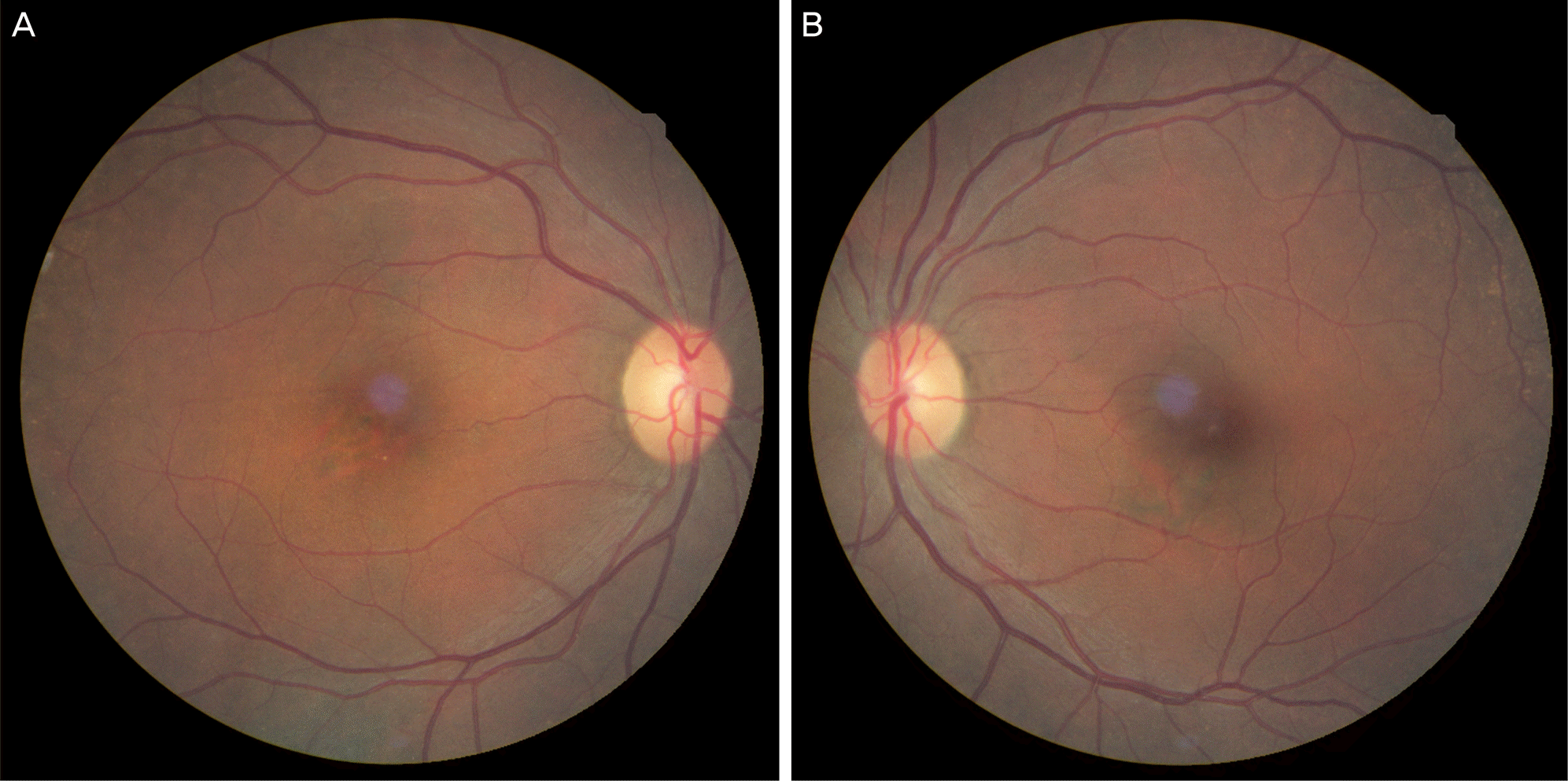 | Figure 1.Color photographs of the right eye (A) shows focal retinal pigment epithelial atrophic change on foveal area. Left eye (B) shows no abnormal sign on foveal area. |
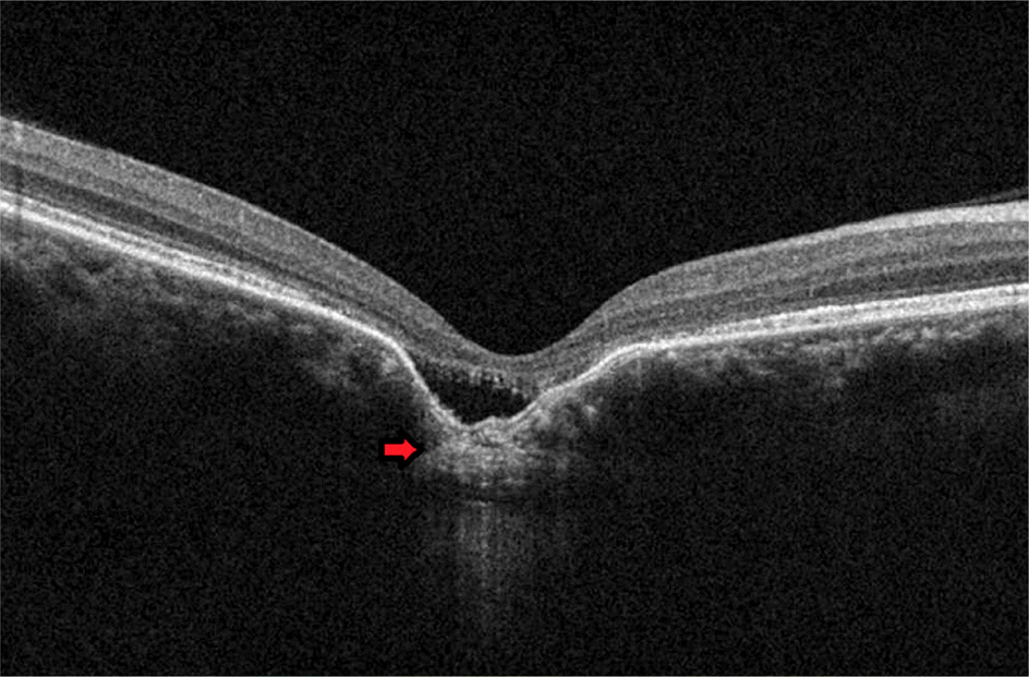 | Figure 2.Focal choroidal excavation with neurosensory retinal detachment is detected on SD-OCT. Hyperreflective un-usual subfoveal choroidal tissue (red arrow) is detected be-neath the excavation. SD-OCT = spectral-domain optical coherence tomography. |
 | Figure 3.Hyperfluorescene is detected on the foveal area and multiple focal hyperfluorescences are detected on perimacular area. Hyperfluorescence is not stained from the early (A) to late (B) phase. Vertically linear hyperfluorescene line is detected from the foveal area caused by retinal pigment epithelial atrophy (C). |
 XML Download