초록
Purpose:
To report a case of retinal blot hemorrhage localized to 1 eye accompanied by Raynaud’s phenomenon.
Case summary:
A 65-year-old female was referred for vision disorder in the right eye. She had been taking an antihypertensive drug since diagnosed with systemic hypertension 2 years previously. On initial examination, her best corrected visual acuity was 20/25 in the right eye and 20/20 in the left eye and intraocular pressure was 15 mm Hg in the right eye and 21 mm Hg in the left eye. Fundus examination showed retinal blot hemorrhage across the entire retina and increased retinal vascular tortuosity. No specific finding was found on visual field examination, transthoracic ultrasonography and carotid Doppler ultrasound. The blood test was positive for antinuclear and anticentromere antibodies, thus she was referred to the rheumatologic department. The patient was diagnosed with primary Raynaud’s phenomenon because no correlation with other rheumatologic diseases was found. Subsequently, she was scheduled for regular follow-ups with a prescription of circulatory stimulant. Five months later, her best corrected visual acuity was 20/20 in the both eyes and retinal blot hemorrhage in the right eye was significantly decreased based on fundus examination.
Conclusions:
In the case of atypical retinal blot hemorrhage without other ophthalmic causes except Raynaud’s phenomenon, the change in retinal circulatory autoregulation associated with the mechanism of retinal blot hemorrhage can be presumed. Therefore, a close examination and history taking should be conducted so that Raynaud’s phenomenon as a pathological factor is not overlooked.
Go to : 
References
1. Cardelli MB, Kleinsmith DM. Raynaud's phenomenon and disease. Med Clin North Am. 1989; 73:1127–41.

4. Wahab A, Allaf A, Belch JJ. Raynaud's phenonemon. Hochberg MC, Silman AJ, Smolen JS, Weinolatt ME, Weimann MH, editors. Rheumatology. 3rd ed.New york: Mosby;2003. p. 1507–12.
5. Wigley FM. Clinical practice. Raynaud's Phenomenon. N Engl J Med. 2002; 347:1001–8.
6. Maricq HR, Weinrich MC, Keil JE, LeRoy EC. Prevalence of Raynaud phenomenon in the general population. A preliminary study by questionnaire. J Chronic Dis. 1986; 39:423–7.
7. Mirbod SM, Yoshida H, Komura Y, et al. Prevalence of Raynaud's phenomenon in different groups of workers operating hand-held vibrating tools. Int Arch Occup Environ Health. 1994; 66:13–22.
8. Olsen N, Nielsen SL. Prevalence of primary Raynaud phenomena in young females. Scand J Clin Lab Invest. 1978; 38:761–4.

9. Koenig M, Joyal F, Fritzler MJ, et al. Autoantibodies and micro-vascular damage are independent predictive factors for the progression of Raynaud's phenomenon to systemic sclerosis: a twen-ty-year prospective study of 586 patients, with validation of pro-posed criteria for early systemic sclerosis. Arthritis Rheum. 2008; 58:3902–12.

10. Riera G, Vilardell M, Vaqué J, et al. Prevalence of Raynaud's phenomenon in a healthy Spanish population. J Rheumatol. 1993; 20:66–9.
11. Salmenson BD, Reisman J, Sinclair SH, Burge D. Macular capil-lary hemodynamic changes associated with Raynaud's phenomenon. Ophthalmology. 1992; 99:914–9.
12. Alexander EL, Firestein GS, Weiss JL, et al. Reversible cold-in-duced abnormalities in myocardial perfusion and function in systemic sclerosis. Ann Intern Med. 1986; 105:661–8.

13. Ellis WW, Baer AN, Robertson RM, et al. Left ventricular dysfunction induced by cold exposure in patients with systemic sclerosis. Am J Med. 1986; 80:385–92.

14. Miller D, Waters DD, Warnica W, et al. Is variant angina the coro-nary manifestation of a generalized vasospastic disorder? N Engl J Med. 1981; 304:763–6.

15. Fahey PJ, Utell MJ, Condemi JJ, et al. Raynaud's phenomenon of the lung. Am J Med. 1984; 76:263–9.

16. Amalric P. Acute choroidal ischaemia. Trans Ophthalmol Soc U K. 1971; 91:305–22.
17. Drance SM, Douglas GR, Wijsman K, et al. Response of blood flow to warm and cold in normal and low-tension glaucoma patients. Am J Ophthalmol. 1988; 105:35–9.

18. Gasser P, Flammer J. Blood-cell velocity in the nailfold capillaries of patients with normal-tension and high-tension glaucoma. Am J Ophthalmol. 1991; 111:585–8.

19. Gasser P, Flammer J, Guthauser U, Mahler F. Do vasospasms pro-voke ocular diseases? Angiology. 1990; 41:213–20.

20. Riva CE, Grunwald JE, Sinclair SH. Laser Doppler measurement of relative blood velocity in the human optic nerve head. Invest Ophthalmol Vis Sci. 1982; 22:241–8.
Go to : 
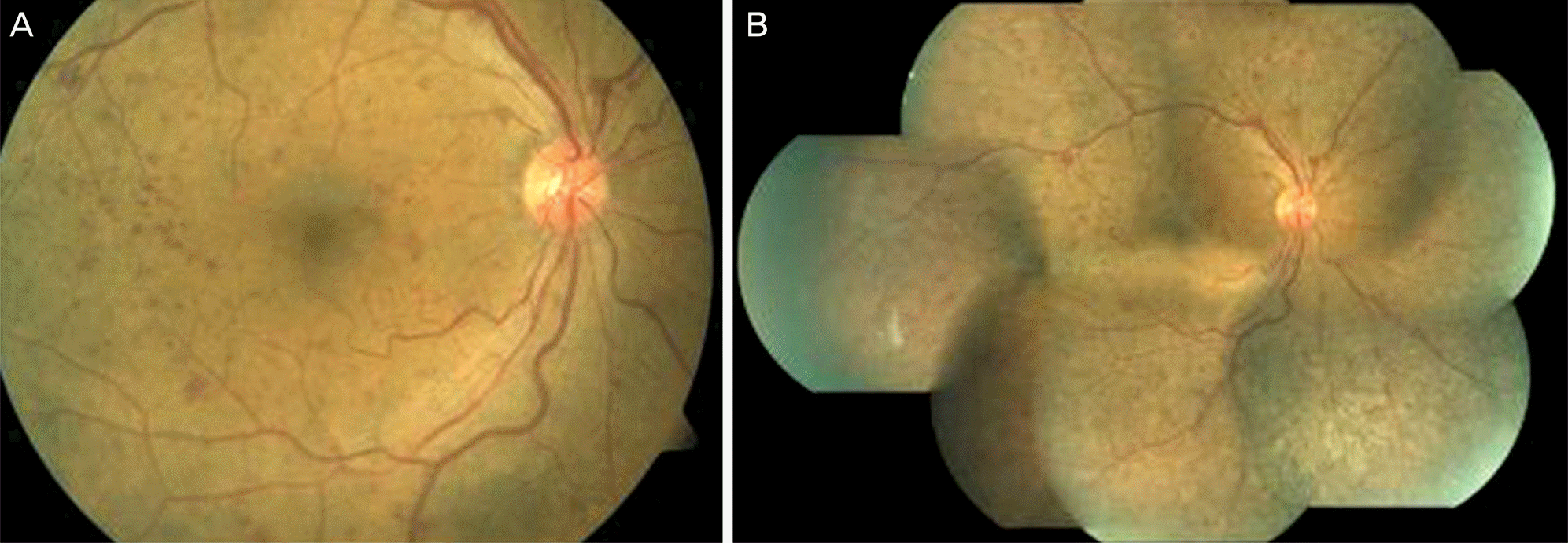
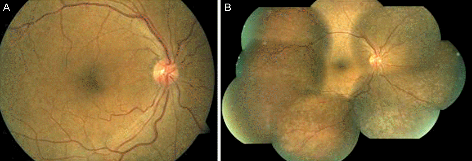
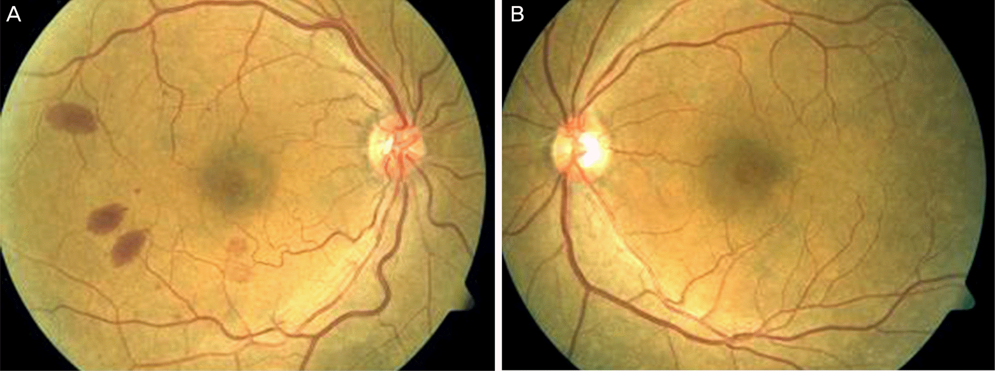 | Figure 1.Fundus photograph at the first visit. (A) Multiple blot hemorrhages and increased vascular tortuosity are seen in the right eye. (B) Fundus photograph of the left eye. |
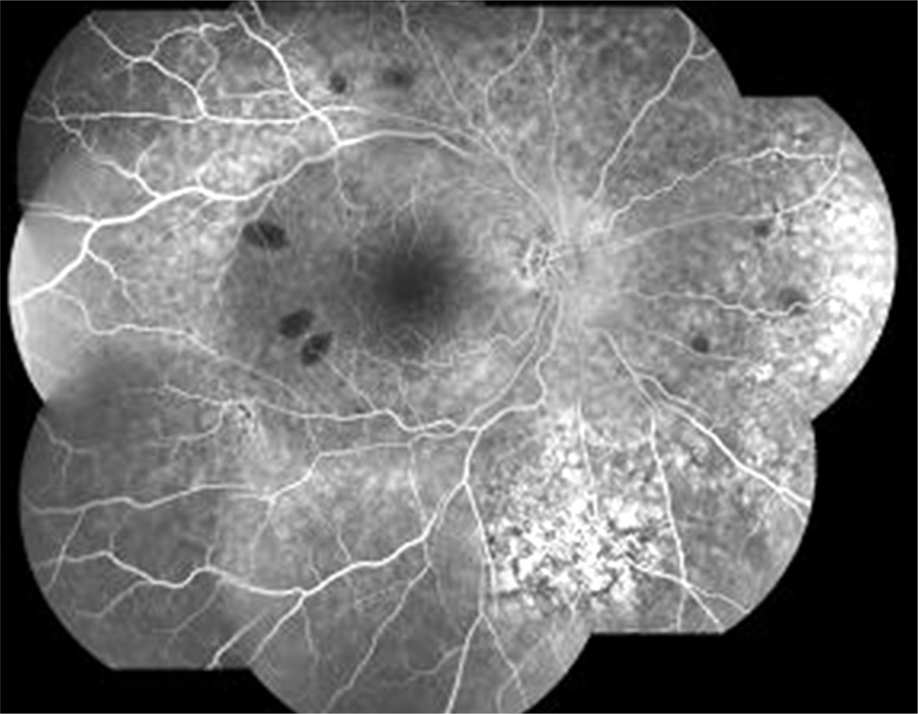 | Figure 2.FAG at the first visit. No specific abnormal finding is seen around the retinal blot hemorrhage. Late leakage was seen in the infero-nasal peripheral retina. FAG = fluorescein angiography. |
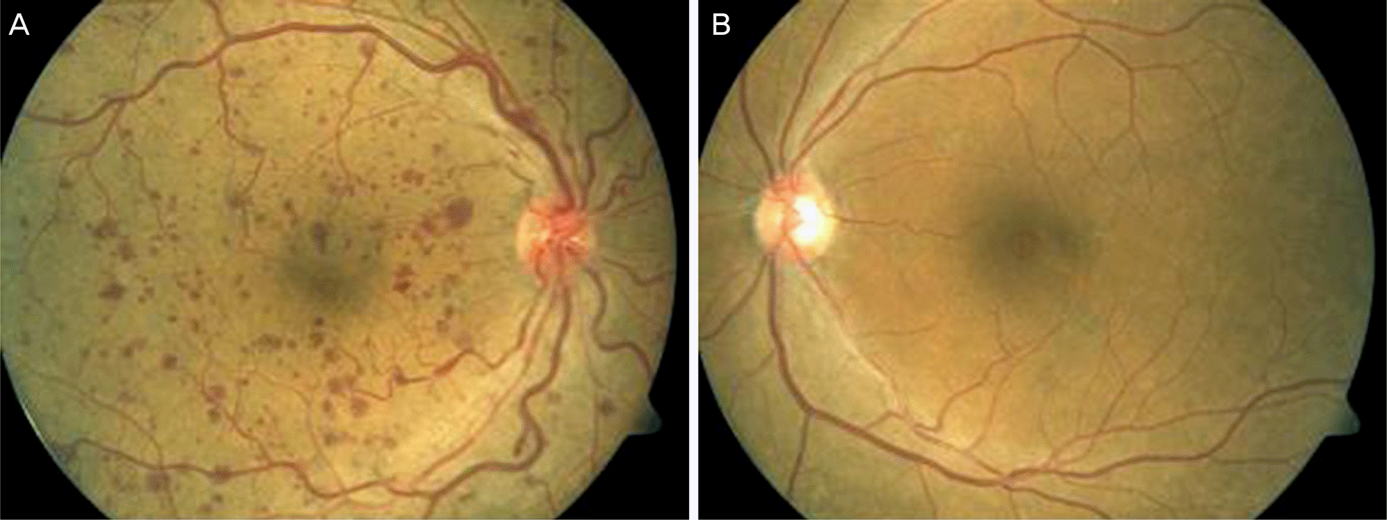 | Figure 3.Photograph taken 8 months after the first visit. (A) It is noted that previous large blot hemorrhages disappeared but many small retinal blot hemorrhages appeared across the whole central retinal area. The degree of vascular tortuosity increased. (B) No interval change is found in the left eye. |
 | Figure 4.FAG taken 8 months after the first visit. No interval change is found except the decrease of the extent of late leakage. FAG = fluorescein angiography. |
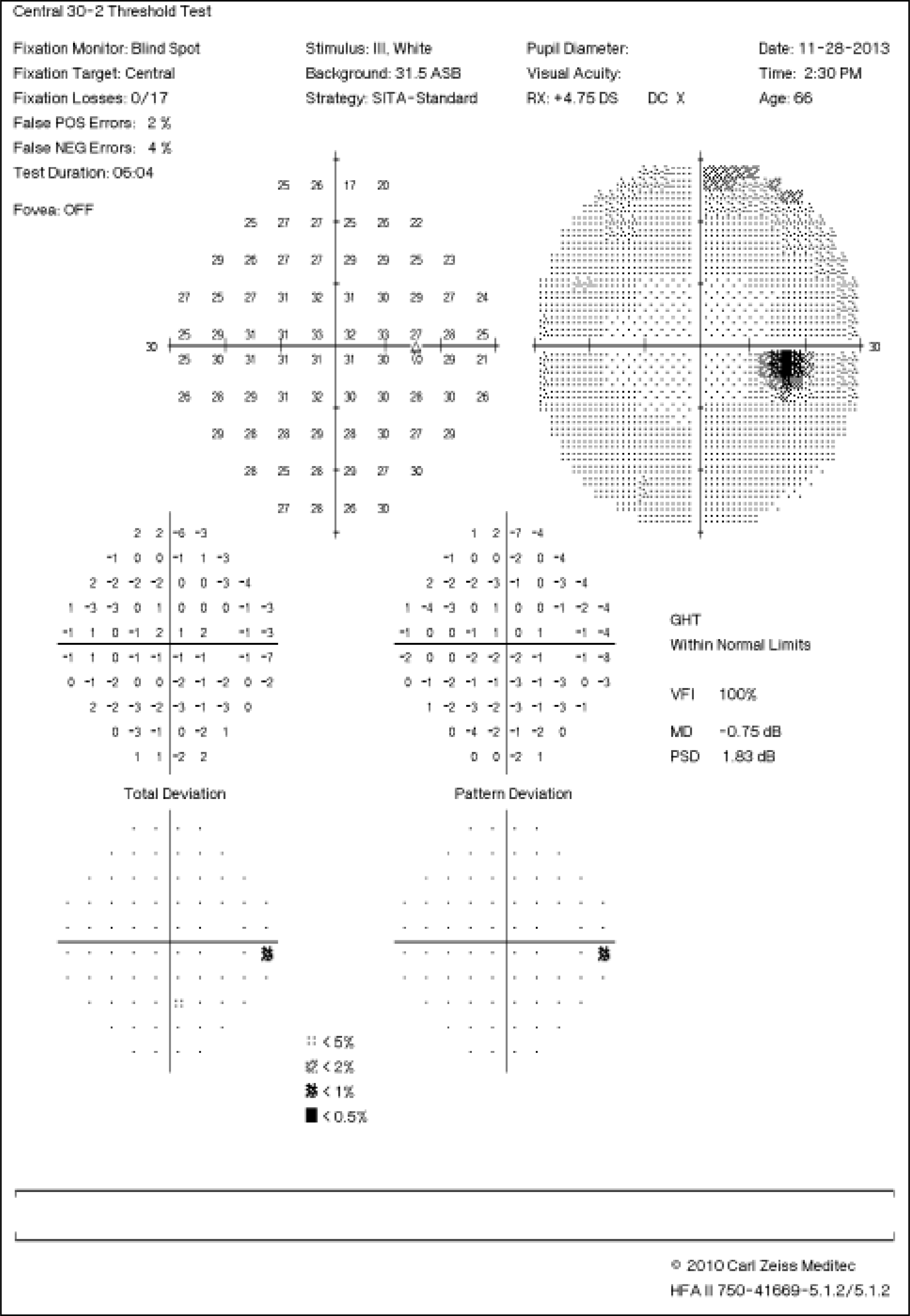 | Figure 5.No specific scotoma or abnormal findings are found on the visual field examination of the right eye. |
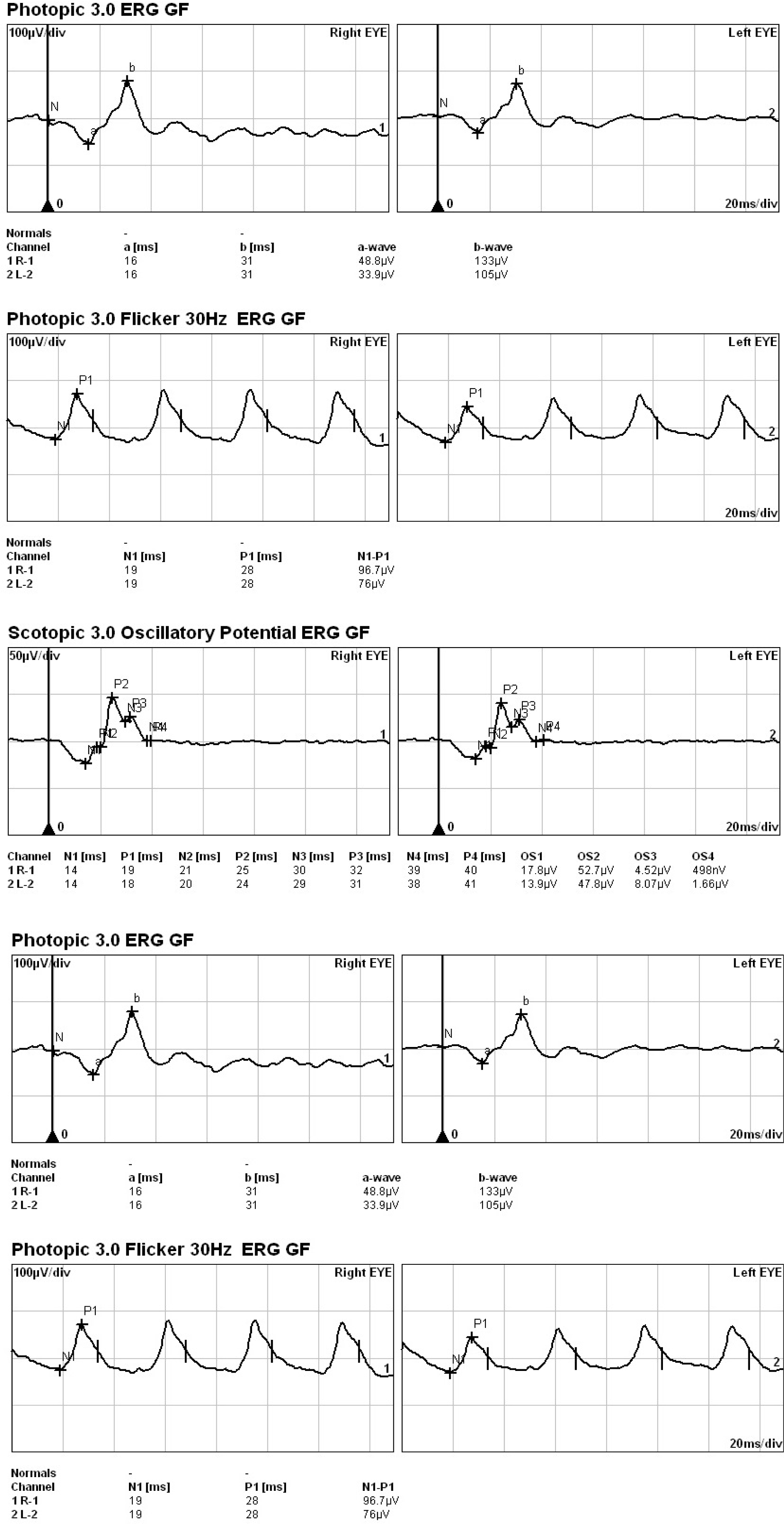 | Figure 6.In scotopic oscilllatory potential ERG, the amplitude of OS3 wave slightly decreased. ERG = electroretinogram; OS3 = oscillatory 3. |
 | Figure 7.In multifocal ERG, the amplitude of P1 wave of both eye decreased. (A) Right eye (B) left eye. ERG = electroretinogram. |


 PDF
PDF ePub
ePub Citation
 Print
Print


 XML Download