초록
Purpose:
To report a case of modified capsular tension ring scleral fixation and in-the-bag toric intraocular lens (IOL) implantation in a pediatric patient with severe crystalline lens subluxation due to homocystinuria.
Case summary:
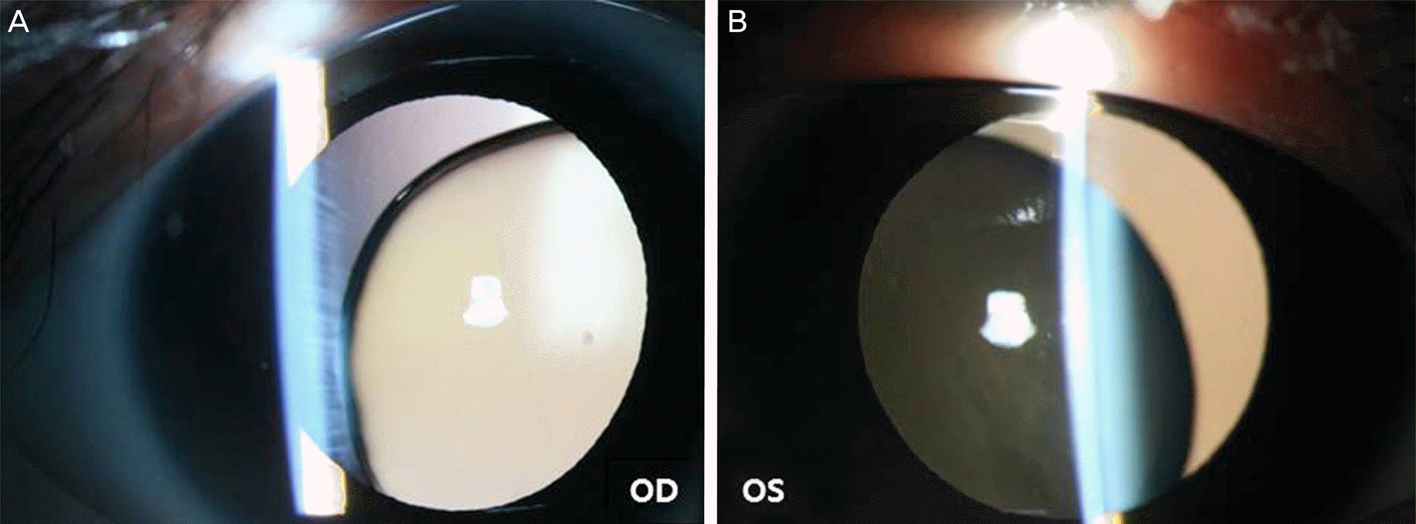
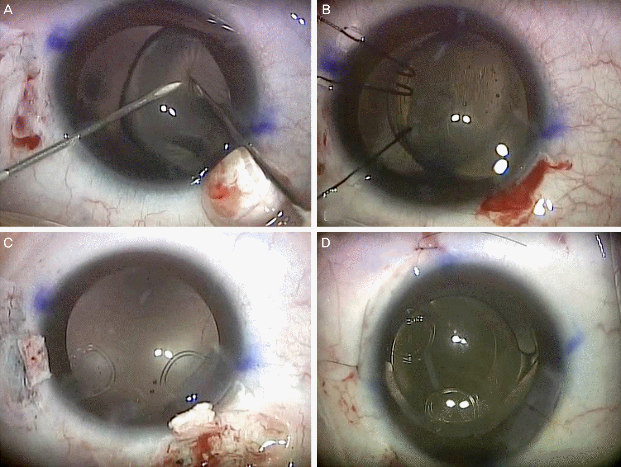
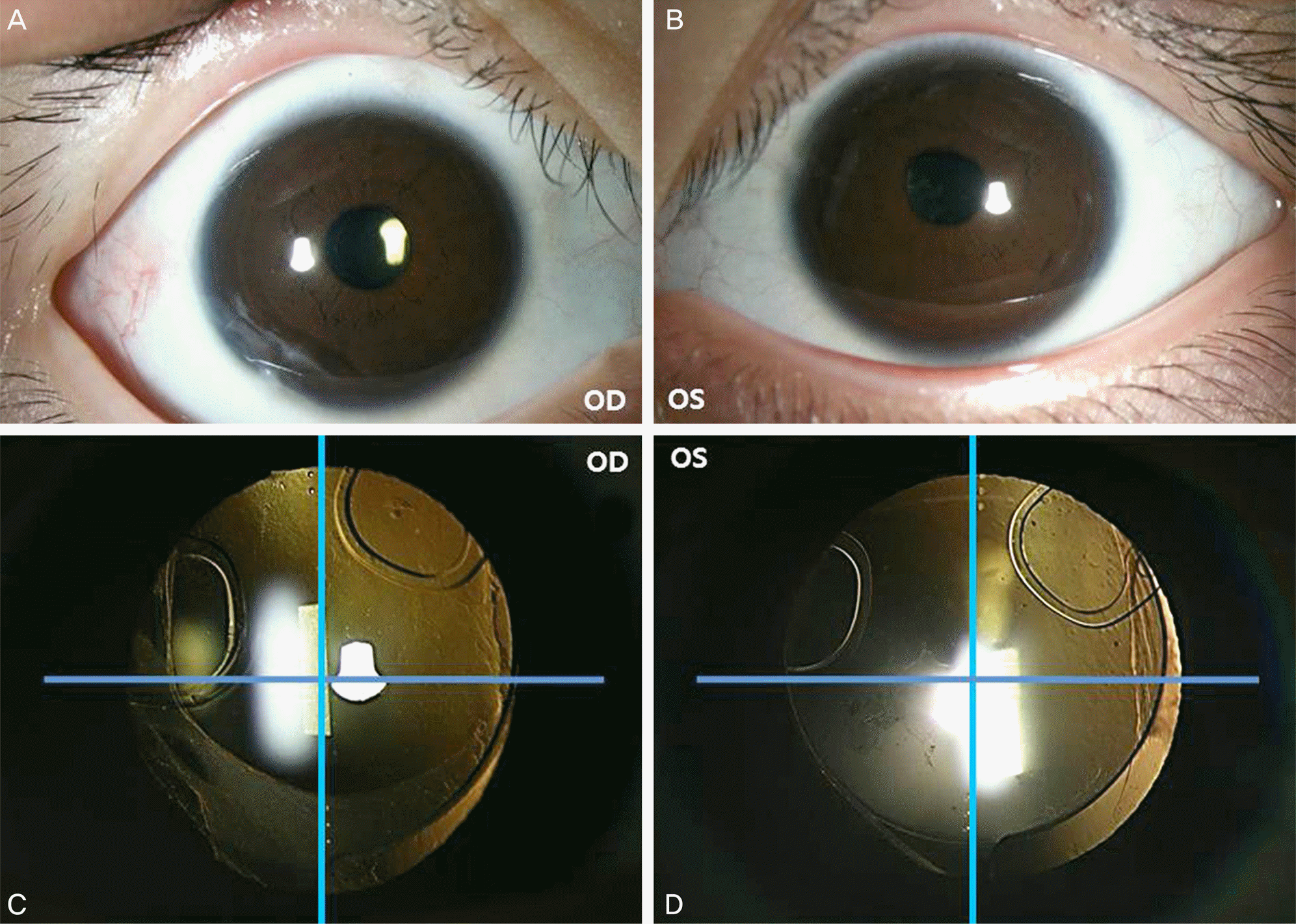
A 9-year-old male diagnosed with homocystinuria and crystalline lens subluxation presented with progressive decrease of visual acuity. Uncorrected distant visual acuity (UDVA) and corrected distant visual acuity were 0.03 and 0.6 in the right eye and 0.01 and 0.5 in the left eye, respectively. Slit-lamp examination showed severe crystalline lens subluxation toward the inferiomedial side in both eyes. Corneal astigmatism in the right eye and left eye was 2.75 diopters (D) and 3.00 D, respectively based on keratometry. A combination of subluxated crystalline lens aspiration, scleral-fixated modified capsular tension ring insertion and in-the-bag toric IOL implantation were performed in both eyes. After continuous curvilinear capsulorhexis, nucleus and cortex of the crystalline lens were removed by irrigation and aspiration. A modified capsular tension ring with 2 fixation hooks (Model 2-L) was inserted into the capsular bag and fixed at the scleral wall. Next, toric IOL was inserted into the capsular bag. UDVA was 0.8 in the right eye and 0.9 in the left eye and 3 months postoperatively, the IOL rotation was less than 3 degrees from intended axis in both eyes.
Go to : 
References
1. Schimke RN, McKusick VA, Huang T, Pollack AD. Homocystinuria. studies of 20 families with 38 affected members. JAMA. 1965; 193:711–9.
2. Mulvihill A, Yap S, O'Keefe M, et al. Ocular findings among patients with late-diagnosed or poorly controlled homocystinuria compared with a screened, well-controlled population. J AAPOS. 2001; 5:311–5.

3. Mudd SH, Skovby F, Levy HL, et al. The natural history of homocystinuria due to cystathionine beta-synthase deficiency. Am J Hum Genet. 1985; 37:1–31.
4. Yap S. Classical homocystinuria: vascular risk and its prevention. J Inherit Metab Dis. 2003; 26:259–65.

5. Bilwani F, Syed NA, Usman M, Khurshid M. Familial homocystinuria. J Coll Physicians Surg Pak. 2005; 15:106–7.
6. Romano PE, Kerr NC, Hope GM. Bilateral ametropic functional amblyopia in genetic ectopia lentis: its relation to the amount of subluxation, an indicator for early surgical management. Binocul Vis Strabismus Q. 2002; 17:235–41.
7. Wu-Chen WY, Letson RD, Summers CG. Functional and structural outcomes following lensectomy for ectopia lentis. J AAPOS. 2005; 9:353–7.

8. Asadi R, Kheirkhah A. Long-term results of scleral fixation of posterior chamber intraocular lenses in children. Ophthalmology. 2008; 115:67–72.

9. Pfeifer V, Morela K. Ectopic lens extraction in children. Coll Antropol. 2001; 25(Suppl):37–41.
10. Chung DY, Chung YT. A case of homocystinuria with ectopia lentis. J Korean Ophthalmol Soc. 1991; 32:110–5.
11. Hoffman RS, Snyder ME, Devgan U, et al. Management of the subluxated crystalline lens. J Cataract Refract Surg. 2013; 39:1904–15.

12. Halpert M, BenEzra D. Surgery of the hereditary subluxated lens in children. Ophthalmology. 1996; 103:681–6.

13. Lam DS, Ng JS, Fan DS, et al. Short-term results of scleral intraocular lens fixation in children. J Cataract Refract Surg. 1998; 24:1474–9.

14. Bardorf CM, Epley KD, Lueder GT, Tychsen L. Pediatric transscleral sutured intraocular lenses: efficacy and safety in 43 eyes followed an average of 3 years. J AAPOS. 2004; 8:318–24.

15. Hoyt CS, Nickel B. Aphakic cystoid macular edema: occurrence in infants and children after transpupillary lensectomy and anterior vitrectomy. Arch Ophthalmol. 1982; 100:746–9.
16. Koenig SB, Ruttum MS, Lewandowski MF, Schultz RO. Pseudophakia for traumatic cataracts in children. Ophthalmology. 1993; 100:1218–24.

17. Gimbel HV, Camoriano GD, Aman-Ullah M. Bilateral Implantation of Scleral-Fixated Cionni Endocapsular Rings and Toric Intraocular Lenses in a Pediatric Patient with Marfan's Syndrome. Case Rep Ophthalmol. 2012; 3:16–23.

18. Trivedi RH, Wilson ME Jr. Single-piece acrylic intraocular lens implantation in children. J Cataract Refract Surg. 2003; 29:1738–43.

19. Trivedi RH, Wilson ME Jr, Bartholomew LR, et al. Opacification of the visual axis after cataract surgery and single acrylic intraocular lens implantation in the first year of life. J AAPOS. 2004; 8:156–64.

20. Kugelberg M, Kugelberg U, Bobrova N, et al. After-cataract in children having cataract surgery with or without anterior vitrectomy implanted with a single-piece AcrySof IOL. J Cataract Refract Surg. 2005; 31:757–62.

21. Sun XY, Vicary D, Montgomery P, Griffiths M. Toric intraocular lenses for correcting astigmatism in 130 eyes. Ophthalmology. 2000; 107:1776–81. discussion 1781-2.

23. Bauer NJ, de Vries NE, Webers CA, et al. Astigmatism management in cataract surgery with the AcrySof toric intraocular lens. J Cataract Refract Surg. 2008; 34:1483–8.

24. Miyake T, Kamiya K, Amano R, et al. Long-term clinical outcomes of toric intraocular lens implantation in cataract cases with preex-isting astigmatism. J Cataract Refract Surg. 2014; 40:1654–60.

25. Thibos LN, Horner D. Power vector analysis of the optical outcome of refractive surgery. J Cataract Refract Surg. 2001; 27:80–5.

26. Cionni RJ, Osher RH, Marques DM, et al. Modified capsular tension ring for patients with congenital loss of zonular support. J Cataract Refract Surg. 2003; 29:1668–73.

Go to : 
 | Figure 1.Slit-lamp photographs showing bilateral inferomedial crystalline lens subluxation through the dilated pupil. (A) Inferomedial crystalline lens subluxation in the right eye. (B) Inferomedial crystalline lens subluxation in the left eye. OD = oculus dexter; OS = oculus sinister. |
 | Figure 2.Intraoperative photographs. (A) Making anterior capsule tear using two 26-gauge needles. (B) Supporting anterior capsule using 3 microhook iris retractors. (C) Fixation of modified capsular tension ring (MCTR) to the sclera. Note that two hooks are not perpendicular. The meridian of second hook is determined by the diameters of MCTR and sulcus, individually. (D) A complex of toric intraocular lens and scleral fixated MCTR is well centered along the intended axis. |
 | Figure 3.Postoperative photohgraphs. (A, B) External photographs shows clear cornea, conjunctiva and round pupil 3 months after operation. (C) Three degrees counterclockwise axis rotation is found in a postoperative 3 months slit-lamp photograph in the right eye. (D) Two degrees clockwise axis rotation is found in a postoperative 3 months slit-lamp photograph in the left eye. OD = oculus dexter; OS = oculus sinister. |
Table 1.
Postoperative clinical results
|
POD # 1 day |
POD # 1 week |
|||||||||
|---|---|---|---|---|---|---|---|---|---|---|
| UDVA | CDVA | MR | SE | IOP | UDVA | CDVA | MR | SE | IOP | |
| OD | 0.5 | - | - | - | 19 | 0.6 | 0.9 | +1.00, -1.50 × 60º | +0.25 | 15 |
| OS | 0.2 | - | - | - | 15 | 0.6 | 0.8 | +0.50, -1.50 ×20º | - 0.25 | 17 |
Table 2.
The distribution of manifest refractive errors and Fourier transformed J0 and J45 values before and after surgery (spectacle plane)
|
Before surgery |
After surgery (POD # 3 months) |
|||||||
|---|---|---|---|---|---|---|---|---|
| M | J0 | J45 | B | M | J0 | J45 | B | |
| OD | -15.25 | 2.11 | 1.77 | 15.50 | -0.13 | -0.31 | 0.54 | 0.64 |
| OS | -17.00 | 2.50 | 0 | 17.18 | -0.13 | 0.59 | 0.21 | 0.64 |


 PDF
PDF ePub
ePub Citation
 Print
Print


 XML Download