초록
Purpose:
Acinetobacter species are common aerobic gram-negative bacterium that contain polymorphisms. Acinetobacter baumannii keratitis has recently received attention, and has various clinical features. Therefore, it is crucial to determine the appro-priate medical treatment for Acinetobacter baumannii keratitis.
Case summary:
There were two infectious crystalline keratitis patients, two other patients that were co-infected with fungus, and the last patient who had the peripheral corneal ulcer type of keratitis.
Conclusions:
Acinetobacter baumannii keratitis demonstrates multiple clinical features. It forms a biofilm that can bring possible resistance to therapy, and it can also co-infect with fungus. In contrast to general bacterial keratitis which occurs in the form of a central corneal ulcer, we found Acinetobacter baumannii to take on the form of a peripheral corneal ulcer in our experiments on the five keratitis patients. Although Acinetobacter species were originally found to be multidrug-resistant, such resistance was not found in our experiments. However, due to the various problems associated with Acinetobacter baumannii, it is always crit-ical for medical staff to take infection of Acinetobacter baumannii into consideration in keratitis patients.
Go to : 
References
1. Eveillard M, Kempf M, Belmonte O, et al. Reservoirs of Acinetobacter baumannii outside the hospital and potential involvement in emerging human community-acquired infections. Int J Infect Dis. 2013; 17:e802–5.

3. Zabel RW, Winegarden T, Holland EJ, Doughman DJ. Acinetobacter corneal ulcer after penetrating keratoplasty. Am J Ophthalmol. 1989; 107:677–8.

4. Crawford PM Jr, Conway MD, Peyman GA. Trauma-induced Acinetobacter lwoffi endophthalmitis with multi-organism recurrence: strategies with intravitreal treatment. Eye (Lond). 1997; 11(Pt 6):863–4.

5. Melki TS, Sramek SJ. Trauma-induced Acinetobacter lwoffi endophthalmitis. Am J Ophthalmol. 1992; 113:598–9.
6. Gopal L, Ramaswamy AA, Madhavan HN, et al. Postoperative endophthalmitis caused by sequestered Acinetobacter calcoaceticus. Am J Ophthalmol. 2000; 129:388–90.

7. Prashanth K, Ranga MP, Rao VA, Kanungo R. Corneal perforation due to Acinetobacter junii: a case report. Diagn Microbiol Infect Dis. 2000; 37:215–7.

8. Khater TT, Jones DB, Wilhelmus KR. Infectious crystalline keratopathy caused by gram-negative bacteria. Am J Ophthalmol. 1997; 124:19–23.

9. Corrigan KM, Harmis NY, Willcox MD. Association of acinetobacter species with contact lens-induced adverse responses. Cornea. 2001; 20:463–6.

10. Kim ST, Lee YC, Heo J, Koh JW. A case of acinetobacter baumannii keratitis after contact lens wearing. J Korean Ophthalmol Soc. 2008; 49:1696–700.

11. Choi JK, Kim IH, Seo JW. A case of keratitis caused by combined infection of multidrug-resistant acinetobacter baumannii and candida parapsilosis. J Korean Ophthalmol Soc. 2012; 53:1167–71.
12. Ruiz J, Núñez ML, Pérez J, et al. Evolution of resistance among clinical isolates of Acinetobacter over a 6-year period. Eur J Clin Microbiol Infect Dis. 1999; 18:292–5.
13. Peleg AY, Seifert H, Paterson DL. Acinetobacter baumannii: emergence of a successful pathogen. Clin Microbiol Rev. 2008; 21:538–82.
14. Zarrilli R, Giannouli M, Tomasone F, et al. Carbapenem resistance in Acinetobacter baumannii: the molecular epidemic features of an emerging problem in health care facilities. J Infect Dev Ctries. 2009; 3:335–41.

15. Gorovoy MS, Stern GA, Hood CI, Allen C. Intrastromal non-inflammatory bacterial colonization of a corneal graft. Arch Ophthalmol. 1983; 101:1749–52.

16. Meisler DM, Langston RH, Naab TJ, et al. Infectious crystalline keratopathy. Am J Ophthalmol. 1984; 97:337–43.

17. Zabel RW, Mintsioulis G, MacDonald I, Tuft S. Infectious crystalline keratopathy. Can J Ophthalmol. 1988; 23:311–4.
18. Kintner JC, Grossniklaus HE, Lass JH, Jacobs G. Infectious crystalline keratopathy associated with topical anesthetic abuse. Cornea. 1990; 9:77–80.

19. Matsumoto A, Sano Y, Nishida K, et al. A case of infectious crystalline keratopathy occurring long after penetrating keratoplasty. Cornea. 1998; 17:119–22.

20. Ormerod LD, Ruoff KL, Meisler DM, et al. Infectious crystalline keratopathy. Role of nutritionally variant streptococci and other bacterial factors. Ophthalmology. 1991; 98:159–69.
21. Elder MJ, Stapleton F, Evans E, Dart JK. Biofilm-related infections in ophthalmology. Eye (Lond). 1995; 9(Pt 1):102–9.

22. Hunts JH, Matoba AY, Osato MS, Font RL. Infectious crystalline keratopathy. The role of bacterial exopolysaccharide. Arch Ophthalmol. 1993; 111:528–30.
23. Costerton JW, Cheng KJ, Geesey GG, et al. Bacterial biofilms in nature and disease. Annu Rev Microbiol. 1987; 41:435–64.

24. Gordon NC, Wareham DW. Multidrug-resistant Acinetobacter baumannii: mechanisms of virulence and resistance. Int J Antimicrob Agents. 2010; 35:219–26.

25. Lee HW, Koh YM, Kim J, et al. Capacity of multidrug-resistant clinical isolates of Acinetobacter baumannii to form biofilm and adhere to epithelial cell surfaces. Clin Microbiol Infect. 2008; 14:49–54.

26. Cevahir N, Demir M, Kaleli I, et al. Evaluation of biofilm pro-duction, gelatinase activity, and mannose-resistant hemagglutination in Acinetobacter baumannii strains. J Microbiol Immunol Infect. 2008; 41:513–8.
27. Vidal R, Dominguez M, Urrutia H, et al. Biofilm formation by Acinetobacter baumannii. Microbios. 1996; 86:49–58.
Go to : 
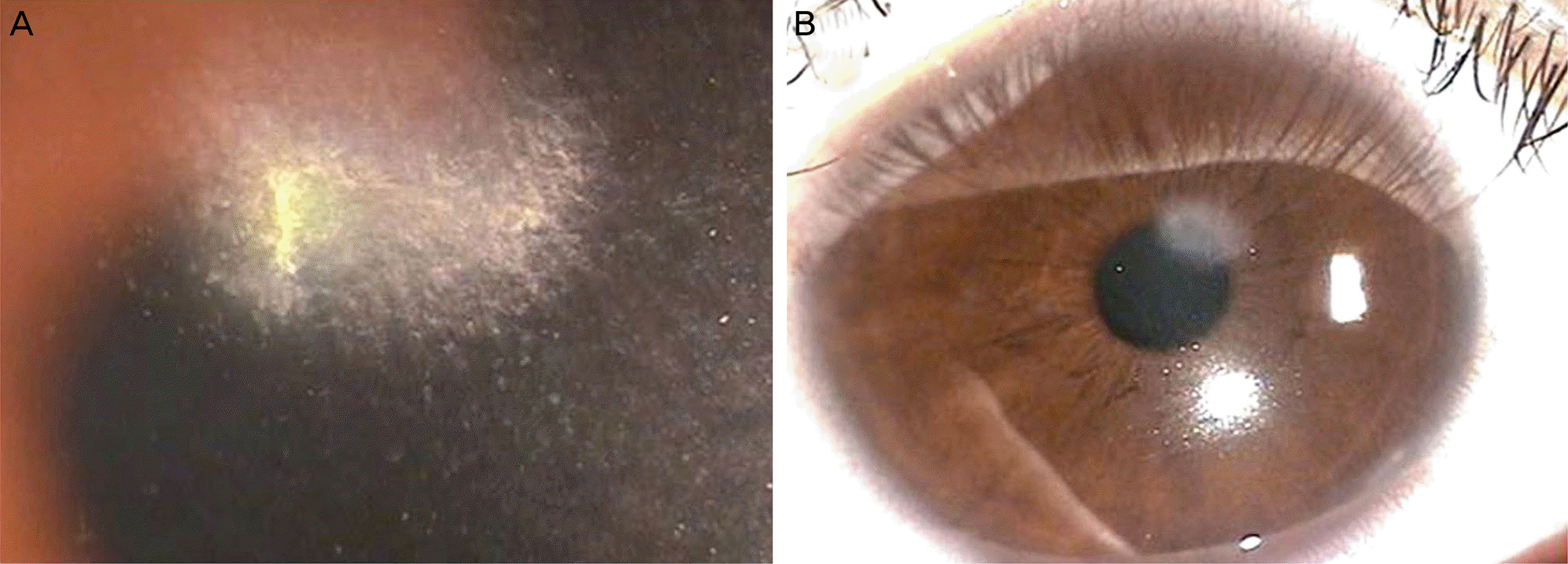 | Figure 1.(A) Slit-lamp photograph shows the presence of crystal-like white stromal infiltrations with epithelial defect at the center. (B) Six month after treatment. |
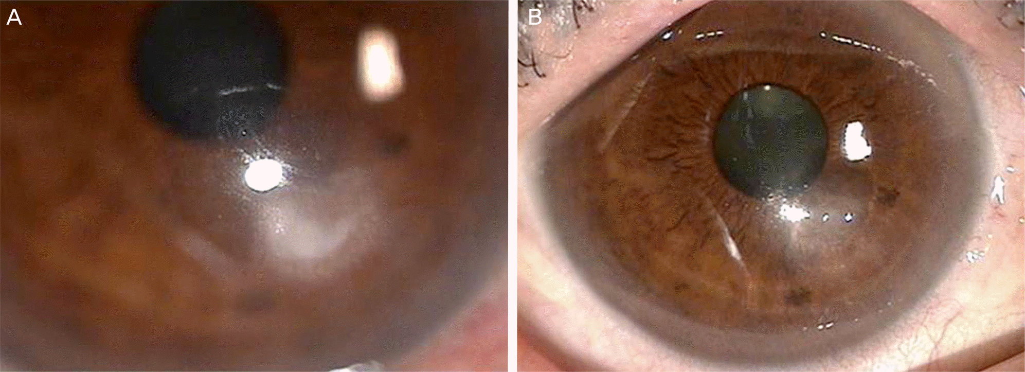 | Figure 2.(A) Corneal infiltration in the anterior stroma with conjunctival injection. (B) Two month after treatment. |
 | Figure 3.Anterior segment optical coherence tomography (AS-OCT) shows relatively well demarcated horizontal infiltration in the anterior stroma (A: patient 1; B: patient 2). |
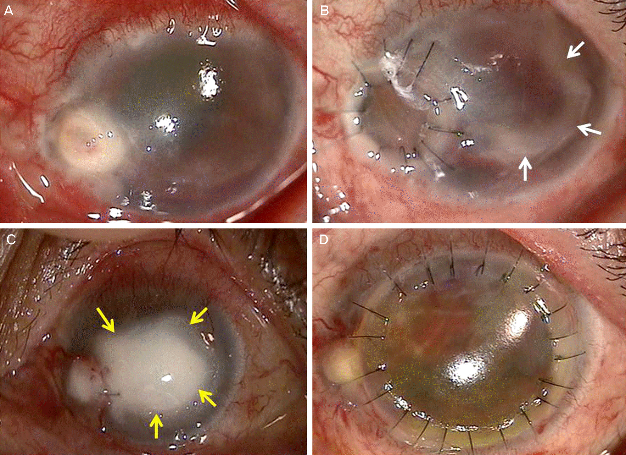 | Figure 4.(A) Corneal ulcer with descemetocele at the nasal area, infiltrations at superior limbus. (B) Two days after therapeutic partial keratoplasty, there is immune ring-like infiltration (white arrows). (C) Stromal necrosis at center of cornea (yellow arrows). (D) After therapeutic keratoplasty. |
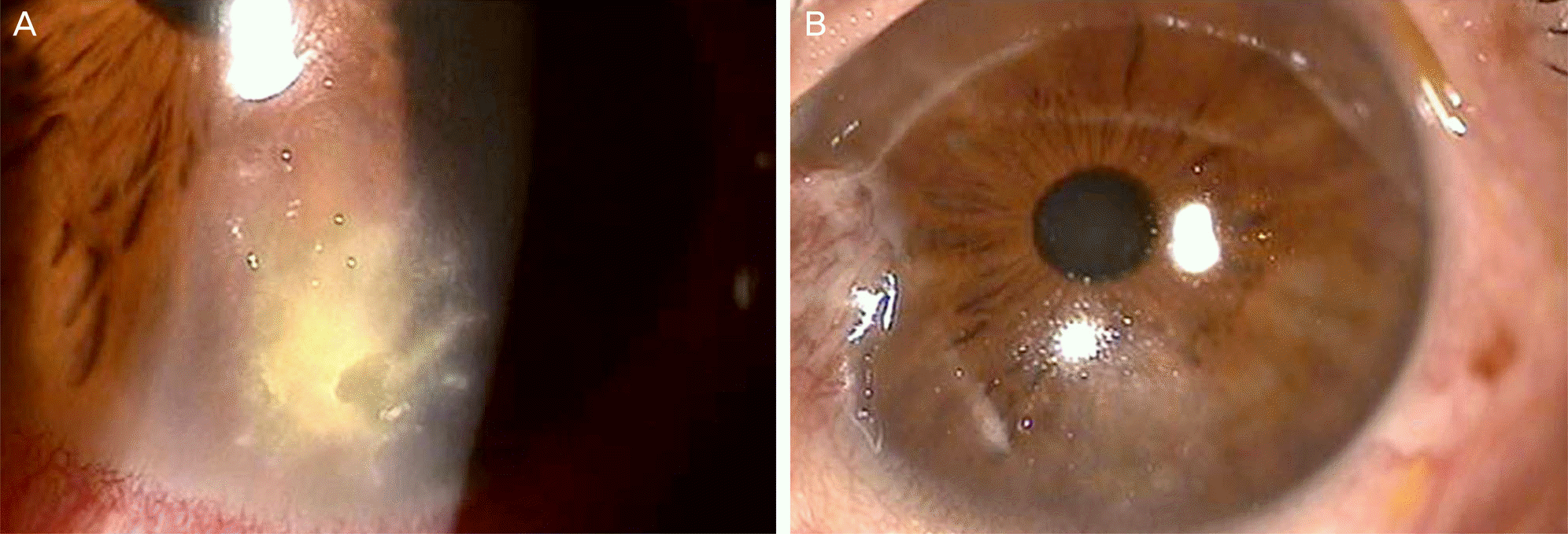 | Figure 5.(A) Corneal infiltration with feathery margin, epithelial defect at the center of infiltration and conjunctival injection. (B) Two month after treatment. |
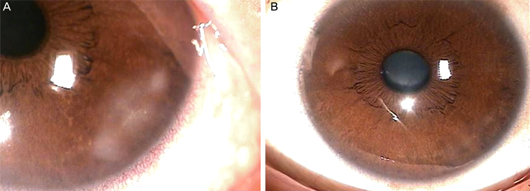 | Figure 6.(A) Peripheral corneal ulcer with stromal infiltration. (B) One month after treatment. |
Table 1.
Data from 5 cases of acinetobacter baumannii keratitis


 PDF
PDF ePub
ePub Citation
 Print
Print


 XML Download