

 PDF
PDF ePub
ePub Citation
 Print
Print


초록
Purpose:
To evaluate the anterior chamber depth (ACD), extent of intraocular lens (IOL) tilt, and decentration and refractive error after transscleral fixation of IOL.
Methods:
We retrospectively reviewed the medical records of 17 cases with transscleral fixation of IOL (6 with aphakia, 5 with IOL dislocation, and 6 with lens subluxation). The acrylic IOL (MN60AC®) was fixated in 12 eyes and the polymethylmethacrylate IOL (CZ70BD®) was fixated in 5 eyes at 1.0 mm posterior from the limbus. We analyzed the ACD, extent of IOL tilt and decentration, manifest refraction, refractive error, higher order aberration, and corneal endothelium at 2 weeks, 1 month and 2 months postoperatively.
Results:
The mean ACD was 3.36 ± 0.11 mm, 3.30 ± 0.12 mm, and 3.27 ± 0.13 mm, the mean extent of IOL tilt was 4.61 ± 0.12°,4.65 ± 0.14°, and 4.60 ± 0.12° and the mean extent of IOL decentration was 0.43 ± 0.01 mm, 0.45 ± 0.01 mm, and 0.45 ± 0.01 mm at 2 weeks, 1 month and 2 months postoperatively, respectively in eyes with transscleral fixation of IOL. The ACD was shallower and the extent of IOL tilt and decentration was greater than with IOL in-the-bag insertion patients. The mean refractive errors were -0.55 ± 0.27 D, -0.63 ± 0.24 D, and -0.69 ± 0.19 D at the same period, respectively.
Conclusions:
Although postoperative refractive error is influenced by surgeon factors such as incision size, distance of fixation suture from limbus, and tightness of suture material, according to our results, an IOL 0.75 D more hyperopic than predicted should be selected in transscleral fixation of IOL at 1.0 mm posterior from the limbus. Additionally, each surgeon should assess their specific results and modify the lens calculations accordingly.
Go to : 
References
1. Hu BV, Shin DH, Gibbs KA, Hong YJ. Implantation of posterior chamber lens in the absence of capsular and zonular support. Arch Ophthalmol. 1988; 106:416–20.

2. Malbran ES, Malbran E Jr, Negri I. Lens guide suture for transport and fixation in secondary IOL implantation after intracapsular extraction. Int Ophthalmol. 1986; 9:151–60.
3. Stark WJ, Gottsch JD, Goodman DF, et al. Posterior chamber intraocular lens implantation in the absence of capsular support. Arch Ophthalmol. 1989; 107:1078–83.
4. Stark WJ, Goodman G, Goodman D, Gottsch J. Posterior chamber intraocular lens implantation in the absence of posterior capsular support. Ophthalmic Surg. 1988; 19:240–3.
5. Lee JH, Oh SY. Corneal endothelial cell loss from suture fixation of a posterior chamber intraocular lens. J Cataract Refract Surg. 1997; 23:1020–2.
6. Kjeka O, Bohnstedt J, Meberg K, Seland JH. Implantation of scler-al-fixated posterior chamber intraocular lenses in adults. Acta Ophthalmol. 2008; 86:537–42.
7. Chang JH, Lee JH. Long-term results of implantation of posterior chamber intraocular lens by suture fixation. Korean J Ophthalmol. 1991; 5:42–6.
8. Lewis JS. Ab externo sulcus fixation. Ophthalmic Surg. 1991; 22:692–5.
9. Lubniewski AJ, Holland EJ, Van Meter WS, et al. Histologic study of eyes with transsclerally sutured posterior chamber intraocular lenses. Am J Ophthalmol. 1990; 110:237–43.
10. Bellucci R, Pucci V, Morselli S, Bonomi L. Secondary implantation of angle-supported anterior chamber and scleral-fixated posterior chamber intraocular lenses. J Cataract Refract Surg. 1996; 22:247–52.
11. Steiner A, Steinhorst UH, Steiner M, et al. [Ultrasound biomicroscopy for localization of artificial lens haptics after transscleral suture fixation]. Ophthalmologe. 1997; 94:41–4.
12. Chang JH, Lee JH. Implantation of posterior chamber intraocular lenses by suture fixation without capsular and zonular support. Korean J Ophthalmol. 1989; 3:90–3.
13. Johnson SM. Results of exchanging anterior chamber lenses with sulcus-fixated posterior chamber IOLs without capsular support in penetrating keratoplasty. Ophthalmic Surg. 1989; 20:465–8.
14. Lee JH, Chang JH. Suture to limbus distances in eyes with a posterior chamber intraocular lens implanted by scleral fixation. J Cataract Refract Surg. 1993; 19:278–83.
15. Holladay JT. Evaluating the intraocular lens optic. Surv Ophthalmol. 1986; 30:385–90.
16. Engren AL, Behndig A. Anterior chamber depth, intraocular lens position, and refractive outcomes after cataract surgery. J Cataract Refract Surg. 2013; 39:572–7.
17. Sasaki K, Sakamoto Y, Shibata T, et al. Measurement of postoperative intraocular lens tilting and decentration using Scheimpflug images. J Cataract Refract Surg. 1989; 15:454–7.
18. Lee JG, Lee JH, Chung H. Factors contributing to retinal detachment after transscleral fixation of posterior chamber intraocular lenses. J Cataract Refract Surg. 1998; 24:697–702.
19. Krause L, Bechrakis NE, Heimann H, et al. Implantation of scleral fixated sutured posterior chamber lenses: a retrospective analysis of 119 cases. Int Ophthalmol. 2009; 29:207–12.
20. Heilskov T, Joondeph BC, Olsen KR, Blankenship GW. Late endophthalmitis after transscleral fixation of a posterior chamber intraocular lens. Arch Ophthalmol. 1989; 107:1427.
21. Teichmann KD, Teichmann IA. The torque and tilt gamble. J Cataract Refract Surg. 1997; 23:413–8.
22. Taskapili M, Gulkilik G, Engin G, et al. Transscleral fixation of a single-piece hydrophilic foldable acrylic intraocular lens. Can J Ophthalmol. 2007; 42:256–61.
23. Oshima Y, Oida H, Emi K. Transscleral fixation of acrylic intraocular lenses in the absence of capsular support through 3.5 mm self-sealing incisions. J Cataract Refract Surg. 1998; 24:1223–9.
24. Kohnen S, Ferrer A, Brauweiler P. Visual function in pseudophakic eyes with poly(methyl methacrylate), silicone, and acrylic intraocular lenses. J Cataract Refract Surg. 1996; 22(Suppl 2):1303–7.
25. Chan CK. An improved technique for management of dislocated posterior chamber implants. Ophthalmology. 1992; 99:51–7.
26. Fass ON, Herman WK. Sutured intraocular lens placement in aphakic post-vitrectomy eyes via small-incision surgery. J Cataract Refract Surg. 2009; 35:1492–7.
27. Duffey RJ, Holland EJ, Agapitos PJ, Lindstrom RL. Anatomic study of transsclerally sutured intraocular lens implantation. Am J Ophthalmol. 1989; 108:300–9.
28. Hayashi K, Hayashi H, Nakao F, Hayashi F. Intraocular lens tilt and decentration, anterior chamber depth, and refractive error after transscleral suture fixation surgery. Ophthalmology. 1999; 106:878–82.
29. Uozato H, Okada Y, Hirai H, Saishin M. What is the tolerable limits of the IOL tilt and decentration. Jpn Rev Clin Ophthalmol. 1988; 82:2308–11.
30. Uthoff D, Teichmann KD. Secondary implantation of scleral-fixated intraocular lenses. J Cataract Refract Surg. 1998; 24:945–50.
31. Bergren RL. Four-point fixation technique for sutured posterior chamber intraocular lenses. Arch Ophthalmol. 1994; 112:1485–7.
32. Almashad GY, Abdelrahman AM, Khattab HA, Samir A. Four-point scleral fixation of posterior chamber intraocular lenses without scleral flaps. Br J Ophthalmol. 2010; 94:693–5.
33. Berger RR, Kenyers AM, Van Coller B, Pretorius CF. Vertical tri-pod fixation (VTF) simplifies transscleral approaches. Ophthalmic Surg. 1995; 26:367–71.
Go to :
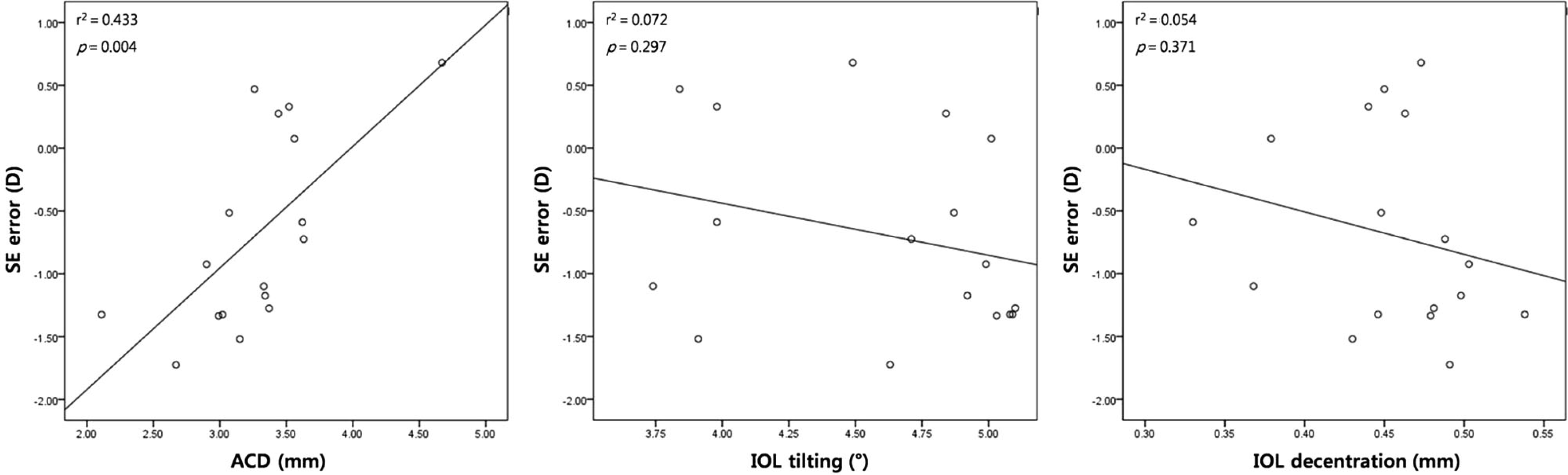 | Figure 1.Scatterplot of correlation between the anterior chamber depth, IOL decentration, and IOL tilting with spherical equivalent (SE) error at postoperative 2 months in eyes with transscleral fixation of IOL. ACD = anterior chamber depth; IOL = intraocular lens. |
Table 1.
Etiologies of transscleral fixation of IOL in patients of group A
| Etiology | Number (%) |
|---|---|
| Aphakia | 6 (35.29) |
| IOL dislocation | 5 (29.41) |
| Lens subluxation | |
| Trauma | 3 (17.65) |
|
Spontaneous |
3 (17.65) |
| Total | 17 |
Table 2.
Preoperative clinical characteristics of each group
| Group A (n = 17) | Group B (n = 39) | p-value | ||
|---|---|---|---|---|
| Sex (M:F) | 12:5 | 10:29 | 0.124 | |
| Age (years) | 53.71 ± 3.80 | 63.26 ± 2.01 | 0.348 | |
| Laterality (right eye:left eye) | 11:6 | 20:19 | 0.353 | |
| UCVA (log MAR) | 1.01 ± 0.09 | 0.51 ± 0.03 | <0.001* | |
| BCVA (log MAR) | 0.44 ± 0.06 | 0.38 ± 0.04 | 0.638 | |
| IOP (mm Hg) | 16.71 ± 1.00 | 14.08 ± 0.48 | 0.021* | |
| Axial length (mm) | 24.65 ± 0.47 | 23.75 ± 0.20 | 0.061 | |
| ACD (mm) | 3.15 ± 0.25 | 3.17 ± 0.07 | 0.922 | |
| Corneal endothelium | Cell density (cells/mm2) | 2,385.06 ± 163.70 | 2,665.46 ± 55.83 | 0.262 |
| Coefficient of variation | 35.12 ± 1.52 | 34.13 ± 0.78 | 0.195 | |
| Hexagonality | 53.65 ± 2.62 | 53.77 ± 1.54 | 0.858 | |
| Keratometry (diopter) | 1.47 ± 0.28 | 1.04 ± 0.10 | 0.260 | |
| Refraction (diopter) | SE | 1.23 ± 2.51 | -0.14 ± 0.49 | 0.864 |
| Cylinder | 1.30 ± 0.15 | 1.17 ± 0.17 | 0.121 |
Pearson's Chi-square test was used for numeric parameters; Mann Whitney U-test was used for continuous parameters; Values are presented as mean ± SD or number; Group A: transscleral fixation of IOL, Group B: phacoemulsification and in-the-bag insertion of IOL.
Table 3.
Comparison of postoperative anterior chamber depth, extent of IOL tilt, and decentration
| Group A (n = 17) | Group B (n = 39) | p-value | ||
|---|---|---|---|---|
| ACD (mm) | Preoperative | 3.15 ± 0.25 | 3.17 ± 0.07 | 0.922 |
| POD #2 weeks | 3.36 ± 0.11 | 4.14 ± 0.10† | <0.001* | |
| POD #1 month | 3.30 ± 0.12 | 4.19 ± 0.09† | <0.001* | |
| POD #2 months | 3.27 ± 0.13 | 4.23 ± 0.10† | <0.001* | |
| IOL tilt (°) | POD #2 weeks | 4.61 ± 0.12 | 1.24 ± 0.04 | <0.001* |
| POD #1 month | 4.65 ± 0.14 | 1.37 ± 0.04 | <0.001* | |
| POD #2 months | 4.60 ± 0.12 | 1.30 ± 0.04 | <0.001* | |
| IOL decentration (mm) | POD #2 weeks | 0.43 ± 0.01 | 0.12 ± 0.01 | <0.001* |
| POD #1 month | 0.45 ± 0.01 | 0.15 ± 0.01 | <0.001* | |
| POD #2 months | 0.45 ± 0.01 | 0.14 ± 0.01 | <0.001* |
Table 4.
Comparison of postoperative refraction (mean spherical and mean cylinder) and spherical equivalent error of each group over time
| Group A (n = 17) | Group B (n = 39) | p-value | ||
|---|---|---|---|---|
| Mean spherical (diopter) | Preoperative | 1.90 ± 2.52 | 0.43 ± 0.51 | 0.872 |
| POD #2 weeks | -0.05 ± 0.12 | -0.37 ± 0.13† | 0.079 | |
| POD #1 month | -0.26 ± 0.20 | -0.51 ± 0.13† | 0.380 | |
| POD #2 months | -0.45 ± 0.18 | -0.50 ± 0.15† | 0.957 | |
| Mean cylinder (diopter) | Preoperative | -1.30 ± 0.15 | -1.17 ± 0.17 | 0.121 |
| POD #2 weeks | -2.34 ± 0.26† | -0.83 ± 0.12† | <0.001* | |
| POD #1 month | -1.93 ± 0.26† | -0.84 ± 0.12† | <0.001* | |
| POD #2 months | -1.63 ± 0.20 | -0.90 ± 0.11 | 0.001* | |
| Spherical equivalent error (diopter) | POD #2 weeks | -0.55 ± 0.27 | -0.03 ± 0.08 | 0.022* |
| POD #1 month | -0.63 ± 0.24 | -0.17 ± 0.07 | 0.022* | |
| POD #2 months | -0.69 ± 0.19 | -0.20 ± 0.08 | 0.018* | |
| Astigmatism (algebraic value)(diopter) | Preoperative | -0.02 ± 0.37 | -0.21 ± 0.26 | 0.867 |
| POD #2 weeks | 2.19 ± 0.33† | -0.22 ± 0.18 | <0.001* | |
| POD #1 month | 1.84 ± 0.29† | -0.17 ± 0.18 | <0.001* | |
| POD #2 months | 1.15 ± 0.35† | -0.32 ± 0.17 | 0.001* |
Table 5.
Comparison of corneal astigmatism by Pentacam® of each group over time
|
Group A (n = 17) |
Group B (n = 39) |
p-value | ||||
|---|---|---|---|---|---|---|
| WTR | ATR | WTR | ATR | |||
| Keratometry (Pentacam®)(diopter) | Preoperative | 0.86 ± 0.16 | 1.15 ± 0.24 | 0.92 ± 0.14 | 1.08 ± 0.14 | 0.673/0.834 |
| POD #2 weeks | 2.01 ± 0.24† | 1.05 ± 0.15 | 1.02 ± 0.15 | 1.14 ± 0.15 | 0.002*/0.842 | |
| POD #1 month | 1.63 ± 0.21 | 1.70 ± 0.61 | 0.91 ± 0.13 | 0.98 ± 0.12 | 0.010*/0.145 | |
| POD #2 months | 1.37 ± 0.16 | 1.30 ± 0.43 | 1.01 ± 0.10 | 1.15 ± 0.16 | 0.067/0.763 | |
Table 6.
Comparison of postoperative outcomes by the type of IOL used in patients with transscleral fixation of IOL
 XML Download