

 PDF
PDF ePub
ePub Citation
 Print
Print


초록
Case summary:
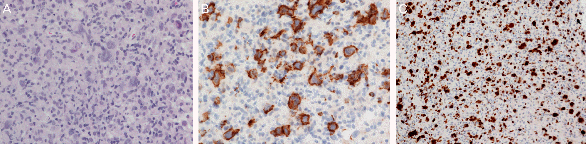
A 57-year-old female presented at our clinic with decreased vision in her right eye 1 month in duration. Slit-lamp examination showed vitreous cells in the right eye. Fundus examination of the right eye revealed an elevated yellowish mass-like lesion at the temporal area and multifocal yellowish patches at the choroidal level. The clinical impression was intraocular lymphoma. We performed diagnostic vitrectomy, but the result was negative. A systemic evaluation revealed enlarged cervical lymph nodes and lymph node biopsy showed diffuse large B-cell lymphoma. The patient was diagnosed as secondary choroidal lymphoma associated with systemic lymphoma and was treated with systemic chemotherapy and 4 injections of intravitreal methotrexate. The patient is scheduled for regular follow-ups.
References
1. Chan CC, Rubenstein JL, Coupland SE, et al. Primary vitreoretinal lymphoma: a report from an International Primary Central Nervous System Lymphoma Collaborative Group symposium. Oncologist. 2011; 16:1589–99.

2. Turaka K, Bryan JS, De Souza S, et al. Vitreoretinal lymphoma: changing trends in diagnosis and local treatment modalities at a single institution. Clin Lymphoma Myeloma Leuk. 2012; 12:412–7.
3. Coupland SE, Damato B. Understanding intraocular lymphomas. Clin Experiment Ophthalmol. 2008; 36:564–78.
4. Apte RS, Al-Abdulla NA, Green WR, et al. Systemic non-Hodgkin B-cell lymphoma encountered as a vanishing choroidal mass. Arch Ophthalmol. 2005; 123:105–9.
5. Leff SR, Shields JA, Augsburger JJ, et al. Unilateral eyelid, conjunctival, and choroidal tumours as initial presentation of diffuse large-cell lymphoma. Br J Ophthalmol. 1985; 69:861–4.
6. Triebenstein O. Ein beitrag zur frage der aleukämischen augen- veränderungen. Klin Monatsbl Augenheilkd. 1920; 64:825–36.
7. Mashayekhi A, Shukla SY, Shields JA, Shields CL. Choroidal lymphoma: clinical features and association with systemic lymphoma. Ophthalmology. 2014; 121:342–51.
8. Shields CL, Arepalli S, Pellegrini M, et al. Choroidal lymphoma shows calm, rippled, or undulating topography on enhanced depth imaging optical coherence tomography in 14 eyes. Retina. 2014; 34:1347–53.
9. Gass JD, Weleber RG, Johnson DR. Non-Hodgkin's lymphoma causing fundus picture simulating fundus flavimaculatus. Retina. 1987; 7:209–14.
10. Rattray KM, Cole MD, Smith SR. Systemic non-Hodgkin's lymphoma presenting as a serpiginous choroidopathy: report of a case and review of the literature. Eye (Lond). 2000; 14(Pt 5):706–10.
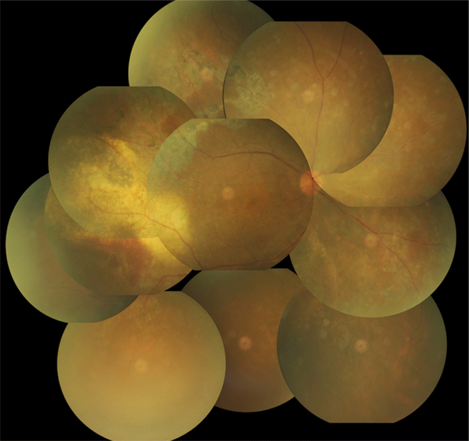
Figure 1.
(A) Fundus photograph reveals a yellowish, elevated choroidal mass in the temporal posterior pole of the right eye, measuring 4.0 × 8.0 disc diameter. Multifocal yellowish choroidal infiltrates are also found. (B) Ultrasonography of the right eye shows a smooth surface with acoustically hollow thickening of the choroid and low to moderate internal reflectivity. (C) Fluorescein angiography of the right eye shows blocked fluorescence corresponding to choroidal mass lesion and multiple punctate hyper-fluorescent lesions intermingling with hypofluorescent lesions at the level of retinal pigment epithelium.
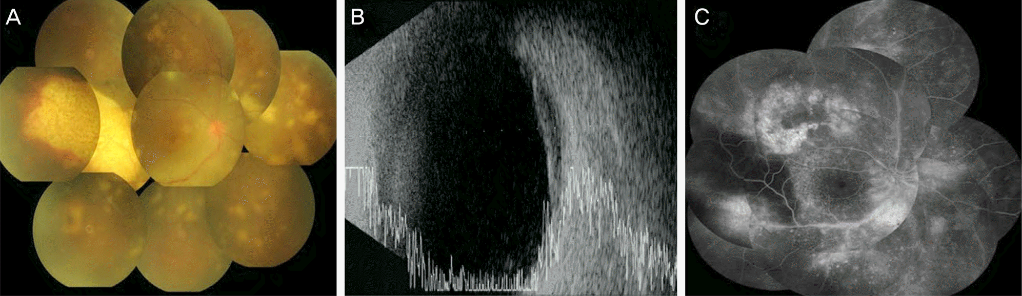
Figure 2.
(A) A vertical optical coherence tomography scan across the central macula (arrow) shows multiple nodular elevations of the retinal pigment epithelium. (B) A horizontal optical coherence tomography scan across the choroidal mass lesion (arrow) shows a dome-shaped large mass with overlying retinal pigment epithelium thinning occupying the choroid, pushing the retina into the vitreous cavity.
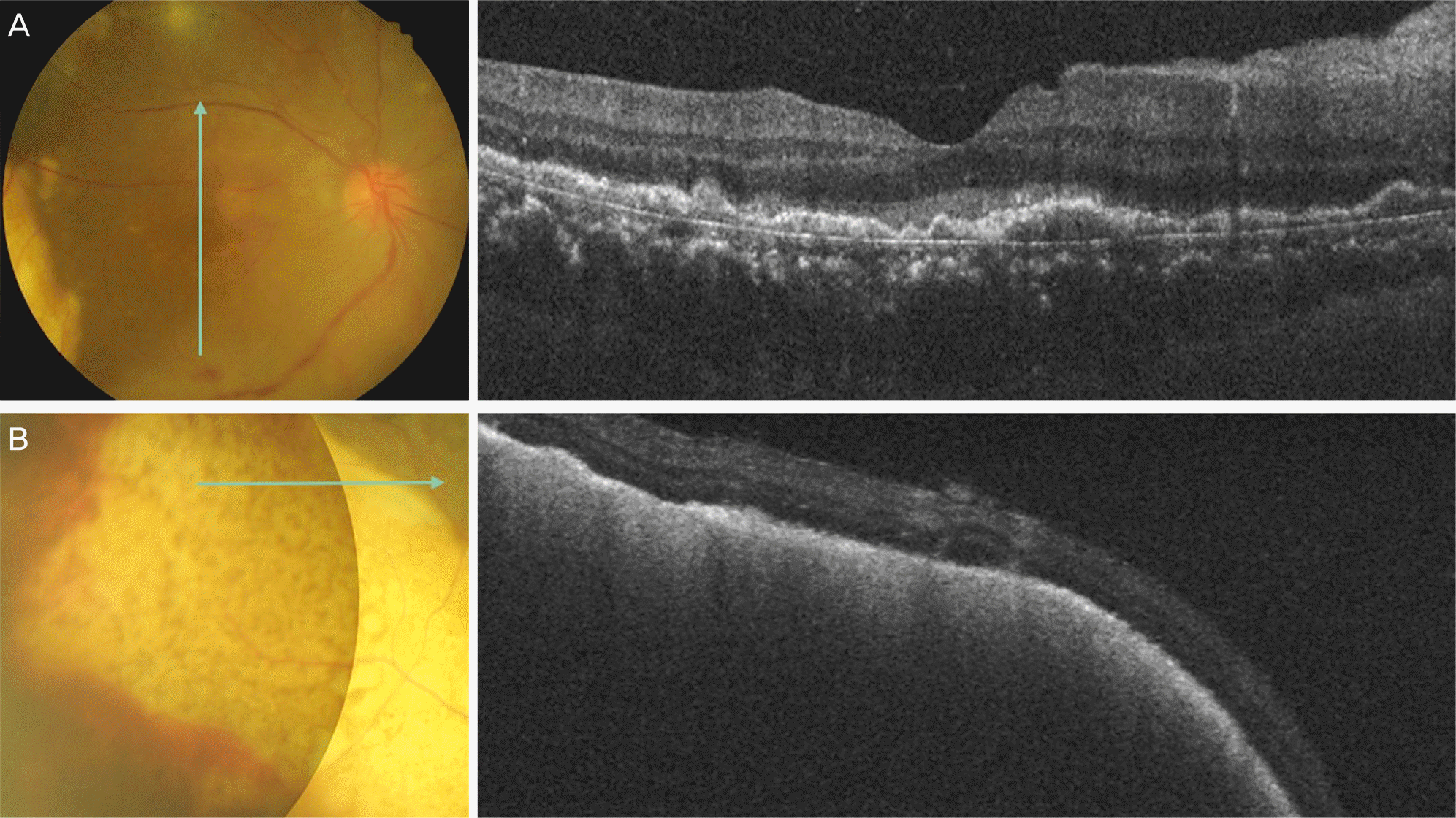
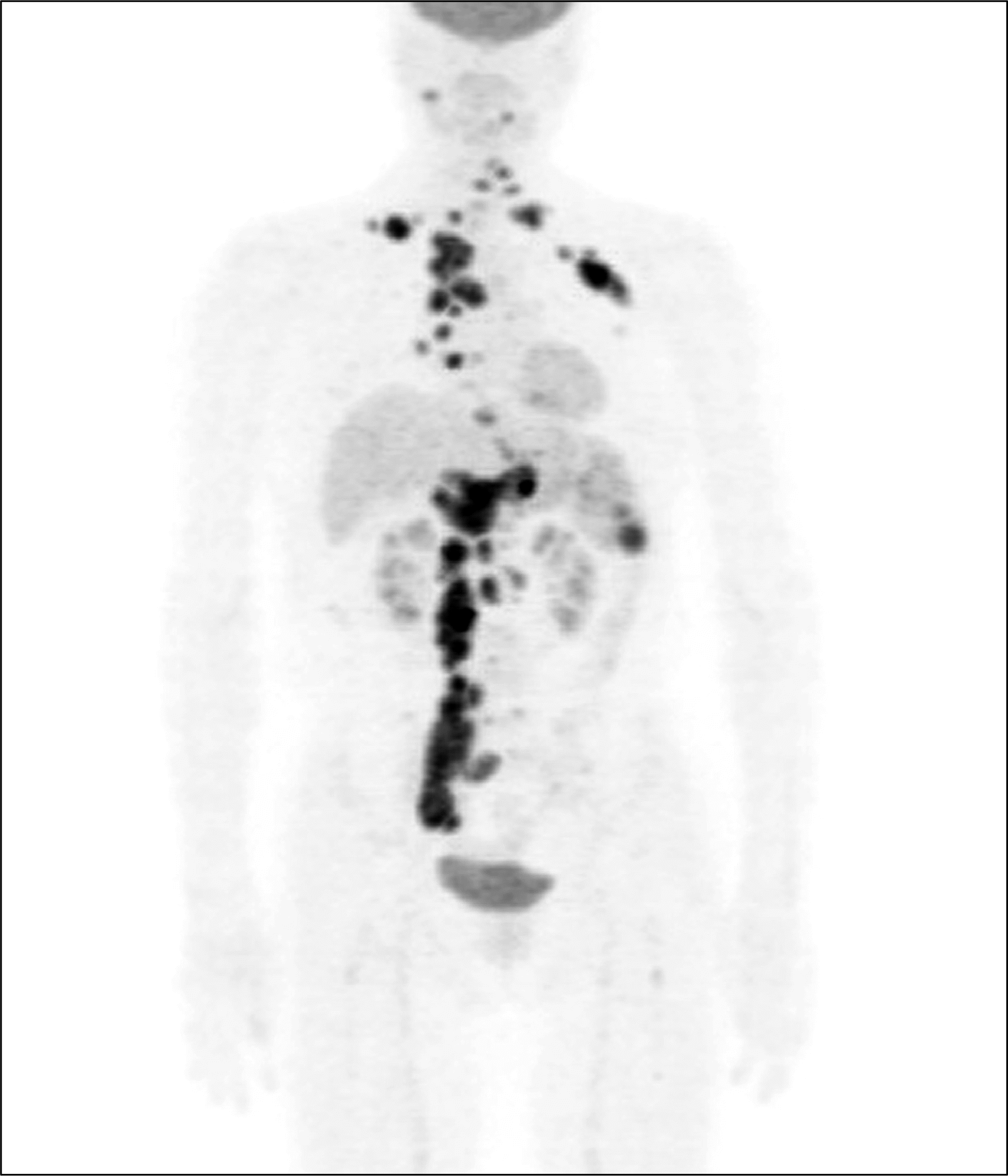
Figure 3.
Positron emission tomography. Probably multiple malignant lesions on both the supraclavicular and neck lymph nodes (LNs), left axillary LNs, internal mammary LNs, car-diophrenic LNs, upper abdominal paraaortic LNs and right iliac LNs and spleen and bone involvements are shown.
 XML Download