초록
Case summary:
An 83-year-old female who had uncontrolled Mooren’s ulcer invading 360 degrees of the limbus with corneal opacity received a 360-degree keratolimbal allograft (KLAL). Another 63-year-old female who had central corneal opacity and corneal neovascularization due to severe limbal cell deficiency with chemical injury received a 360-degree KLAL. During the average 17.5 months of follow-up, both eyes were tectonically maintained without severe graft rejection.
References
1. Tsai RJ, Tseng SC. Human allograft limbal transplantation for corneal surface reconstruction. Cornea. 1994; 13:389–400.

2. Son MG, Tchah HW. Combined surgery of the penetrating keratoplasty and the limbal cell transplantation. J Korean Ophthalmol Soc. 1997; 38:1112–20.
3. Lee DI, Kim KW, Kim JC. A case of autologous tragal perichon-drium graft in a patient with Mooren's ulcer. J Korean Ophthalmol Soc. 2014; 55:437–42.

4. Holland EJ, Schwartz GS, Daya SM, Djalilian A. Surgical techniques for ocular surface reconstruction. Krachmer JH, Mannis MJ, Holland EJ, editors. Cornea. 3rd ed. Elsevier;2011; v. 2:chap. 155. 1728–44.

5. Holland EJ, Schwartz GS. The evolution of epithelial transplantation for severe ocular surface disease and a proposed classification system. Cornea. 1996; 15:549–56.

7. Kwitko S, Marinho D, Barcaro S, et al. Allograft conjunctival transplantation for bilateral ocular surface disorders. Ophthalmology. 1995; 102:1020–5.

9. Holland EJ. Epithelial transplantation for the management of severe ocular surface disease. Trans Am Ophthalmol Soc. 1996; 94:677–743.

11. Turgeon PW, Nauheim RC, Roat MI, et al. Indications for keratoepithelioplasty. Arch Ophthalmol. 1990; 108:233–6.

12. Tsubota K, Toda I, Saito H, et al. Reconstruction of the corneal epithelium by limbal allograft transplantation for severe ocular surface disorders. Ophthalmology. 1995; 102:1486–96.

13. Yoon JT, Tchah HW. The surgical outcomes of limbal transplantation - long term follow up study. J Korean Ophthalmol Soc. 1999; 40:1793–803.
14. Kaçmaz RO, Kempen JH, Newcomb C, et al. Cyclosporine for ocular inflammatory diseases. Ophthalmology. 2010; 117:576–84.

Figure 1.
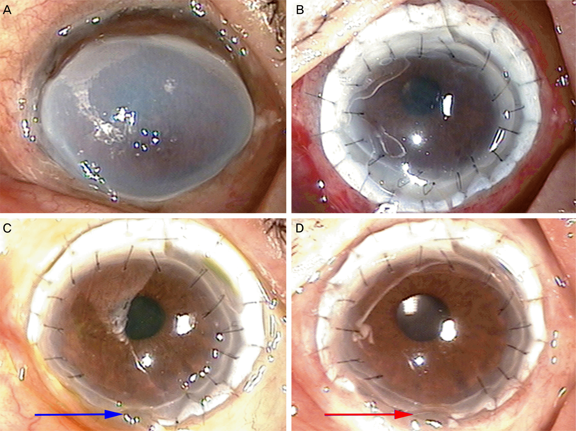
Slit lamp photograph shows a 83-year-old female with Mooren’s ulceration. (A) Peripheral corneal ulceration involving 360-degree limbus with accompanying corneal opacity was noted. (B) 1 day after surgery, well adapted keratolimbal allograft was noted. (C, D) 6 months after sugery, there was no signs of graft failure and corneal transparency was maintained. 19 months after surgery, inferior corneal thinning (blue arrow) occurred at 6 months after surgery but remained stable (red arrow). Visual improvement from hand-motion to 0.3 (best corrected visual acuity) was noted.

Figure 2.
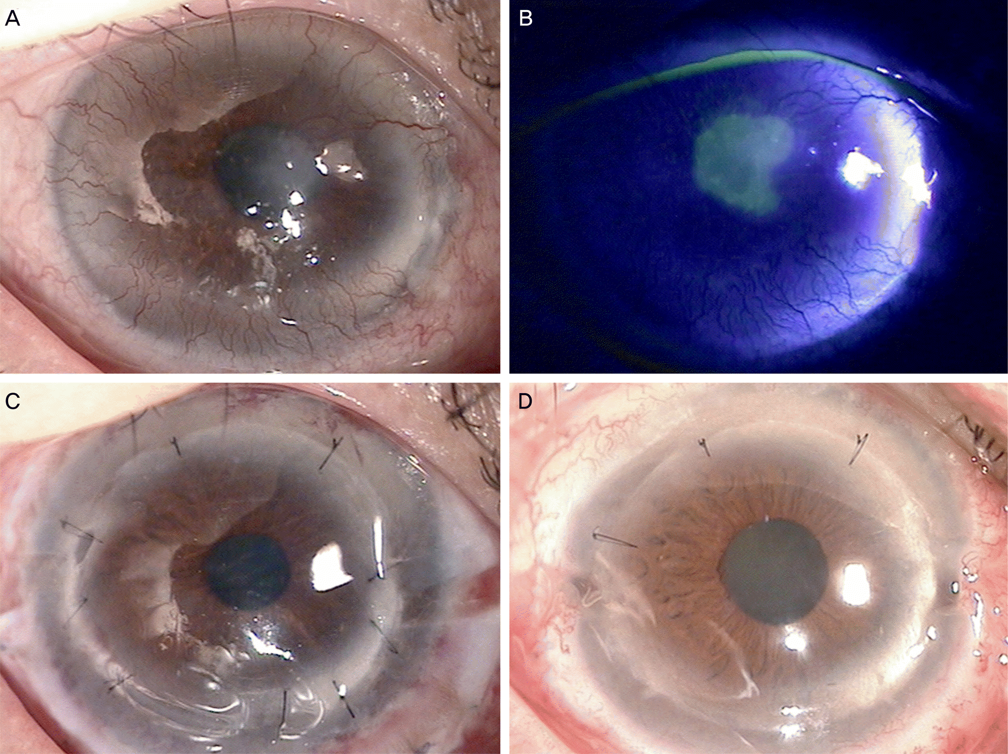
Slit lamp photograph shows a 63-year-old female who has severe limbal cell deficiency after chemical burn injury of the cornea. (A, B) Peripheral corneal neovascularization involving 360-degree and central corneal epithelial defects and opacity were noted. (C) 1 day after surgery, well adapted keratolimbal allograft was noted. (D) 10 months after sugery, there was no sign of graft failure and corneal transparency was maintained. Visual improvement from 0.02 to 0.6 (best corrected visual acuity) was noted.

Figure 3.
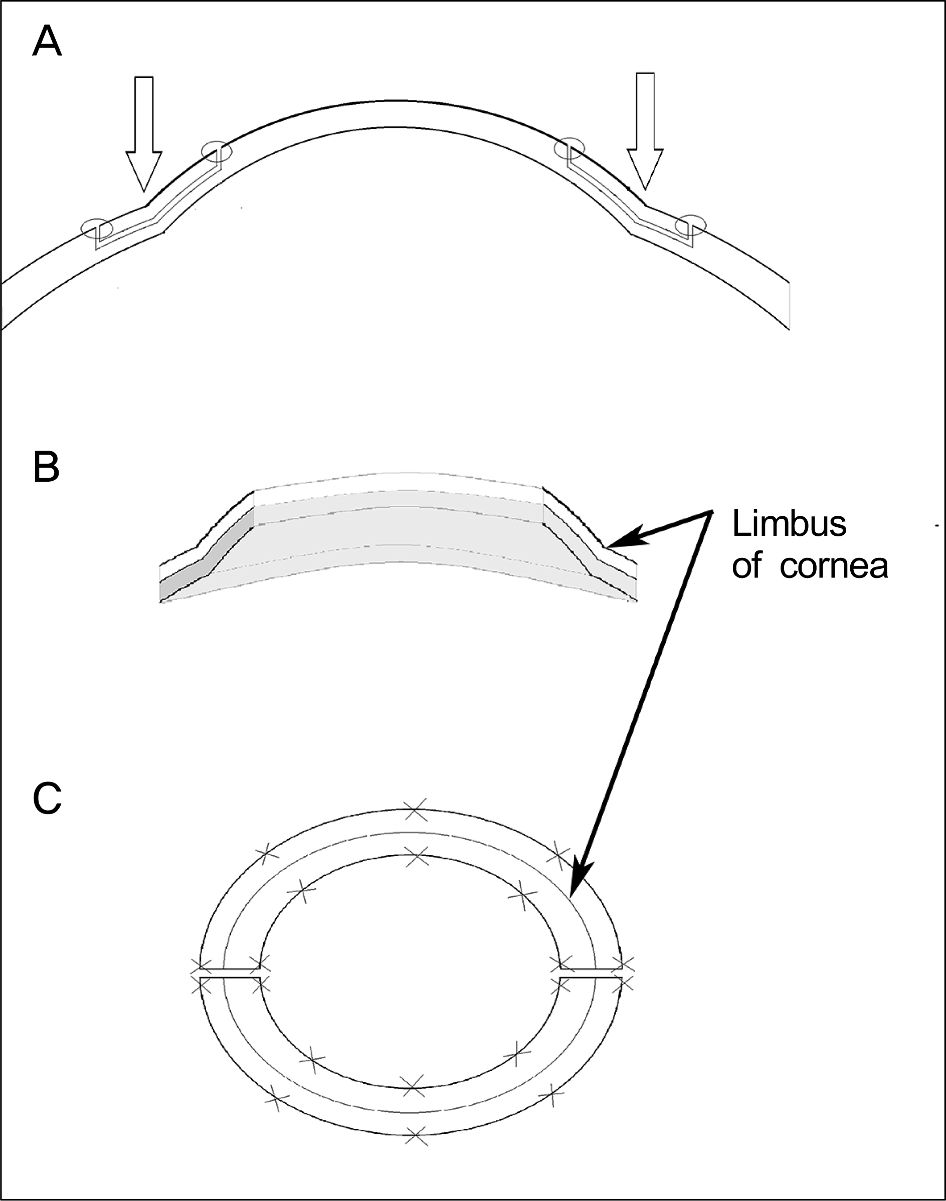
Schematic diagram of keratolimbal allograft procedure. Donor’s keratolimbal allograft lenticules are provided from 2 cadaver corneoscleral rims with the central cornea removed by trephination (C). Posterior part (2/3 thickness) of keratolimbal allograft lenticules (grey area) are removed (B). Conjunctival peritomy for 360 degrees and ten-ectomy is performed and the conjunctival recession is performed on the recipient. Abnormal corneal epithelium and abnormal recipient keratolimbal area (anterior 1/3 thickness) are removed. Keratolimbal lenticules (blank arrow) are secured to the recipient limbus using 10-0 nylon interrupted sutures (A, C). After applying therapeutic lens to the cornea, operation is done.


 PDF
PDF ePub
ePub Citation
 Print
Print


 XML Download