초록
Purpose:
To compare the outcomes of phacovitrectomy and phacoemulsification after vitrectomy for treatment of rhegmatogenous retinal detachment (RRD).
Methods:
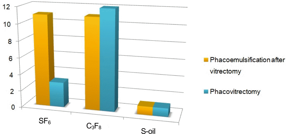
We performed a retrospective comparative analysis of 39 consecutive eyes with phakic primary RRD followed up for more than 6 months. The patients were divided into phacoemulsifcation after vitrectomy and phacovitrectomy groups. The main outcome measures were the best corrected visual acuity (BCVA), anatomical success rate and postoperative complications.
Results:
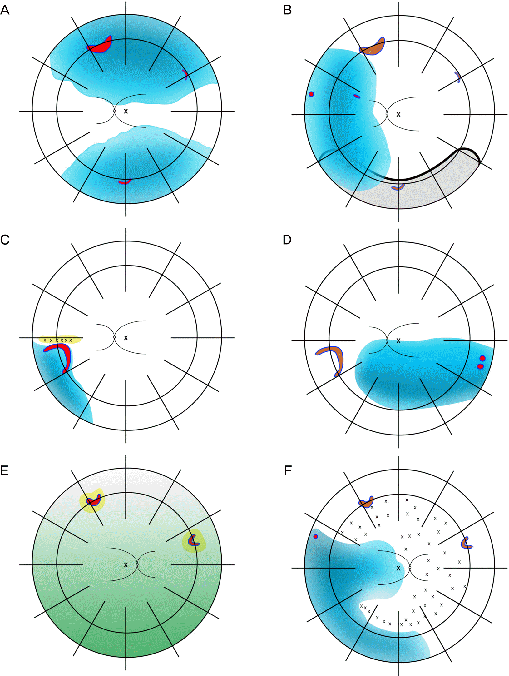
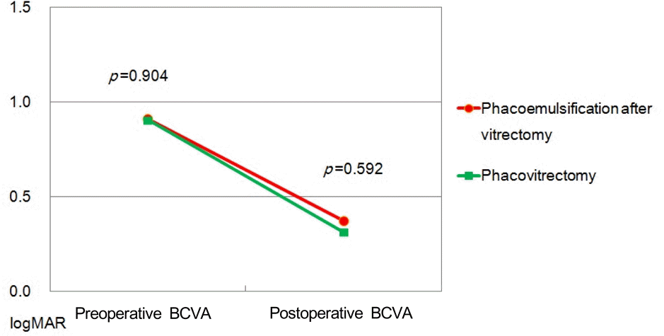
The mean age was 54.17 years in the phacoemulsifcation after vitrectomy group (n = 23) and 56.69 years in the phacovitrectomy group (n = 16; p = 0.031). The log MAR BCVA improved in both groups with no statistically significant difference between the 2 groups ( p = 0.592). The anatomical success rate after initial surgical intervention was 100% in both groups. Retinal detachment recurred in 3 eyes in the phacoemulsifcation after vitrectomy group; caused by new retinal tear.
Conclusions:
The new RRD rate in phacoemulsification after vitrectomy group was higher than in the phacovitrectomy group. Due to the retrospective and limited data in this study, whether simultaneous combined cataract surgery with retinal detachment surgery should be recommended to reduce RRD risk is inconclusive and further larger, prospectively designed studies are nec-essary to confirm the present findings.
Go to : 
References
1. Schaal S, Sherman MP, Barr CC, Kaplan HJ. Primary retinal detachment repair: comparison of 1-year outcomes of four surgical techniques. Retina. 2011; 31:1500–4.
2. Díaz Lacalle V, Orbegozo Gárate FJ, Martinez Alday N, et al. Phacoemulsification cataract surgery in vitrectomized eyes. J Cataract Refract Surg. 1998; 24:806–9.
3. Szijarto Z, Haszonits B, Biró Z, Kovacs B. Phacoemulsification on previously vitrectomized eyes: results of a 10-year-period. Eur J Ophthalmol. 2007; 17:601–4.
4. Cole CJ, Charteris DG. Cataract extraction after retinal detachment repair by vitrectomy: visual outcome and complications. Eye (Lond). 2009; 23:1377–81.
5. Braunstein RE, Airiani S. Cataract surgery results after pars plana vitrectomy. Curr Opin Ophthalmol. 2003; 14:150–4.

6. Ahfat FG, Yuen CH, Groenewald CP. Phacoemulsification and intraocular lens implantation following pars plana vitrectomy: a prospective study. Eye (Lond). 2003; 17:16–20.

7. Demetriades AM, Gottsch JD, Thomsen R, et al. Combined phacoemulsification, intraocular lens implantation, and vitrectomy for eyes with coexisting cataract and vitreoretinal pathology. Am J Ophthalmol. 2003; 135:291–6.

8. Foster RE, Lowder CY, Meisler DM, et al. Combined extracapsular cataract extraction, posterior chamber intraocular lens implantation, and pars plana vitrectomy. Ophthalmic Surg. 1993; 24:446–52.

9. Gu BY, Sagong M, Chang WH. Phacovitrectomy versus vitrectomy only for primary rhegmatogenous retinal detachment repair. J Korean Ophthalmol Soc. 2011; 52:537–43.

10. Koh TH, Choi MJ, Cho SW, et al. Scleral buckling and primary vitrectomy in simple rhegmatogenous retinal detachment. J Korean Ophthalmol Soc. 2010; 51:366–71.

11. Heimann H, Zou X, Jandeck C, et al. Primary vitrectomy for rhegmatogenous retinal detachment: an analysis of 512 cases. Graefes Arch Clin Exp Ophthalmol. 2006; 244:69–78.

12. Mendrinos E, Dang-Burgener NP, Stangos AN, et al. Primary vitrectomy without scleral buckling for pseudophakic rhegmatogenous retinal detachment. Am J Ophthalmol. 2008; 145:1063–70.

13. McDermott ML, Puklin JE, Abrams GW, Eliott D. Phacoemulsification for cataract following pars plana vitrectomy. Ophthalmic Surg Lasers. 1997; 28:558–64.

14. Grusha YO, Masket S, Miller KM. Phacoemulsification and lens implantation after pars plana vitrectomy. Ophthalmology. 1998; 105:287–94.

15. Chung TY, Chung H, Lee JH. Combined surgery and sequential surgery comprising phacoemulsification, pars plana vitrectomy, and intraocular lens implantation: comparison of clinical outcomes. J Cataract Refract Surg. 2002; 28:2001–5.
16. Smith M, Raman SV, Pappas G, et al. Phacovitrectomy for primary retinal detachment repair in presbyopes. Retina. 2007; 27:462–7.

17. Byon IS, Pak KY, Lee SM, et al. Lens-save versus phacoemulsification with intraocular lens implantation in primary vitrectomy for phakic rhegmatogenous retinal detachment. J Korean Ophthalmol Soc. 2013; 54:449–55.

18. Tuft SJ, Minassian D, Sullivan P. Risk factors for retinal detachment after cataract surgery: a case-control study. Ophthalmology. 2006; 113:650–6.
19. Pastor JC. Proliferative vitreoretinopathy: an overview. Surv Ophthalmol. 1998; 43:3–18.
Go to : 
 | Figure 3.Fundus findings before vitrectomy surgery (A, C, E) and after cataract surgery (B, D, F) of three patients with retinal redetachment after cataract surgery. E: Two horseshoe tears are observed but definite retinal detachment border is not identified due to vitreous hemorrhage. Blue: detached retina; red: new retinal tear; brown: old retinal tear; green: vitreous hemorrhage. |
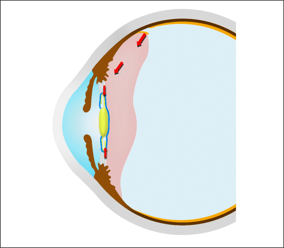
 | Figure 4.New retinal tear caused by traction (red arrow) on any remaining anterior hyaloids. |
Table 1.
Demographics and clinical data of patients
Table 2.
Severity of retinal detachment of two groups
Table 3.
Postoperative BCVA and complications of two groups
Table 4.
Summary of redetachment cases


 PDF
PDF ePub
ePub Citation
 Print
Print


 XML Download