

 PDF
PDF ePub
ePub Citation
 Print
Print


초록
Purpose:
To evaluate the tear meniscus in aqueous tear-deficient dry eye patients using Fourier-domain optical coherence tomography (FD-OCT) and to investigate the clinical usefulness of tear meniscus values.
Methods:
The present study included 79 aqueous tear-deficient dry eyes and 50 normal eyes. Tear meniscus height (TMH), tear meniscus depth (TMD), and tear meniscus area (TMA) were imaged using FD-OCT and measured with computer calipers. Schirmer’s test, tear break-up time, and corneal fluorescein staining were also performed and the correlations between the tests were analyzed. Additionally, the diagnostic power of tear meniscus values was compared using area under the receiver operating characteristic curve (AUROC).
Results:
Tear meniscus values were significantly decreased in the aqueous tear-deficient dry eye group ( p < 0.05). TMH, TMD, and TMA were positively correlated with Schirmer’s test and tear break-up time ( p < 0.05), and TMH and TMD were negatively correlated with corneal fluorescein staining in the aqueous tear-deficient dry eye group ( p < 0.05). The AUROCs of TMH, TMD, and TMA were 0.978, 0.788, and 0.957, respectively.
Conclusions:
Tear meniscus values measured using FD-OCT were significantly lower in aqueous tear-deficient dry eyes and were correlated with Schirmer’s test, tear break-up time, and corneal fluorescein staining. Tear meniscus measurements obtained using FD-OCT can be useful clinical parameters for the diagnosis and treatment of aqueous tear-deficient dry eye.
Go to : 
References
1. The definition and classification of dry eye disease: report of the Definition and Classification Subcommittee of the International Dry Eye WorkShop (2007). Ocul Surf. 2007; 5:75–92.
2. Tiffany JM. Surface tension in tears. Arch Soc Esp Oftalmol. 2006; 81:363–6.
3. Holly FJ. Physical chemistry of the normal and disordered tear film. Trans Ophthalmol Soc U K. 1985; 104(Pt 4):374–80.
4. Yokoi N, Bron AJ, Tiffany JM, et al. Relationship between tear vol-ume and tear meniscus curvature. Arch Ophthalmol. 2004; 122:1265–9.

5. Mainstone JC, Bruce AS, Golding TR. Tear meniscus measurement in the diagnosis of dry eye. Curr Eye Res. 1996; 15:653–61.
6. Yokoi N, Bron AJ, Tiffany JM, Kinoshita S. Reflective meniscom-etry: a new field of dry eye assessment. Cornea. 2000; 19(3 Suppl):S37–43.
7. Wang J, Aquavella J, Palakuru J, Chung S. Repeated measurements of dynamic tear distribution on the ocular surface after instillation of artificial tears. Invest Ophthalmol Vis Sci. 2006; 47:3325–9.
8. Wang J, Palakuru JR, Aquavella JV. Correlations among upper and lower tear menisci, noninvasive tear break-up time, and the Schirmer test. Am J Ophthalmol. 2008; 145:795–800.
9. Ibrahim OM, Dogru M, Takano Y, et al. Application of visante optical coherence tomography tear meniscus height measurement in the diagnosis of dry eye disease. Ophthalmology. 2010; 117:1923–9.
10. Nguyen P, Huang D, Li Y, et al. Correlation between optical coherence tomography-derived assessments of lower tear meniscus parameters and clinical features of dry eye disease. Cornea. 2012; 31:680–5.
11. Tung CI, Perin AF, Gumus K, Pflugfelder SC. Tear meniscus dimensions in tear dysfunction and their correlation with clinical parameters. Am J Ophthalmol. 2014; 157:301–10.e1.
12. Qiu X, Gong L, Sun X, Jin H. Age-related variations of human tear meniscus and diagnosis of dry eye with Fourier-domain anterior segment optical coherence tomography. Cornea. 2011; 30:543–9.
13. Sahai A, Malik P. Dry eye: prevalence and attributable risk factors in a hospital-based population. Indian J Ophthalmol. 2005; 53:87–91.
14. Calonge M, Diebold Y, Sáez V, et al. Impression cytology of the ocular surface: a review. Exp Eye Res. 2004; 78:457–72.
15. Nelson JD, Shimazaki J, Benitez-del-Castillo JM, et al. The international workshop on meibomian gland dysfunction: report of the definition and classification subcommittee. Invest Ophthalmol Vis Sci. 2011; 52:1930–7.
16. Bron AJ, Evans VE, Smith JA. Grading of corneal and conjunctival staining in the context of other dry eye tests. Cornea. 2003; 22:640–50.
17. Song JS, Hyon JY, Lee D, et al. Current practice pattern for dry eye patients in South Korea: a multicenter study. Korean J Ophthalmol. 2014; 28:115–21.
18. Hyon JY, Kim HM, Lee D, et al. Korean guidelines for the diagnosis and management of dry eye: development and validation of clinical efficacy. Korean J Ophthalmol. 2014; 28:197–206.
19. Halberg GP, Berens C. Standardized Schirmer tear test kit. Am J Ophthalmol. 1961; 51:840–2.
20. Clinch TE, Benedetto DA, Felberg NT, Laibson PR. Schirmer's test. A closer look. Arch Ophthalmol. 1983; 101:1383–6.
21. Maurice D. The Charles Prentice award lecture 1989: the physiol-ogy of tears. Optom Vis Sci. 1990; 67:391–9.
22. Pflugfelder SC, Tseng SC, Sanabria O, et al. Evaluation of subjective assessments and objective diagnostic tests for diagnosing tear-film disorders known to cause ocular irritation. Cornea. 1998; 17:38–56.
23. Kojima T, Ishida R, Dogru M, et al. A new noninvasive tear stability analysis system for the assessment of dry eyes. Invest Ophthalmol Vis Sci. 2004; 45:1369–74.
24. Zhou S, Li Y, Lu AT, et al. Reproducibility of tear meniscus measurement by Fourier-domain optical coherence tomography: a pilot study. Ophthalmic Surg Lasers Imaging. 2009; 40:442–7.
25. Keech A, Flanagan J, Simpson T, Jones L. Tear meniscus height determination using the OCT2 and the RTVue-100. Optom Vis Sci. 2009; 86:1154–9.
26. Hanada AL, de Souza EN Jr, Moribe I, Cruz AA. Comparison of palpebral fissure obliquity in three different racial groups. Ophthal Plast Reconstr Surg. 2001; 17:423–6.
27. Lemp MA, Crews LA, Bron AJ, et al. Distribution of aqueous-defi-cient and evaporative dry eye in a clinic-based patient cohort: a retrospective study. Cornea. 2012; 31:472–8.
28. Patel S, Bevan R, Farrell JC. Diurnal variation in precorneal tear film stability. Am J Optom Physiol Opt. 1988; 65:151–4.
29. Shen M, Wang J, Tao A, et al. Diurnal variation of upper and lower tear menisci. Am J Ophthalmol. 2008; 145:801–6.
Go to :
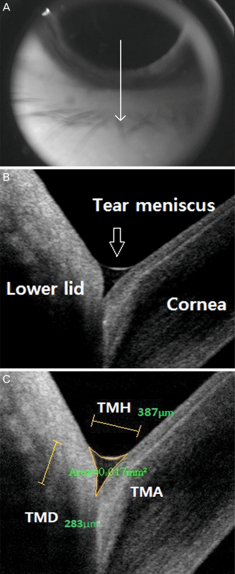 | Figure 1.(A) Image illustrating the 6 mm vertical scan path (arrowed line) of the optical coherence tomography beam to image the lower tear meniscus. (B) An optical coherence tomography vertical line scan cross-sectional image of the lower tear meniscus (arrow). (C) Optical coherence tomography image of the lower tear meniscus showing the tear meniscus height, depth and area. Measurements of TMH, TMD, TMA were performed using RTVue-100 software. TMH = tear meniscus height; TMD = tear meniscus depth; TMA = tear meniscus area. |
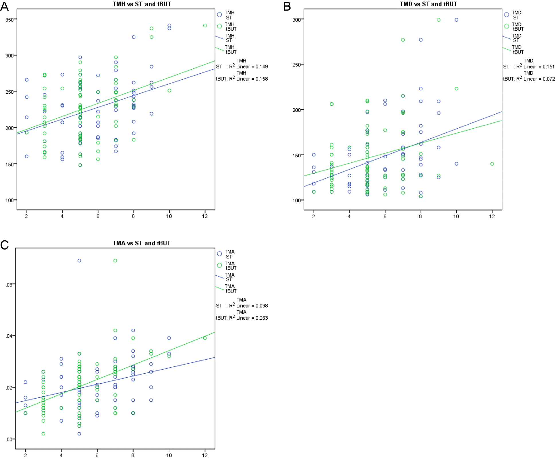 | Figure 2.(A) Scatter plot showing relationship between TMH versus Schirmer’s test (ST) and tear break-up time (tBUT) in aqueous tear-deficient dry eye patients. (B) Scatter plot showing relationship between TMD versus ST and tBUT in aqueous tear-deficient dry eye patients. (C) Scatter plot showing relationship between TMA versus ST and tBUT in aqueous tear-deficient dry eye patients. TMH = tear meniscus height; TMD = tear meniscus depth; TMA = tear meniscus area. |
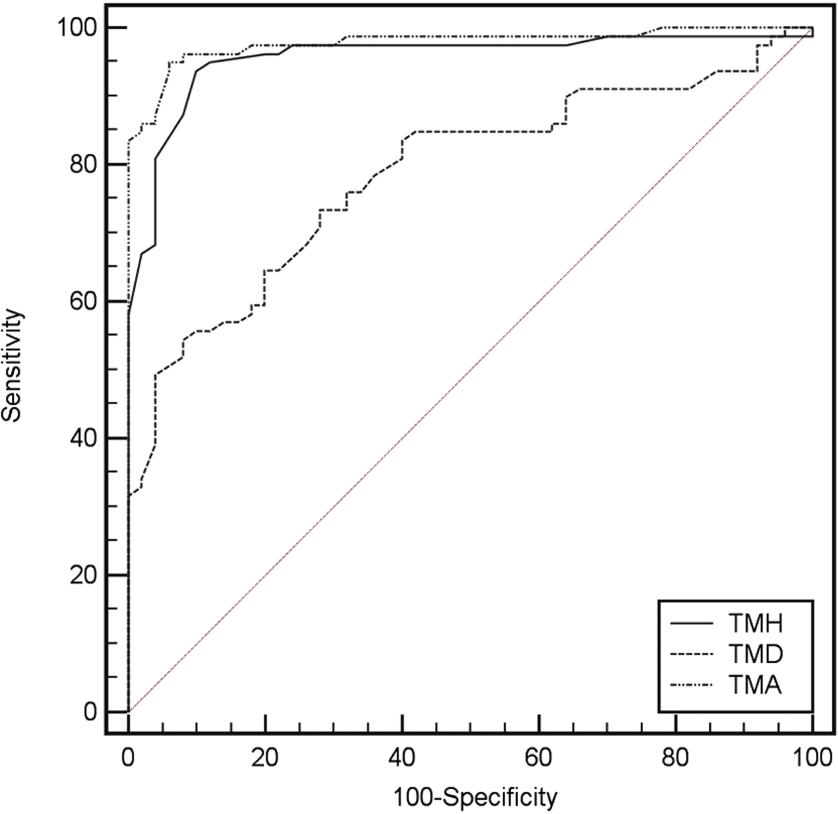 | Figure 3.Receiver operator characteristic curves for TMH, TMD, and TMA. TMH = tear meniscus height; TMD = tear meniscus depth; TMA = tear meniscus area. |
Table 1.
Average results of tear parameters in the aqueous tear-deficient dry eye patients and normal subjects
Table 2.
Correlation between tear meniscus dimensions in aqueous deficient dry eye patients and normal subjects
| OCT parameters | Conventional tests |
Aqueous tear-deficient dry eye (n = 79) |
Normal (n = 50) |
||
|---|---|---|---|---|---|
| p-value*s | p-value | p-value* | p-value | ||
| TMH | Schirmer | 0.386 | 0.000 | 0.850 | 0.000 |
| tBUT | 0.397 | 0.000 | 0.735 | 0.000 | |
| Staining | -0.289 | 0.010 | -0.817 | 0.000 | |
| TMD | Schirmer | 0.389 | 0.000 | 0.453 | 0.008 |
| tBUT | 0.269 | 0.017 | 0.255 | 0.152 | |
| Staining | -0.248 | 0.028 | -0.448 | 0.009 | |
| TMA | Schirmer | 0.312 | 0.005 | 0.662 | 0.000 |
| tBUT | 0.513 | 0.000 | 0.549 | 0.001 | |
| Staining | -0.168 | 0.139 | -0.706 | 0.000 | |
 XML Download