Abstract
Purpose
The treatment for reverse amblyopia is to discontinue occlusion therapy with most cases showing improvement of visual acuity in the dominant eye. Herein, we report a case of reverse amblyopia after monocular cataract surgery which was refractory to treatment and showed strabismus.
Case summary
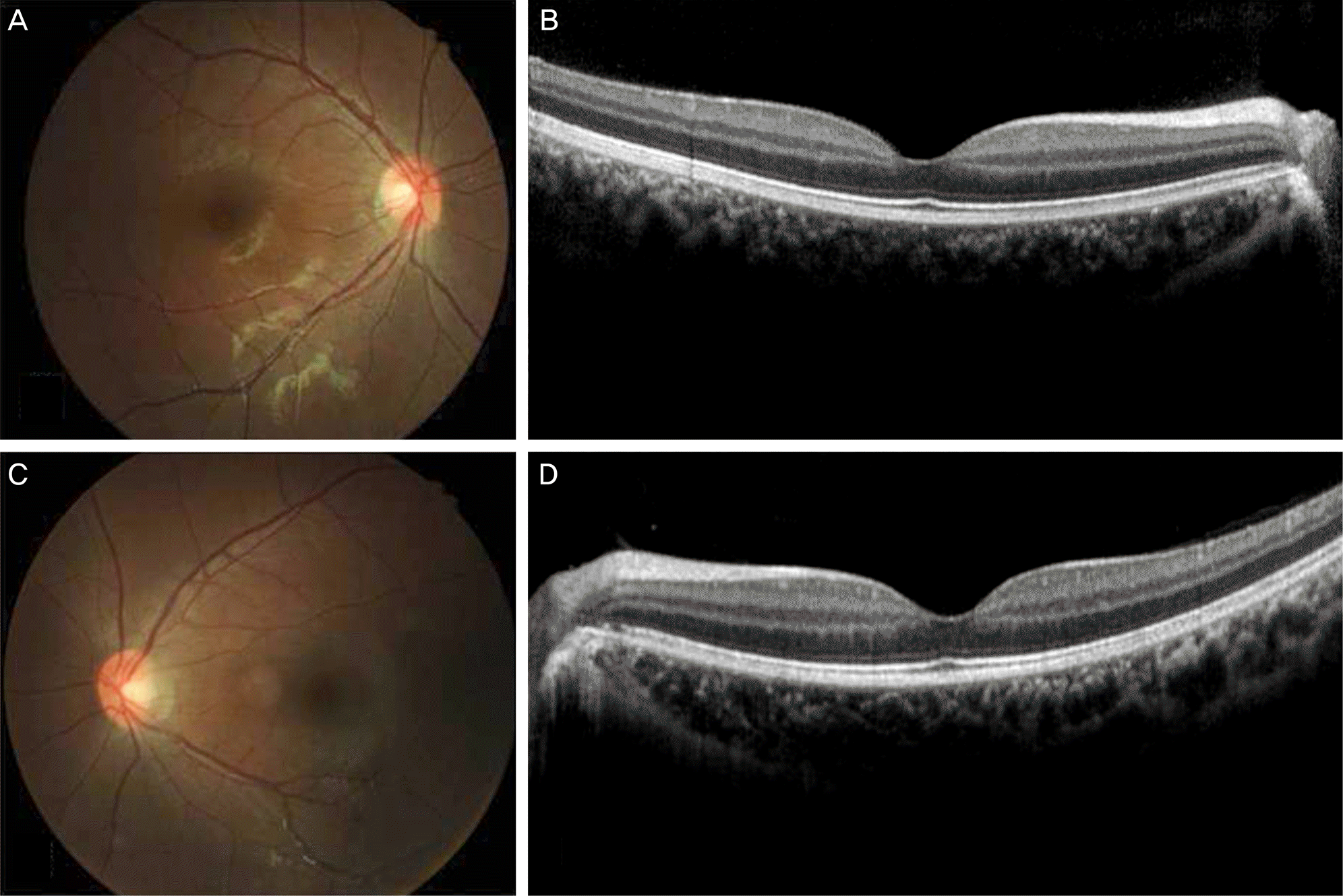
A 3-month-old female was diagnosed with congenital cataract in her left eye and underwent aspiration of lenses, posterior capsulectomy, and anterior vitrectomy. After the surgery, her mother performed strict 6:1 occlusion therapy on her right eye as prescribed. The best corrected visual acuity measured for the first time at the age of 32 months was 1.70 in the right eye and 0.52 in the left eye and the patient was referred to the Pediatric Ophthalmology clinic. At that time, eccentric fixation with slight exotropia was observed. With the diagnosis of reverse amblyopia in the right eye, the occlusion therapy was postponed for several months, however, visual acuity in the right eye did not recover after 4 months. After the age of 3 years, she was treated with left eye occlusion therapy, but the vision was still low and eccentric fixation was observed. At the age of 5 years she was continuously treated with left eye occlusion and the eccentric fixation improved, and at 6 years of age, a secondary intraocular lens implantation was performed. At 9 years of age, the patient underwent lateral rectus recession and medial rectus resection in the right eye for the treatment of exotropia.
Conclusions
In the case of monocular congenital cataract, occlusion therapy should be prescribed after surgical treatment. However, because reverse amblyopia which is refractory to treatment can occur, the fixation pattern should be monitored care-fully and the occlusion duration controlled appropriately.
Go to : 
References
1. Simons K. Amblyopia characterization, treatment, and prophylaxis. Surv Ophthalmol. 2005; 50:123–66.

2. von Noorden GK. Binocular vision and ocular motility. 6th ed.St. Louis: Mosby;2002. p. 246–97.
3. Korean Association for Pediatric Ophthalmology and Strabismus. Current concepts in strabismus. 2nd ed.Naewae Haksool;2008. p. 403–7.
4. Scott WE, Stratton VB, Fabre J. Full-time occlusion therapy for amblyopia. Am Orthopt J. 1980; 30:125–30.

5. Foley-Nolan A, McCann A, O'Keefe M. Atropine penalisation versus occlusion as the primary treatment for amblyopia. Br J Ophthalmol. 1997; 81:54–7.

6. Pediatric Eye Disease Investigator Group. A randomized trial of atropine vs. patching for treatment of moderate amblyopia in children. Arch Ophthalmol. 2002; 120:268–78.
7. Simons K, Stein L, Sener EC. . Full-time atropine, intermittent atropine, and optical penalization and binocular outcome in treatment of strabismic amblyopia. Ophthalmology. 1997; 104:2143–55.

9. Simons K. Preschool vision screening: rationale, methodology and outcome. Surv Ophthalmol. 1996; 41:3–30.

10. Oliver M, Gotesman N, Shimshoni M. Compliance and results of treatment for amblyopia in children more than 8 years old. CHILDREN. 1986; 197:75.

11. Searle A, Norman P, Harrad R, Vedhara K. Psychosocial and clinical determinants of compliance with occlusion therapy for am-blyopic children. Eye (Lond). 2002; 16:150–5.

12. Kim YH, Choi MY. The prospective comparison of the efficacy of intermittent atropine penalization and parttime occlusion therapy. J Korean Ophthalmol Soc. 2008; 49:958–66.

Go to : 


 PDF
PDF ePub
ePub Citation
 Print
Print


 XML Download