

 PDF
PDF ePub
ePub Citation
 Print
Print


Abstract
Purpose
We intended to investigate the clinical features and success rates of surgery in premature children with exotropia.
Methods
Twenty-two exotropia children with a gestational age of less than 37 weeks, or a birth weight of less than 2,000 g were included. The children had also been clinically observed for at least six months after surgery for exotropia. This study analyzed the results of their surgery in terms of gestational age, birth weight, ages at the first examination, and surgery, visual acuity, the angle of exodeviation at the first examination and the maximum angle of exodeviation before surgery, the frequency of accom-panied strabismus, and surgical results. Surgical success was defined as postoperative angle of deviation less than 10 PD. The patients were divided into success and failure groups, and a comparative analysis was performed between the 2 groups.
Results
The mean maximum angle of exodeviation before surgery was 28.4 ± 8.2 PD at far distance and 28.5 ± 8.3 PD at near distance. The mean angle of exodeviation at last visit after surgery was decreased to 8.0 ± 14.2 PD at far distance and 9.1 ± 14.8 PD at near distance. The success rate of surgery was measured to be 59.1% at the last visit. Recurrence was a major cause of failure, which occurred in eight cases (36.4%). The angle of exodeviation at the first examination and the maximum angle of exo-deviation in the failure group were higher than in the success group with statistical significance. The incidence of vertical strabismus before or after the surgery was 45.6% (10 cases) and there was no statistically significant difference between the 2 groups.
Conclusions
The premature children with exotropia had a relatively low surgical success rate and the major cause of the surgical failure was the recurrence of exotropia. The degree of the angle of deviation at the first examination and the maximum angle of exodeviation before surgery was an influential factor on the surgical results.
References
1. Keith CG, Kitchen WH. Ocular morbidity in infants of very low birth weight. Br J Ophthalmol. 1983; 67:302–5.

2. Cooke RW. Preterm mortality and morbidity over 25 years. Arch Dis Child Fetal Neonatal Ed. 2006; 91:F293–4.
3. Gallo JE, Lennerstrand G. A population-based study of ocular ab-normalities in premature children aged 5 to 10 years. Am J Ophthalmol. 1991; 111:539–47.
4. Pott JW, Van Hof-van Duin J, Heersema DJ. . Strabismus in very low birth weight and/or very preterm children: discrepancy between age of onset and start of treatment. Eur J Pediatr. 1995; 154:225–9.
5. McGinnity FG, Halliday HL. Perinatal predictors of ocular morbidity in school children who were very low birthweight. Paediatr Perinat Epidemiol. 1993; 7:417–25.
6. Lim KH, Yu YS, Chang BL. Clinical features of strabismus in premature baby. J Korean Ophthalmol Soc. 1997; 38:485–90.
7. Lim KH, Yu YS, Chang BL. Incidence and risk factors of strabismus in premature baby. J Korean Ophthalmol Soc. 1998; 39:1250–4.
8. Holmström G, Larsson E. Long-term follow-up of visual functions in prematurely born children--a prospective population-based study up to 10 years of age. J AAPOS. 2008; 12:157–62.
9. O'Connor AR, Stephenson TJ, Johnson A. . Change of refractive state and eye size in children of birth weight less than 1701 g. Br J Ophthalmol. 2006; 90:456–60.
10. Larsson EK, Rydberg AC, Holmström GE. A population-based study of the refractive outcome in 10-year-old preterm and full-term children. Arch Ophthalmol. 2003; 121:1430–6.
11. Bae JH, Choi DG. Refractive errors, amblyopia and strabismus in 3-year-old premature children. J Korean Ophthalmol Soc. 2010; 51:1385–91.
12. The Committee for the Classification of Retinopathy of Prematurity. An international classification of retinopathy of prematurity. Arch Ophthalmol. 1984; 102:1130–4.
13. The International Committee for the Classification of the Late Stages of Retinopathy of Prematurity. An international classification of retinopathy of prematurity. II. The classification of retinal detachment. Arch Ophthalmol. 1987; 105:906–12.
14. Wright KW, Ryan SJ. Color atlas of ophthalmic surgery: Strabismus. Philadelphia: Lippincott;1991. p. 241–3.
15. Archer SM, Sondhi N, Helveston EM. Strabismus in infancy. Ophthalmology. 1989; 96:133–7.
16. Kitchen WH, Ryan MM, Rickards A. . A longitudinal study of very low-birthweight infants. IV: An overview of performance at eight years of age. Dev Med Child Neurol. 1980; 22:172–88.
17. Koole FD, Bax PP, Samsom JF, van der Lei J. Ocular examination in nine-month-old infants with very low birthweights. Ophthalmic Paediatr Genet. 1990; 11:89–94.
18. Schaffer DB, Johnson L, Quinn GE. . Vitamin E and retinop-athy of prematurity. Follow-up at one year. Ophthalmology. 1985; 92:1005–11.
19. Cho YA, Shin HS, Joo HS, Jung HR. Surgical treatment of inter-mittent exotropia. J Korean Ophthalmol Soc. 1987; 28:1315–22.
20. Kwak MS, Kwon JY, Kim SY. A clinical study on exodeviation. J Korean Ophthalmol Soc. 1994; 35:95–102.
21. Lim HT, Jin YH. Concomitant hypertropia with intermittent exotropia. J Korean Ophthalmol Soc. 2001; 42:459–63.
22. Maly E. Frequency and natural history of retinopathy of prematurity (ROP). A prospective study in a Swedish city 1986-1990. Acta Ophthalmol Suppl. 1993; (210):52–5.
23. Ben-Sira I, Nissenkorn I, Weinberger D. . Long-term results of cryotherapy for active stages of retinopathy of prematurity. Ophthalmology. 1986; 93:1423–8.
24. Schaffer DB, Quinn GE, Johnson L. Sequelae of arrested mild retinopathy of prematurity. Arch Ophthalmol. 1984; 102:373–6.
25. Gibson NA, Fielder AR, Trounce JQ, Levene MI. Ophthalmic findings in infants of very low birthweight. Dev Med Child Neurol. 1990; 32:7–13.
26. Page JM, Schneeweiss S, Whyte HE, Harvey P. Ocular sequelae in premature infants. Pediatrics. 1993; 92:787–90.
27. Park SJ, Chang BL. Stabismus, amblyopia and refractive errors in patients with cerebral palsy. J Korean Ophthalmol Soc. 1999; 40:2898–903.
28. Hiles DA. Results of strabismus therapy in cerebral palsied children. Am Orthopt J. 1975; 25:46–55.
29. Yu YS, Kim SM, Kwon JY. . Preschool vision screening in Korea: preliminary study. J Korean Ophthalmol Soc. 1991; 32:1092–6.
30. Rhee KO, Rhee KI, Rhee KS. Preschool vision screening for amblyopia and refractive errors in Taejon. J Korean Ophthalmol Soc. 1999; 40:1375–84.
31. Kim KI, Ahn SK, Koo BS, Kim SJ. Preschool vision screening for 3 to 6-year old children in Seoul. J Korean Ophthalmol Soc. 2002; 43:714–27.
32. Pratt-Johnson JA, Barlow JM, Tillson G. Early surgery in inter-mittent exotropia. Am J Ophthalmol. 1977; 84:689–94.
33. Hardesty HH, Boynton JR, Keenan JP. Treatment of intermittent exotropia. Arch Ophthalmol. 1978; 96:268–74.
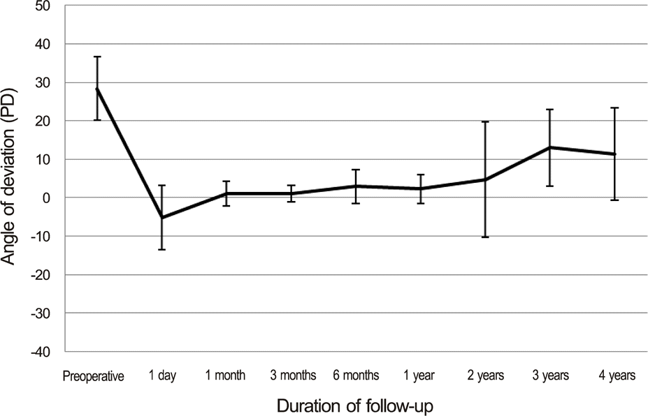
Figure 2.
Change of angle of deviation during the follow-up after surgery in 3 groups. A: patients who maintained post-operative angle of deviation less than 10 prism diopter (PD) at last visit; B: patients of recurred exotropia at last visit; C: patients of consecutive esotropia at last visit.
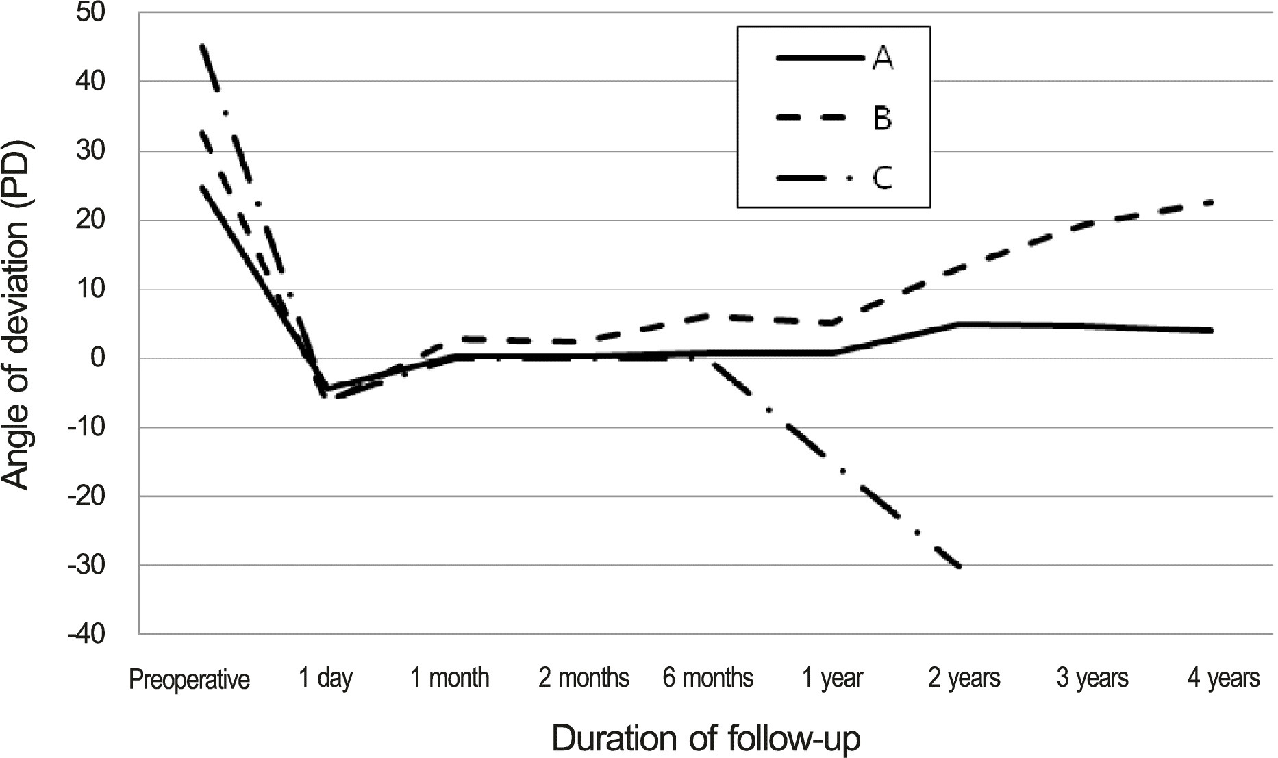
Table 1.
Surgical outcome of exotropia according to retinopathy of prematurity
|
Retinopathy of prematurity (n) |
||
|---|---|---|
| (+) | (-) | |
| Surgical success (n) | 6 | 7 |
| Surgical failure (n) | 4 | 5 |
Table 2.
Surgical outcome of exotropia according to operation for retinopathy of prematurity
|
Operation for retinopathy of prematurity (n) |
||
|---|---|---|
| (+) | (-) | |
| Surgical success (n) | 0 | 6 |
| Surgical failure (n) | 2 | 2 |
Table 3.
Associated central nervous system abnormalities
| Central nervous system abnormalities | Number of patients |
|---|---|
| Intraventricular hemorrhage | 2 |
| Germinal matrix hemorrhage | 1 |
| Cerebral palsy | 3 |
| Seizure | 1 |
| Encephalomalacia | 1 |
Table 4.
The demographic and baseline characteristics of premature children with exotropia
Table 5.
Comparison of preoperative and postoperative data between surgical success group and surgical failure group
| Surgical success group (n = 13) | Surgical failure group (n = 9) | p-value | |
|---|---|---|---|
| Sex (M:F) | 7:6 | 3:6 | 0.42∗ |
| Gestational age (weeks, range) | 31.4 ± 2.8 (25 to 36) | 31.8 ± 4.2 (25 to 36) | 0.56† |
| Birth weight (gram, range) | 1491.5 ± 420.6 (690 to 2,100) | 1552.0 ± 457.7 (882 to 2,270) | 0.95† |
| Age of diagnosis (years, range) | 4.4 ± 3.7 (1 to 12) | 2.9 ± 1.3 (1 to 4) | 0.74† |
| Age at surgery (years, range) | 7.1 ± 2.3 (5 to 12) | 5.1 ± 2.0 (2 to 9) | 0.06† |
| Follow-up (months, range) | 22.2 ± 16.7 (6 to 48) | 26.7 ± 16.5 (6 to 48) | 0.56† |
| Angle of exodeviation at first examination (PD, range) | |||
| Near | 20.4 ± 4.8 (10 to 25) | 30.6 ± 10.4 (15 to 50) | 0.01† |
| Far | 19.2 ± 4.5 (10 to 25) | 30.0 ± 10.6 (15 to 50) | 0.01† |
| Angle of exodeviation at operation (PD, range) | |||
| Near | 25.4 ± 4.8 (15 to 30) | 33.0 ± 10.4 (12 to 50) | 0.02† |
| Far | 24.6 ± 4.3 (15 to 30) | 33.9 ± 9.6 (20 to 50) | 0.02† |
| Postoperative angle of deviation at last visit (PD, range) | |||
| Near | 3.1 ± 4.2 (0 to 10) | 17.8 ± 20.2 (-30 to 40) | 0.00† |
| Far | 2.6 ± 3.6 (0 to 10) | 15.8 ± 19.9 (-30 to 40) | 0.00† |
| Vertical strabismus (n) | 5 | 5 | 0.67∗ |
| Amblyopia (n) | 2 | 3 | 0.61∗ |
Table 6.
Comparison of degree of refractive errors between surgical success group and surgical failure group
| Surgical success group (diopter) | Surgical failure group (diopter) | p-value | |
|---|---|---|---|
| Myopia | -0.71 ± 0.10 | -0.75 ± 0.00 | 0.86∗ |
| Emmetropia | +1.26 ± 0.50 | +1.17 ± 0.74 | 0.38∗ |
| Hyperopia | +3.50 ± 0.00 | +4.50 ± 0.00 | 0.67∗ |
| Astigmatism | 1.59 ± 0.38 | 1.95 ± 0.62 | 0.17∗ |
 XML Download