Journal List > J Korean Ophthalmol Soc > v.56(12) > 1010170

Kim, Lee, and Ahn: Comparison of Ocular Pulse Amplitude Measured Using Dynamic Contour Tonometry and Ocular Blood Flow Analyzer

Abstract

Purpose

To compare ocular pulse amplitude (OPA) measured using dynamic contour tonometry (DCT) and ocular blood flow analyzer (BFA).

Methods

Thirty-five eyes of 35 patients were enrolled in this cross-sectional and retrospective study. OPA was measured using DCT. Pulse amplitude (PA) and pulsatile ocular blood flow were measured using BFA.

Results

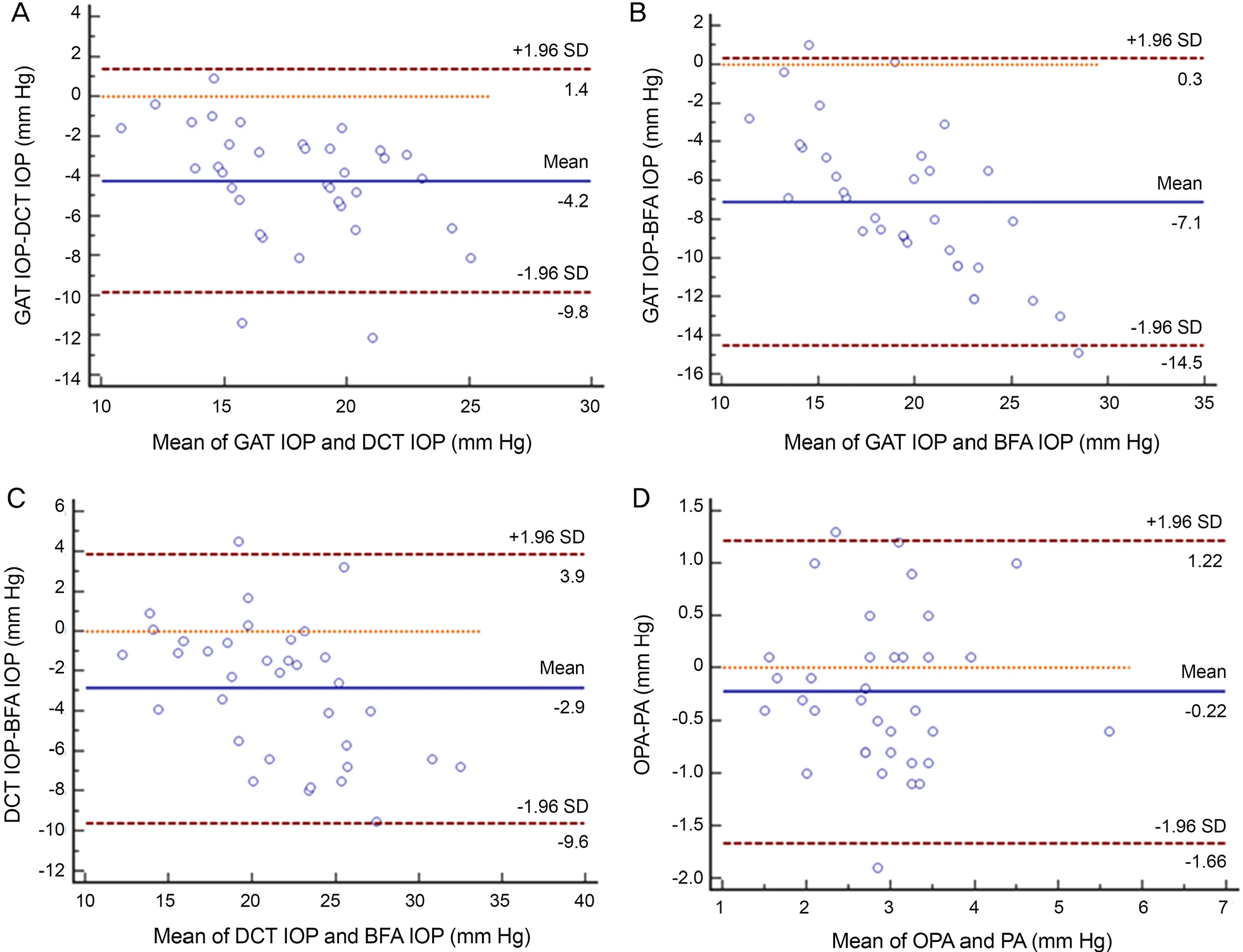
OPA measured using DCT (2.79 ± 0.89 mm Hg) was not significantly different from PA measured with BFA (3.02 ±0.90 mm Hg; p = 0.082) and both were significantly correlated (r = 0.663, p < 0.001). Mean difference ± limit of agreement was -0.22 ± 1.44 mm Hg between OPA and PA. OPA correlated significantly with intraocular pressure (IOP) measured using Goldmann applanation tonometry (r = 0.330, p = 0.047) but not PA (r = 0.057, p = 0.745). Both PA and OPA did not show sig-nificant correlation with the spherical equivalent of refractive error and central corneal thickness.

Conclusions

Although both OPA and PA measure IOP fluctuation and are not significantly different, they showed different rela-tionships with IOP.

References

1. Fontana L, Poinoosawmy D, Bunce CV. . Pulsatile ocular blood flow investigation in asymmetric normal tension glaucoma and normal subjects. Br J Ophthalmol. 1998; 82:731–6.
crossref
2. Kerr J, Nelson P, O'Brien C. A comparison of ocular blood flow in untreated primary open-angle glaucoma and ocular hypertension. Am J Ophthalmol. 1998; 126:42–51.
crossref
3. Kerr J, Nelson P, O'Brien C. Pulsatile ocular blood flow in primary open-angle glaucoma and ocular hypertension. Am J Ophthalmol. 2003; 136:1106–13.
crossref
4. Kergoat H. Using the POBF as an index of interocular blood flow effects during unilateral vascular stress. Vision Res. 1997; 37:1085–9.
crossref
5. Centofanti M, Migliardi R, Bonini S. . Pulsatile ocular blood flow during pregnancy. Eur J Ophthalmol. 2002; 12:276–80.
crossref
6. Grieshaber MC, Katamay R, Gugleta K. . Relationship between ocular pulse amplitude and systemic blood pressure measurements. Acta Ophthalmol. 2009; 87:329–34.
crossref
7. Choi J, Lee J, Park SB. . Factors affecting ocular pulse ampli-tude in eyes with open angle glaucoma and glaucoma-suspect eyes. Acta Ophthalmol. 2012; 90:552–8.
crossref
8. Kaufmann C, Bachmann LM, Robert YC, Thiel MA. Ocular pulse amplitude in healthy subjects as measured by dynamic contour tonometry. Arch Ophthalmol. 2006; 124:1104–8.
crossref
9. Stalmans I, Harris A, Fieuws S. . Color doppler imaging and ocular pulse amplitude in glaucomatous and healthy eyes. Eur J Ophthalmol. 2009; 19:580–7.
crossref
10. De Moraes CG, Reis AS, Cavalcante AF. . Choroidal ex-pansion during the water drinking test. Graefes Arch Clin Exp Ophthalmol. 2009; 247:385–9.
crossref
11. Aykan U, Erdurmus M, Yilmaz B, Bilge AH. Intraocular pressure and ocular pulse amplitude variations during the Valsalva maneuver. Graefes Arch Clin Exp Ophthalmol. 2010; 248:1183–6.
crossref
12. Dastiridou AI, Ginis HS, De Brouwere D. . Ocular rigidity, oc-ular pulse amplitude, and pulsatile ocular blood flow: the effect of intraocular pressure. Invest Ophthalmol Vis Sci. 2009; 50:5718–22.
crossref
13. Kanngiesser HE, Kniestedt C, Robert YC. Dynamic contour ton-ometry: presentation of a new tonometer. J Glaucoma. 2005; 14:344–50.
14. Silver DM, Farrell RA. Validity of pulsatile ocular blood flow measurements. Surv Ophthalmol. 1994; 38:Suppl:S72-80.
crossref
15. Razminia M, Trivedi A, Molnar J. . Validation of a new for-mula for mean arterial pressure calculation: the new formula is su-perior to the standard formula. Catheter Cardiovasc Interv. 2004; 63:419–25.
crossref
16. Sehi M, Flanagan JG, Zeng L. . Relative change in diurnal mean ocular perfusion pressure: a risk factor for the diagnosis of primary open-angle glaucoma. Invest Ophthalmol Vis Sci. 2005; 46:561–7.
crossref
17. Kaufmann C, Bachmann LM, Thiel MA. Comparison of dynamic contour tonometry with goldmann applanation tonometry. Invest Ophthalmol Vis Sci. 2004; 45:3118–21.
crossref
18. Hsu SY, Sheu MM, Hsu AH. . Comparisons of intraocular pressure measurements: Goldmann applanation tonometry, non-contact tonometry, Tono-Pen tonometry, and dynamic contour tonometry. Eye (Lond). 2009; 23:1582–8.
crossref
19. Ouyang PB, Li CY, Zhu XH, Duan XC. Assessment of intraocular pressure measured by Reichert Ocular Response Analyzer, Goldmann Applanation Tonometry, and Dynamic Contour Tonometry in healthy individuals. Int J Ophthalmol. 2012; 5:102–7.
20. Jimenez-Roman J, Gil-Carrasco F, Martinez A. . Comparison of Goldmann applanation and dynamic contour tonometry in a pop-ulation of Mexican open-angle glaucoma patients. Int Ophthalmol. 2013; 33:221–5.
crossref
21. Lee J, Lee CH, Choi J. . Comparison between dynamic contour tonometry and Goldmann applanation tonometry. Korean J Ophthalmol. 2009; 23:27–31.
crossref
22. Realini T, Weinreb RN, Hobbs G. Correlation of intraocular pres-sure measured with goldmann and dynamic contour tonometry in normal and glaucomatous eyes. J Glaucoma. 2009; 18:119–23.
crossref
23. Haustein M, Spoerl E, Boehm AG. The effect of acetazolamide on different ocular vascular beds. Graefes Arch Clin Exp Ophthalmol. 2013; 251:1389–98.
crossref
24. Zinkernagel MS, Ebneter A. Acetazolamide influences ocular pulse amplitude. J Ocul Pharmacol Ther. 2009; 25:141–4.
crossref
25. Breusegem C, Fieuws S, Zeyen T, Stalmans I. The effect of trabe-culectomy on ocular pulse amplitude. Invest Ophthalmol Vis Sci. 2010; 51:231–5.
crossref
26. Plange N, Rennings C, Herr A. . Ocular pulse amplitude before and after cataract surgery. Curr Eye Res. 2012; 37:115–9.
crossref
27. Dastiridou AI, Ginis H, Tsilimbaris M. . Ocular rigidity, ocular pulse amplitude, and pulsatile ocular blood flow: the effect of axial length. Invest Ophthalmol Vis Sci. 2013; 54:2087–92.
crossref

Figure 1.
Agreement between intraocular pressure and ocular pulse amplitude measured by different devices. (A) Bland-Altman plot showing the inter-instrument repeatability of IOP measurements by DCT and GAT. (B) Bland-Altman plot showing the inter-instru-ment repeatability of IOP measurements by ocular BFA and GAT. (C) Bland-Altman plot showing the inter-instrument repeatability of IOP measurements by BFA and DCT. (D) Bland-Altman plot showing the inter-instrument repeatability of OPA measurements by DCT with measurements using BFA. GAT = Goldman applanation tonometry; IOP = intraocular pressure; DCT = dynamic contour tonometry; BFA = ocular blood flow analyzer; OPA = ocular pulse amplitude; PA = pulse amplitude measured by BFA; SD = standard deviation.
jkos-56-1906f1.tif
Table 1.
Descriptive statistics of enrolled subjects
Parameter Mean ± SD Minimum Median value Maximum
Age (years) 47.0 ± 10.4 31 45 71
Male/female ratio 17/18 - - -
MAP (mm Hg) 94.2 ± 10.1 70.3 94.7 115.7
PP (mm Hg) 50.3 ± 10.9 33 50 75
MOPP (mm Hg) 47.0 ± 6.8 33.9 46.7 60.2
PR (beats per minute) 77.8 ± 11.4 54 76 108
CCT (μ m) 545.84 ± 38.06 458.0 543.0 629.3
SE (diopters) -2.036 ± -2.790 -9.50 -0.50 1.50
GAT IOP (mm Hg) 15.8 ± 3.17 10 15 21
DCT IOP (mm Hg) 20.01 ± 4.21 11.6 20.6 29.1
OPA (mm Hg) 2.79 ± 0.89 1.3 2.7 5.3
BFA IOP (mm Hg) 22.89 ± 5.78 12.8 22.9 35.9
PA (mm Hg) 3.02 ± 0.90 1.5 3.1 5.9
PV (μ L) 4.92 ± 1.75 1.8 4.9 9.5
POBF (μ L/sec) 12.21 ± 3.83 6.2 12.6 21.8

Values are presented as mean ± SD unless otherwise indicated. SD = standard deviation; MAP = mean arterial pressure; PP = pulse pressure; MOPP = mean ocular perfusion pressure; PR = pulse rate; CCT = central corneal thickness; SE = spherical equivalent; GAT = Goldmann applanation tonometry; IOP = intraocular pressure; DCT = dynamic contour tonometry; OPA = ocular pulse amplitude; BFA = ocular blood flow analyzer; PA = pulse amplitude measured by BFA; PV = pulse volume measured by BFA; POBF = pulsatile ocular blood flow measured by BFA.

Table 2.
Comparison of IOP differences measured by 3 different devices and correlations between IOP measurements
Compared parameters Mean difference (± standard deviation) p-value* LOA (mm Hg) Range (mm Hg) Correlation between two parameters p-value
GAT IOP-DCT IOP -4.2 ± 2.9 <0.001 -9.8, 1.4 11.2 0.733 <0.001
GAT IOP-BFA IOP -7.1 ± 3.8 <0.001 -14.5, 0.3 14.8 0.795 <0.001
DCT IOP-BFA IOP -2.9 ± 3.4 <0.001 -9.6, 3.9 13.6 0.808 <0.001
OPA-PA -0.22 ± 0.74 0.082 -1.66, 1.22 2.88 0.663 <0.001

Values are presented as mean ± SD unless otherwise indicated. IOP = intraocular pressure; LOA = limit of agreement; GAT = Goldmann applanation tonometry; DCT = dynamic contour tonometry; BFA = ocular blood flow analyzer; OPA = ocular pulse amplitude measured by DCT; PA = pulse amplitude measured by BFA.* Paired t-test; Pearson correlation coefficient.

Table 3.
Correlations between IOP or OPA-derived parameters and systemic or other ocular parameters (Pearson correlation co-efficient R and p-value in parenthesis)
Age CCT SE
GAT IOP -0.211 (0.224) 0.296 (0.047) -0.278 (0.106)
DCT IOP -0.004 (0.980) 0.235 (0.174) -0.268 (0.120)
BFA IOP -0.258 (0.134) 0.144 (0.409) -0.900 (0.079)
OPA -0.031 (0.861) -0.146 (0.404) 0.154 (0.376)
PA 0.123 (0.482) -0.165 (0.344) 0.308 (0.072)
POBF 0.209 (0.228) -0.339 (0.047) 0.431 (0.010)

IOP = intraocular pressure; OPA = ocular pulse amplitude measured by DCT; CCT = central corneal thickness; SE = spherical equivalent; GAT = Goldmann applanation tonometry; DCT = dynamic contour tonometry; BFA = ocular blood flow analyzer; PA = pulse amplitude measured by BFA; POBF = pulsatile ocular blood flow measured by BFA.

TOOLS
Similar articles