Abstract
Purpose
Various forms and degrees of morphological changes in the lower eyelid are presented in this study. A compre-hensive, individualized clinical approach based on sound anatomical principles and lower lid stability is needed. We evaluated morphological outcomes of lower blepharoplasty by measuring lower eyelid position.
Methods
One hundred sixty-eight eyes underwent lower blepharoplasty between August 2009 and August 2014. The mean pa-tient age was 62.1 ± 13 years and mean follow-up period was 6.0 ± 10.2 months. Lower eyelid position of 84 consecutive primary lower blepharoplasty patients (52 females and 32 males) was analyzed using digital images and measured with standardized data points using the Image J Program.
Results
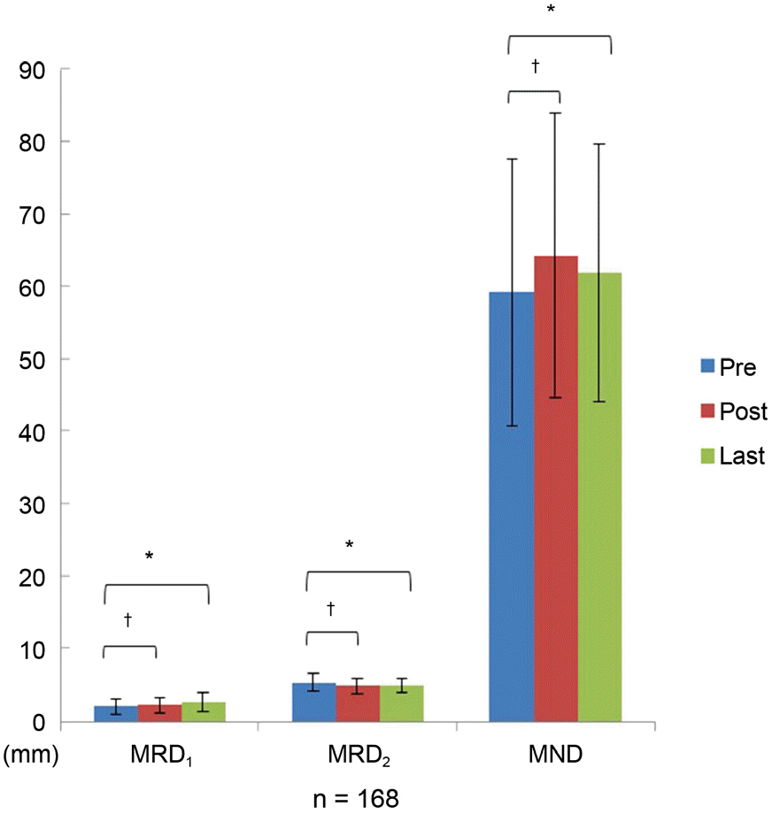
Of the 168 individualized lower eyelid blepharoplasties analyzed, margin reflex distance 2 (MRD2) decreased from 5.4 ± 1.1 mm to 4.9 ± 0.8 mm ( p = 0.005) and marginal nose distance (MND) increased from 59.2 ± 18.3 mm to 61.9 ± 17.6 mm ( p < 0.001). Complications after lower blepharoplasty were not observed.
Go to : 
References
1. Nishihira T, Ohjimi H, Eto A. A new digital image analysis system for measuring blepharoptosis patients’ upper eyelid and eyebrow positions. Ann Plast Surg. 2014; 72:209–13.

2. Macdonald KI, Mendez AI, Hart RD, Taylor S. Eyelid and brow asymmetry in patients evaluated for upper lid blepharoplasty. J Otolaryngol Head Neck Surg. 2014; 43:36.

3. Coombes AG, Sethi CS, Kirkpatrick WN. . A standardized dig-ital photography system with computerized eyelid measurement analysis. Plast Reconstr Surg. 2007; 120:647–56.

4. Chi MJ, Park MS, Baek SH. The effect of transconjunctival lower blepharoplasty combined with pinch skin excision technique. J Korean Ophthalmol Soc. 2007; 48:755–60.
5. Jacono AA, Moskowitz B. Transconjunctival versus trans-cutaneous approach in upper and lower blepharoplasty. Facial Plast Surg. 2001; 17:21–8.

6. Zarem HA, Resnick JI. Expanded applications for transconjunctival lower lid blepharoplasty. Plast Reconstr Surg. 1999; 103:1041–3. discussion 1044-5.

7. Nassif PS. Lower blepharoplasty: transconjunctival fat repositioning. Facial Plast Surg Clin North Am. 2005; 13:553–9. vi.

9. Ko SJ, Kim SD. Involutional ectropion repair with the modified medial spindle and the lateral tarsal strip procedure. J Korean Ophthalmol Soc. 2012; 53:187–92.

10. Nowinski TS, Anderson RL. The medial spindle procedure for in-volutional medial ectropion. Arch Ophthalmol. 1985; 103:1750–3.

11. Levine MR. Manual of Oculoplastic Surgery, 4th ed. Thorofare: SLACK Inc.,. 2010; 173–82.
12. White WL, Woog JJ. Lower eyelid malpositions. In: Albert DM, Jacobiec FA, eds. Principles and Practice of Ophthalmology, 2nd ed. Philadelphia: WB Saunders,. 1994; 596–693.
13. Wright KA. Interactive ophthalmology on CD-ROM-textbook and review, 1st ed. Baltimore: Williams & Wilkins,. 1998; 5191–326.
14. Bashour M, Harvey J. Causes of involutional ectropion and en-tropion-age-related tarsal changes are the key. Ophthal Plast Reconstr Surg. 2000; 16:131–41.

15. Stefanyszyn MA, Hidayat AA, Flanagan JC. The histopathology of involutional ectropion. Ophthalmology. 1985; 92:120–7.

16. Seo HR, Ahn HB. Morphological changes of the eyelid according to age. J Korean Ophthalmol Soc. 2009; 50:1461–7.

17. Ji JY, Kim YD. Acellular dermal allograft for the correction of eye-lid retraction. J Korean Ophthalmol Soc. 2005; 46:1–9.
19. Kim SY, Shin SJ, Yang SW, Han SH. Microscopic anatomy of the lower eyelid in Koreans. J Korean Ophthalmol Soc. 2006; 47:292–6.
Go to : 
 | Figure 1.Examples of patients before and after individualized lower blepharoplasty. Case 1, 54- year-old female underwent skin ex-cision, orbicularis tightening and fat removal. (A) Before surgery, (B) after surgery. Case 2, 78-year-old male underwent orbicularis muscle tightening and lateral canthal suspension. (C) Before surgery, (D) after surgery. Case 3, 59-year-old male underwent skin excision and minimal fat removal. (E) Before surgery, (F) after surgery. Case 4, 58-year-old male underwent fat transposition. (G) Before surgery, (H) after surgery. |
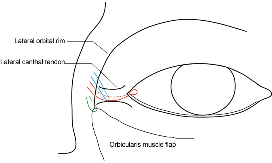
 | Figure 2.Schematic drawing of surgery for correction of right lower eyelid laxity. Blue line indicates lateral canthopexy, red line indicates lateral canthal suspension and green line in-dicates orbicularis flap suspension (Bold line: superficial lay-er; dotted line: deep layer). |
 | Figure 3.Measurement of the clinical parameters for the low-er lid position. MRD1, IPF, MRD2 and MND were analyzed with the Image J program (NIH, Bethesda, MD, USA). IPF = interpalpebral fissure; MND = marginal nose distance; MRD1 = marginal reflex distance 1; MRD2 = marginal reflex distance 2. |
 | Figure 4.Periorbital parameters after lower blepharoplasty us-ing individualized technique. Pre = pre-operation; Post = post-operation; MRD1 = marginal reflex distance 1; MRD2 = marginal reflex distance 2; MND = marginal nose distance. * Paired t-test, p < 0.05 compared to last follow-up; † Paired t-test, p < 0.05 compared to post 1 month. |
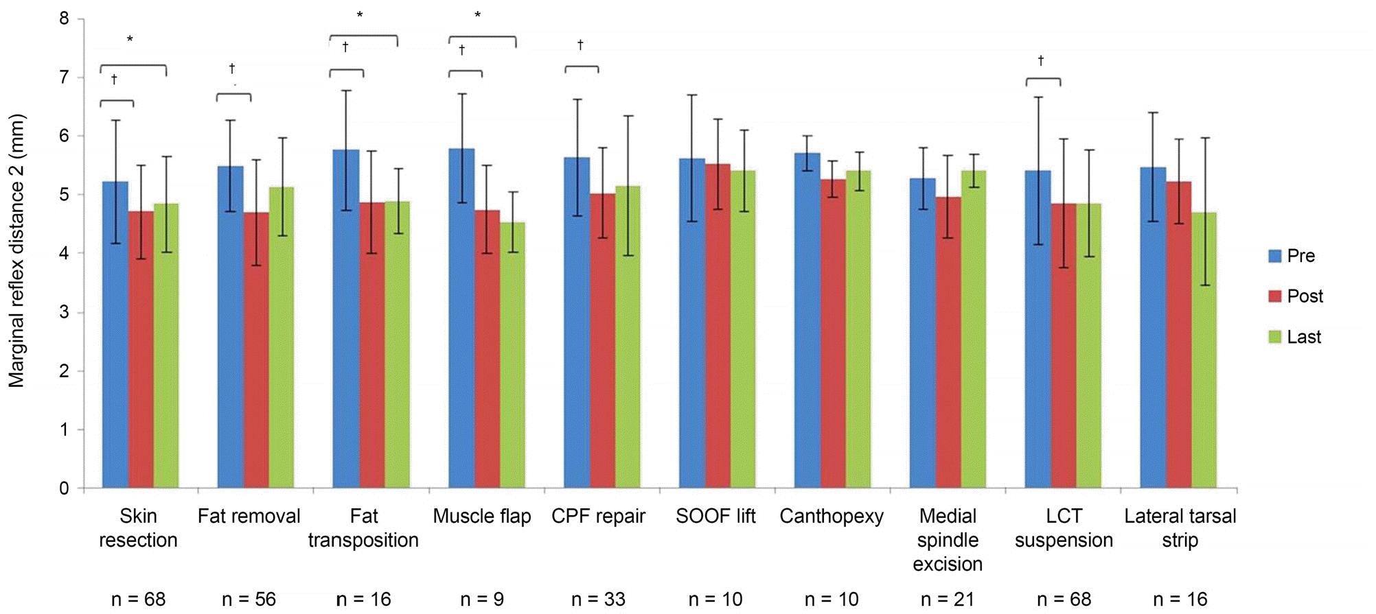
 | Figure 5.Lower eyelid position presenting marginal reflex distance 2 of the lower eyelid following lower blepharoplasty using in-dividualized technique. Pre = pre-operation; Post = post-operation; CPF = capsulopalpebral fascia; SOOF = suborbicularis oculi fat; LCT = lateral canthal tendon. * Wilcoxon signed rank test: p < 0.05 compared to the last follow-up; † Wilcoxon signed rank test: p < 0.05 compared to postoperative 1 month. |
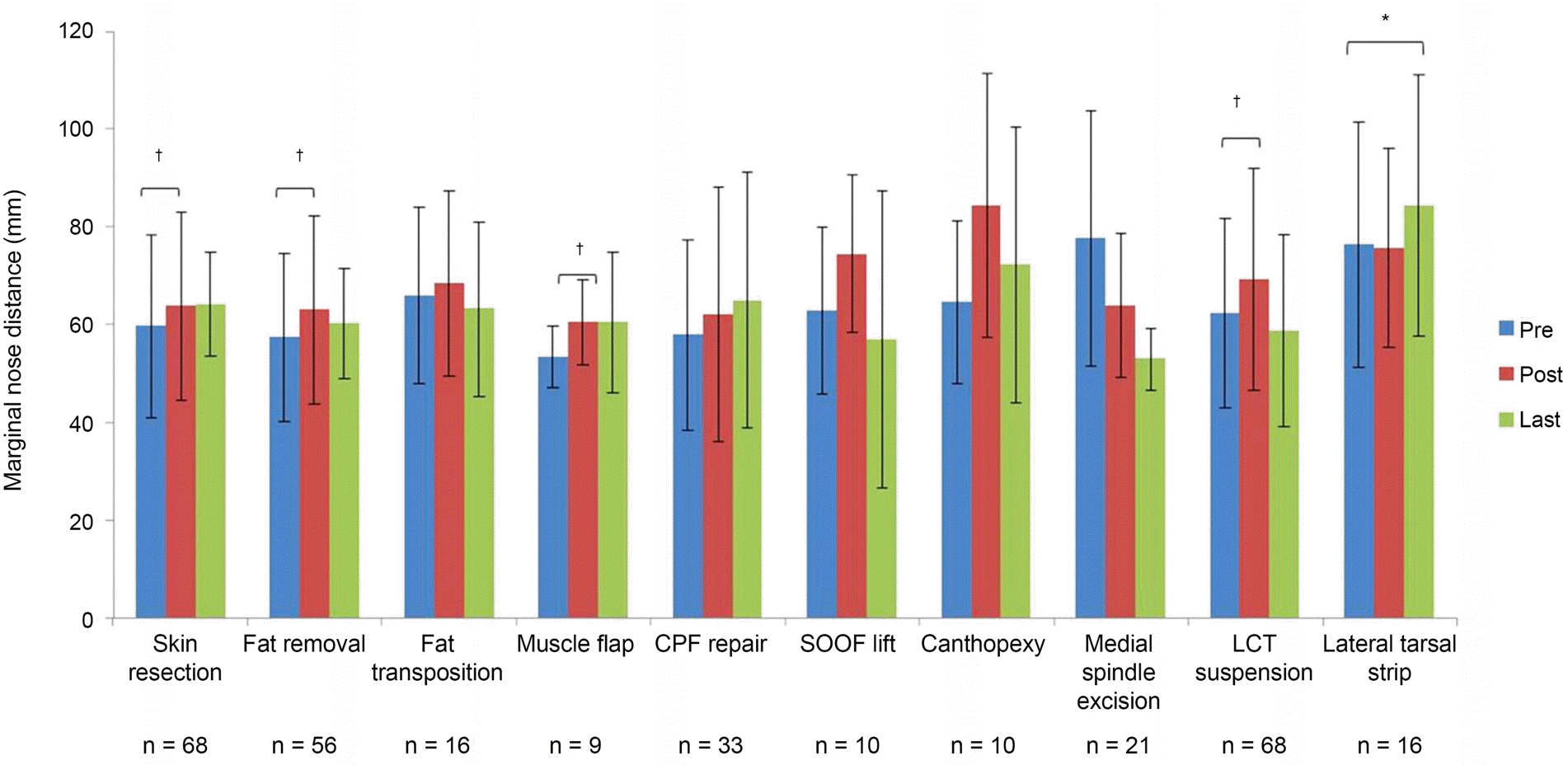
 | Figure 6.Lower eyelid position presenting marginal nose distance of the lower eyelid following lower blepharoplasty using in-dividualized technique. Pre = pre-operation; Post = post-operation; CPF = capsulopalpebral fascia; SOOF = suborbicularis oculi fat; LCT = lateral canthal tendon, * Wilcoxon signed rank test: p < 0.05 compared to last follow-up; † Wilcoxon signed rank test: p < 0.05 compared to postoperative 1 month. |
Table 1.
Clinical parameters after lower blepharoplasty in total patients (n = 168)


 PDF
PDF ePub
ePub Citation
 Print
Print


 XML Download