Abstract
Purpose
To assess changes in ganglion cell-inner plexiform layer (GCIPL) thickness after cataract surgery using spectral-do-main optical coherence tomography (OCT).
Methods
Forty-three eyes of 33 patients, who underwent cataract surgery were imaged with spectral-domain OCT before and after surgery to measure peripapillary retinal nerve fiber layer (RNFL) and GCIPL thickness, signal strength (SS), quadrant, 12 clock-hour RNFL thickness and sectoral GCIPL thickness.
Results
The postoperative SS, RNFL and GCIPL thickness were higher than before surgery ( p < 0.05). Multivariate analysis showed that endothelial cell count and preoperative SS were significantly correlated with SS changes in RNFL parameters and preoperative SS was significantly correlated with SS changes in GCIPL parameters. Univariate analysis indicated that age was significantly correlated with RNFL thickness changes in RNFL parameters and no factor was correlated with GCIPL thickness in GCIPL parameters ( p < 0.05).
Go to : 
References
1. Schuman JS, Hee MR, Puliafito CA. . Quantification of nerve fiber layer thickness in normal and glaucomatous eyes using opti-cal coherence tomography. Arch Ophthalmol. 1995; 113:586–96.

3. Hee MR, Izatt JA, Swanson EA. . Optical coherence tomog-raphy of the human retina. Arch Ophthalmol. 1995; 113:325–32.

4. Guedes V, Schuman JS, Hertzmark E. . Optical coherence to mography measurement of macular and nerve fiber layer thickness in normal and glaucomatous human eyes. Ophthalmology. 2003; 110:177–89.
5. Wollstein G, Schuman JS, Price LL. . Optical coherence to-mography longitudinal evaluation of retinal nerve fiber layer thick-ness in glaucoma. Arch Ophthalmol. 2005; 123:464–70.

6. Sakata LM, Deleon-Ortega J, Sakata V, Girkin CA. Optical coher-ence tomography of the retina and optic nerve-a review. Clin Experiment Ophthalmol. 2009; 37:90–9.
7. Leung CK, Cheung CY, Weinreb RN. . Evaluation of retinal nerve fiber layer progression in glaucoma: a study on optical co-herence tomography guided progression analysis. Invest Ophthal-mol Vis Sci. 2010; 51:217–22.

8. Leung CK, Cheung CY, Weinreb RN. . Retinal nerve fiber lay-er imaging with spectral-domain optical coherence tomography: a variability and diagnostic performance study. Ophthalmology. 2009; 116:1257–63. 1263.e1-2.
9. Lee JR, Jeoung JW, Choi J. . Structure-function relationships in normal and glaucomatous eyes determined by time- and spec-tral-domain optical coherence tomography. Invest Ophthalmol Vis Sci. 2010; 51:6424–30.

10. Garas A, Vargha P, Holló G. Reproducibility of retinal nerve fiber layer and macular thickness measurement with the RTVue-100 op-tical coherence tomograph. Ophthalmology. 2010; 117:738–46.

11. Zeimer R, Asrani S, Zou S. . Quantitative detection of glau-comatous damage at the posterior pole by retinal thickness mapping. A pilot study. Ophthalmology. 1998; 105:224–31.
12. Ishikawa H, Stein DM, Wollstein G. . Macular segmentation with optical coherence tomography. Invest Ophthalmol Vis Sci. 2005; 46:2012–7.

13. Tan O, Li G, Lu AT. . Mapping of macular substructures with optical coherence tomography for glaucoma diagnosis. Ophthal-mology. 2008; 115:949–56.

14. Seong M, Sung KR, Choi EH. . Macular and peripapillary reti-nal nerve fiber layer measurements by spectral domain optical co-herence tomography in normal-tension glaucoma. Invest Ophthal-mol Vis Sci. 2010; 51:1446–52.

15. Stein DM, Wollstein G, Ishikawa H. . Effect of corneal drying on optical coherence tomography. Ophthalmology. 2006; 113:985–91.

16. Savini G, Zanini M, Barboni P. Influence of pupil size and cataract on retinal nerve fiber layer thickness measurements by Stratus OCT. J Glaucoma. 2006; 15:336–40.

17. Smith M, Frost A, Graham CM, Shaw S. Effect of pupillary dilata-tion on glaucoma assessments using optical coherence tomography. Br J Ophthalmol. 2007; 91:1686–90.

18. El-Ashry M, Appaswamy S, Deokule S, Pagliarini S. The effect of phacoemulsification cataract surgery on the measurement of reti-nal nerve fiber layer thickness using optical coherence tomo-graphy. Curr Eye Res. 2006; 31:409–13.

19. Sánchez-Cano A, Pablo LE, Larrosa JM, Polo V. The effect of pha-coemulsification cataract surgery on polarimetry and tomography measurements for glaucoma diagnosis. J Glaucoma. 2010; 19:468–74.

20. Mwanza JC, Bhorade AM, Sekhon N. . Effect of cataract and its removal on signal strength and peripapillary retinal nerve fiber layer optical coherence tomography measurements. J Glaucoma. 2011; 20:37–43.

21. Lee KS, Kim YM, Kim JH. . Changes in optic nerve parameter measurements on spectral-domain optical coherence tomography, after cataract surgery. J Korean Ophthalmol Soc. 2013; 54:1573–80.

22. Nakatani Y, Higashide T, Ohkubo S. . Effect of cataract and its removal on ganglion cell complex thickness and peripapillary reti-nal nerve fiber layer thickness measurements by fourier-domain optical coherence tomography. J Glaucoma. 2013; 22:447–55.

23. Firat PG, Ozsoy E, Demirel S. . Evaluation of peripapillary ret-inal nerve fiber layer, macula and ganglion cell thickness in am-blyopia using spectral optical coherence tomography. Int J Ophthalmol. 2013; 6:90–4.
24. Kim NR, Lee ES, Seong GJ. . Comparing the ganglion cell complex and retinal nerve fibre layer measurements by Fourier do-main OCT to detect glaucoma in high myopia. Br J Ophthalmol. 2011; 95:1115–21.

25. Park SJ, Moon YS, Kim NR. Difference of GCIPL thickness of diabetes and normal eyes in spectral domain OCT. J Korean Ophthalmol Soc. 2014; 55:1476–80.

26. Kim WJ, Kim KN, Kim CS. Comparison of diagnostic power among OCT parameters according to peripapillary atrophy in high myopic glaucoma. J Korean Ophthalmol Soc. 2013; 54:1844–55.

27. Lee HS, Park YS, Park SW. Change in the mGC-IPL in patients with a history of APAC according to SD-OCT. J Korean Ophthalmol Soc. 2014; 55:1167–73.

28. Budenz DL, Anderson DR, Varma R. . Determinants of normal retinal nerve fiber layer thickness measured by Stratus OCT. Ophthalmology. 2007; 114:1046–52.

29. Ray R, Stinnett SS, Jaffe GJ. Evaluation of image artifact produced by optical coherence tomography of retinal pathology. Am J Ophthalmol. 2005; 139:18–29.

30. Modjtahedi S, Chiou C, Modjtahedi B. . Comparison of mac-ular thickness measurement and segmentation error rate between stratus and fourier-domain optical coherence tomography. Ophthalmic Surg Lasers Imaging. 2010; 41:301–10.

31. von Jagow B, Ohrloff C, Kohnen T. Macular thickness after un-eventful cataract surgery determined by optical coherence tomo-graphy. Graefes Arch Clin Exp Ophthalmol. 2007; 245:1765–71.

32. Stifter E, Sacu S, Benesch T, Weghaupt H. Impairment of visual acuity and reading performance and the relationship with cataract type and density. Invest Ophthalmol Vis Sci. 2005; 46:2071–5.

33. Aydin A, Wollstein G, Price LL. . Optical coherence tomog-raphy assessment of retinal nerve fiber layer thickness changes af-ter glaucoma surgery. Ophthalmology. 2003; 110:1506–11.

34. Liu L, Zou J, Huang H. . The influence of corneal astigmatism on retinal nerve fiber layer thickness and optic nerve head parameter measurements by spectral-domain optical coherence tomography. Diagn Pathol. 2012; 7:55.

35. Cagini C, Fiore T, Iaccheri B. . Macular thickness measured by optical coherence tomography in a healthy population before and after uncomplicated cataract phacoemulsification surgery. Curr Eye Res. 2009; 34:1036–41.

36. Biro Z, Balla Z, Kovacs B. Change of foveal and perifoveal thick-ness measured by OCT after phacoemulsification and IOL implantation. Eye (Lond). 2008; 22:8–12.

37. Kusbeci T, Eryigit L, Yavas G, Inan UU. Evaluation of cystoid macular edema using optical coherence tomography and fundus fluorescein angiography after uncomplicated phacoemulsification surgery. Curr Eye Res. 2012; 37:327–33.

Go to : 
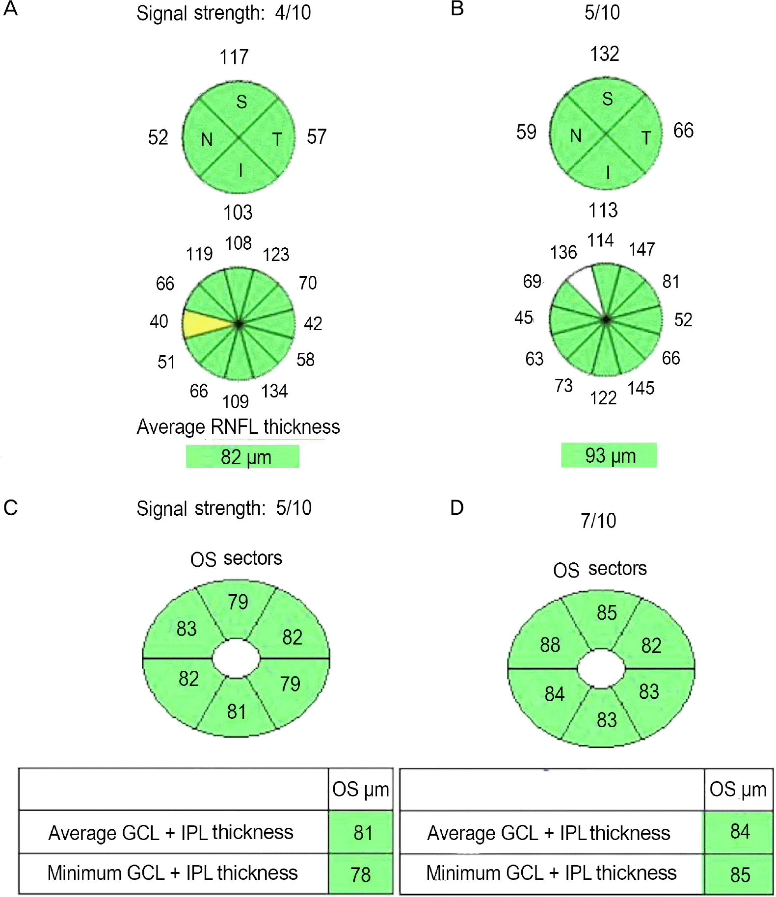
 | Figure 1.Changes of RNFL and GCIPL thickness before (A, C) and after (B, D) cataract surgery in the left eye of a 69-year-old man. Signal strength, quadrant, 12 clock-hour, average RNFL thickness, sectoral, aver-age, minimum GCIPL thickness were higher than before surgery. Color codes of 9, 11 clock-hour RNFL thickness were changed after surgery. RNFL = retinal nerve fiber layer; GCIPL = ganglion cell-inner plexiform layer; S = superior; N = nasal; I = inferior; T = temporal; OS = oculus sinister; GCL = gan-glion cell layer; IPL = inner plexiform layer. |
Table 1.
Clinical characteristics of patients
Table 2.
Comparison of RNFL parameter between preoperative, postoperative data measured
Table 3.
Comparison of GCIPL parameter between preoperative, postoperative data measured *
Table 4.
Factors associated with SS changes in RNFL parameter
Table 5.
Factors associated with SS changes in GCIPL parameter
Table 6.
Factors associated with average RNFL thickness changes in RNFL parameter
Table 7.
Factors associated with average GCIPL thickness changes in GCIPL parameter
Table 8.
Factors associated with SS changes


 PDF
PDF ePub
ePub Citation
 Print
Print


 XML Download