

 PDF
PDF ePub
ePub Citation
 Print
Print


Abstract
Purpose
To compare ocular biometry and refractive results measured using conventional applanation ultrasonography and 3 different optical interferometries, Lenstar LS900®, AL-Scan® and OA-2000®.
Methods
The biometries of 31 cataractous eyes were measured using ultrasonography, Lenstar LS900®, AL-Scan® or OA-2000®. The axial length, anterior chamber depth and keratometry were measured. The SRK/T formula was used to calculate intraocular lens power. Two months after cataract surgery, the refractive outcome was determined and results from the 4 differ-ent biometry methods were compared.
Results
Axial lengths were 23.39 ± 0.95 mm, 23.42 ± 0.98 mm, 23.43 ± 0.98 mm and 23.44 ± 0.98 mm measured using ultra-sonography, Lenstar LS900®, AL-Scan® and OA-2000®, respectively with no statistically significant differences observed ( p = 0.996). The anterior chamber depth and keratometry were 3.14 ± 0.41 mm, 3.10 ± 0.38 mm and 3.13 ± 0.39 mm ( p = 0.936) and 44.41 ± 1.52 D, 44.54 ± 1.57 D and 44.44 ± 1.52 D ( p = 0.937) for Lenstar LS900®, AL-Scan® and OA-2000® respectively. There were no statistically significant differences between the 3 optical devices. The mean absolute error of the 4 different devices were not statistically significant ( p = 0.722).
References
1. Giers U, Epple C. Comparison of A-scan device accuracy. J Cataract Refract Surg. 1990; 16:235–42.

2. Tehrani M, Krummenauer F, Blom E, Dick HB. Evaluation of the practicality of optical biometry and applanation ultrasound in 253 eyes. J Cataract Refract Surg. 2003; 29:741–6.
3. Holzer MP, Mamusa M, Auffarth GU. Accuracy of a new partial coherence interferometry analyser for biometric measurements. Br J Ophthalmol. 2009; 93:807–10.
4. Cruysberg LP, Doors M, Verbakel F. . Evaluation of the Lenstar LS 900 non-contact biometer. Br J Ophthalmol. 2010; 94:106–10.
5. Kaswin G, Rousseau A, Mgarrech M. . Biometry and intra-ocular lens power calculation results with a new optical biometry device: comparison with the gold standard. J Cataract Refract Surg. 2014; 40:593–600.
6. Huang J, Savini G, Li J. . Evaluation of a new optical biometry device for measurements of ocular components and its comparison with IOLMaster. Br J Ophthalmol. 2014; 98:1277–81.
7. Grajciar B, Pircher M, Hitzenberger CK. . High sensitive measurement of the human axial eye length in vivo with Fourier domain low coherence interferometry. Opt Express. 2008; 16:2405–14.
8. Aristodemou P, Knox Cartwright NE, Sparrow JM, Johnston RL. Formula choice: Hoffer Q, Holladay 1, or SRK/T and refractive outcomes in 8108 eyes after cataract surgery with biometry by partial coherence interferometry. J Cataract Refract Surg. 2011; 37:63–71.
9. Olsen T. Sources of error in intraocular lens power calculation. J Cataract Refract Surg. 1992; 18:125–9.
10. Haigis W, Lege B, Miller N, Schneider B. Comparison of im-mersion ultrasound biometry and partial coherence interferometry for intraocular lens calculation according to Haigis. Graefes Arch Clin Exp Ophthalmol. 2000; 238:765–73.
11. Hoffer KJ, Shammas HJ, Savini G. Comparison of 2 laser instru-ments for measuring axial length. J Cataract Refract Surg. 2010; 36:644–8.
12. Santodomingo-Rubido J, Mallen EA, Gilmartin B, Wolffsohn JS. A new non-contact optical device for ocular biometry. Br J Ophthalmol. 2002; 86:458–62.
13. Lam AK, Chan R, Pang PC. The repeatability and accuracy of axial length and anterior chamber depth measurements from the IOLMaster. Ophthalmic Physiol Opt. 2001; 21:477–83.
14. Szalai E, Berta A, Hassan Z, Módis L Jr. Reliability and repeat-ability of swept-source Fourier-domain optical coherence tomog-raphy and Scheimpflug imaging in keratoconus. J Cataract Refract Surg. 2012; 38:485–94.
15. Kim SI, Kang SJ, Oh TH. . Accuracy of ocular biometry and postoperative refraction in cataract patients with AL-Scan(R). J Korean Ophthalmol Soc. 2013; 54:1688–93.
16. Shin JW, Seong M, Kang MH. . Comparison of ocular bio-metry and postoperative refraction in cataract patients between Lenstar(R) and IOL Master(R). J Korean Ophthalmol Soc. 2012; 53:833–8.
17. Shin JA, Chung SK. Comparison of the refractive results measured by ultrasound and partial coherence interferometers. J Korean Ophthalmol Soc. 2013; 54:723–7.
18. Kwag JY, Choi SH. Comparison of ocular biometry measured by ultrasound and two kinds of partial coherence interferometers. J Korean Ophthalmol Soc. 2011; 52:169–74.
Figure 1.
Bland-Altman plots showing the agreement between axial length (mm) obtained by 4 different divicies. (A) A-scan and Lenstar LS900®, (B) A-scan and AL-Scan®, (C) A-scan and OA-2000®, (D) Lenstar LS900® and AL-Scan®, (E) Lenstar LS900® and OA-2000®, (F) AL-Scan® and OA-2000®. 95% limits of agreement for axial length difference are -0.23~0.17 mm, -0.26~0.17 mm, -0.25~0.14 mm, -0.10~0.06 mm, -0.08~0.03 mm and -0.10~0.80 mm.
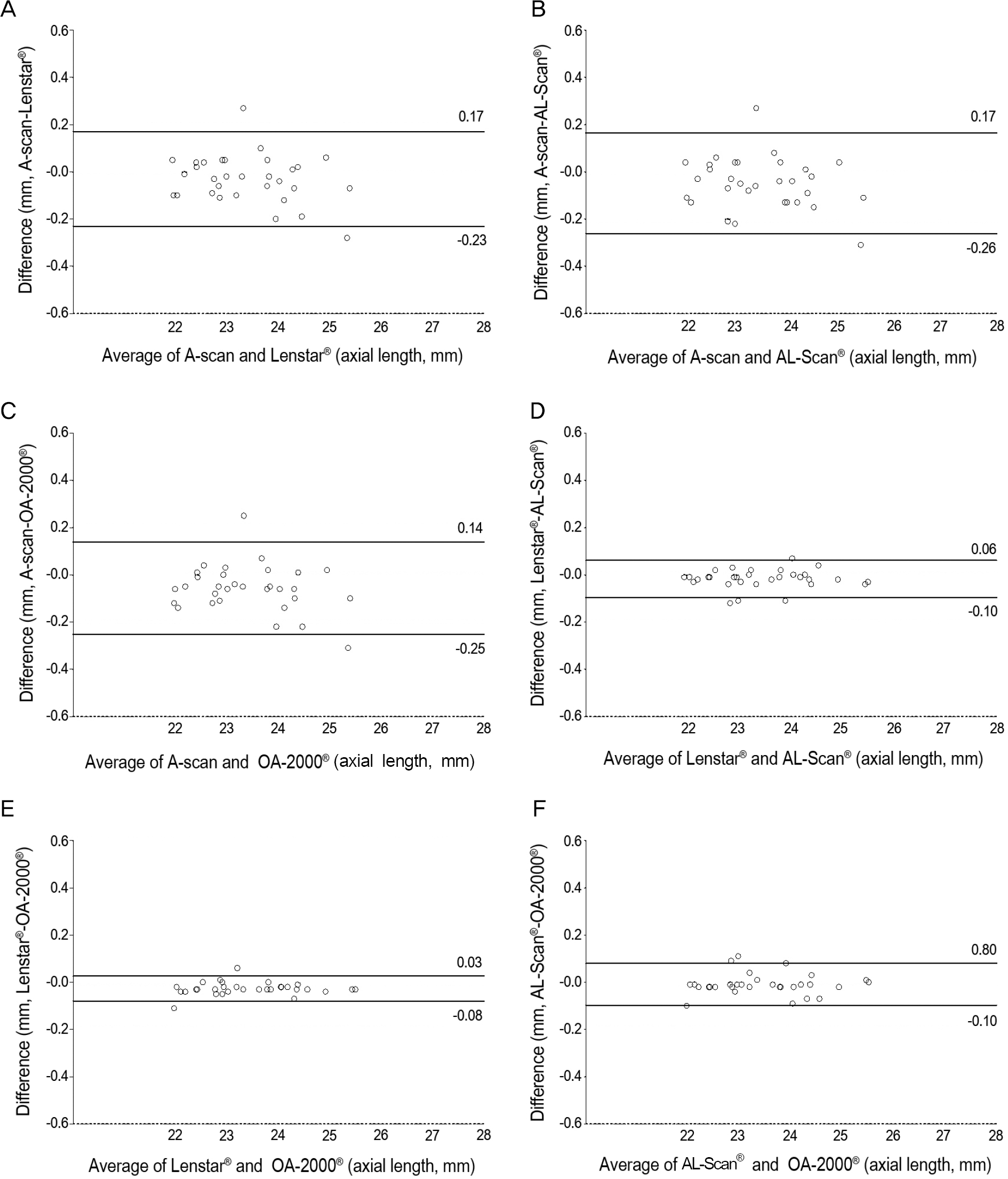
Figure 2.
Bland-Altman plots showing the agreement between anterior chamber depth (mm) obtained by 3 different optical divicies. (A) Lenstar LS900® and AL-Scan®, (B) Lenstar LS900® and OA-2000®, (C) AL-Scan® and OA-2000®. 95% limits of agreement for anterior chamber depth difference are -0.17~0.24 mm, -0.20~0.21 mm and -0.22~0.16 mm.

Figure 3.
Bland-Altman plots showing the agreement between keratometry (D) obtained by 3 different optical divicies. (A) Lenstar LS900® and AL-Scan®, (B) Lenstar LS900® and OA-2000®, (C) AL-Scan® and OA-2000®. 95% limits of agreement for keratometry difference are -0.85~0.58 D, -0.67~0.61 D and -0.58~ 0.79 D.
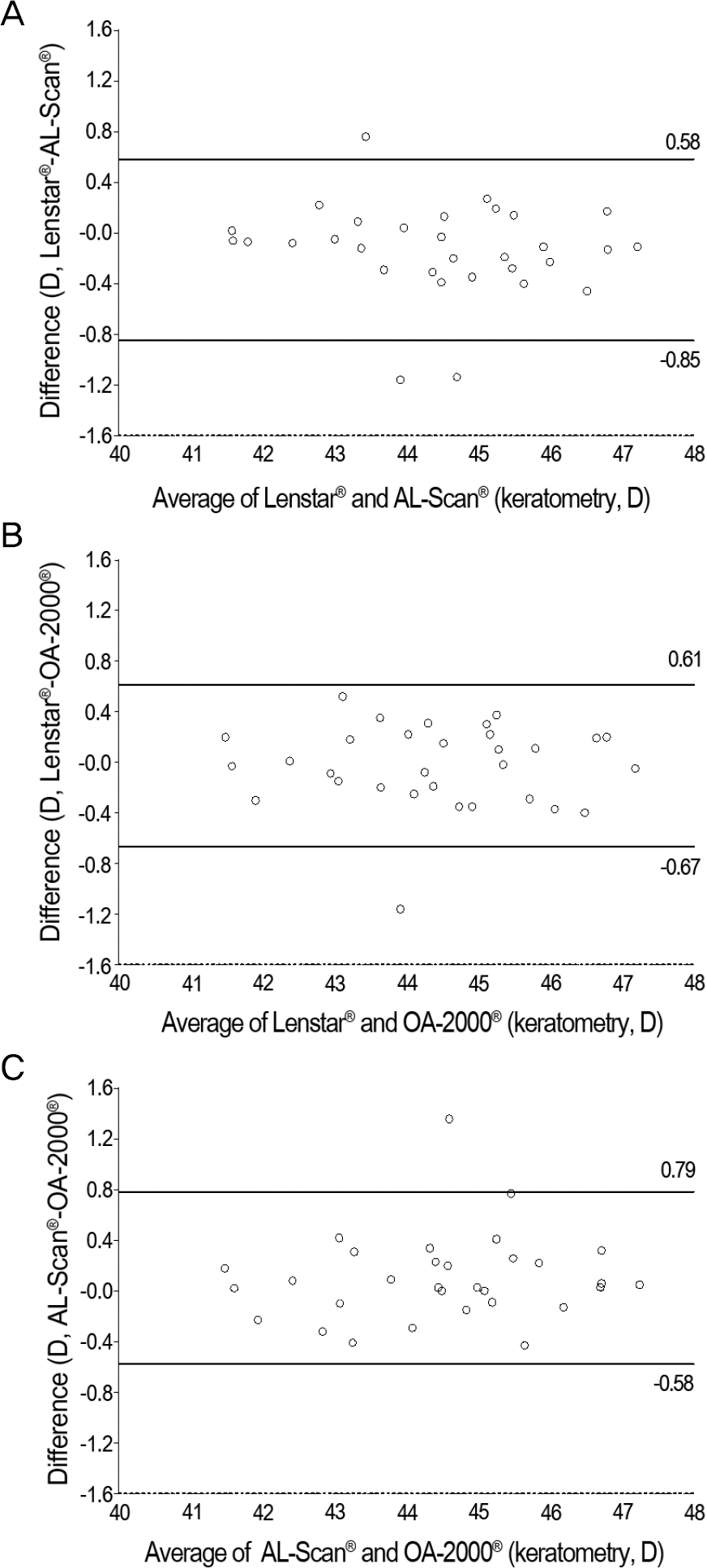
Table 1.
Biometry mearsurements by A-scan, Lenstar LS900®, AL-Scan® and OA-2000®
| A-scan | Lenstar LS900® | AL-Scan® | OA-2000® | p-value* | |
|---|---|---|---|---|---|
| AL (mm) | 23.39 ± 0.95 | 23.42 ± 0.98 | 23.43 ± 0.98 | 23.44 ± 0.98 | 0.996 |
| ACD (mm) | - | 3.14 ± 0.41 | 3.10 ± 0.38 | 3.13 ± 0.39 | 0.936 |
| Mean K (D) | - | 44.41 ± 1.52 | 44.54 ± 1.57 | 44.44 ± 1.52 | 0.937 |
Table 2.
Refractive results: comparison of A-scan, Lenstar LS900®, AL-Scan® and OA-2000®
Table 3.
Technical information of three optical interferometries
OLCR = optical low coherence reflectometry; PCI = partial coherence interferometry; AL = axial length; ACD = anterior chamber depth; CCT = central corneal thickness; LT = lens thickness; KM = corneal curvature; WTW = White to white; PS = pupil size; RT = retinal thickness; US = ultrasound; IOL = intraocular lens; LASIK = laser-assisted in-situ keratomileusis; SRK = Sanders/Retzlaff/Kraff; T = third-generation; PL = post-LASIK.
 XML Download