

 PDF
PDF ePub
ePub Citation
 Print
Print


Abstract
Purpose
To compare the anterior segment parameters including precorneal tear film thickness (PTFT) using Pentacam®(Oculus, Wetzlar, Germany) between normal control and dry eye groups and to examine the relationships between the PTFT and other parameters for dry eye.
Methods
The present study included 23 normal controls (31 eyes) and 25 patients with dry eyes (31 eyes). We compared meas-urements including PTFT, corneal thickness and astigmatism using Pentacam® and analyzed the correlations among the PTFT and fluorescein tear break-up time (FBUT), Schirmer I test (without anesthesia), and ocular surface disease index (OSDI).
Results
The mean PTFT in dry eyes (21.1 ± 2.0 μ m) was significantly thinner than in normal eyes (37.6 ± 2.0 μ m; p < 0.01). In the dry eye group, the corneal thickness was thicker than in the normal eye group but there were no clinically significant differences. The dry eye group experienced more frequent and severe corneal astigmatism compared with the normal group. OSDI scores showed a weak negative correlation with objective clinical measures of dry eye (FBUT, Schirmer I test) but was not statistically significant. However, OSDI was statistically significantly negatively correlated with PTFT (r = -0.46, p < 0.01). The PTFT showed a weak positive correlation with FBUT and Schirmer I test without statistical significance.
Go to : 
References
1. Koh S, Maeda N. Wavefront sensing and the dynamics of tear film. Cornea. 2007; 26:(9 Suppl 1). S41–5.

2. Wang J, Palakuru JR, Aquavella JV. Correlations among upper and lower tear menisci, noninvasive tear break-up time, and the Schirmer test. Am J Ophthalmol. 2008; 145:795–800.
3. Seo SG. The effect of artificial tear application on central corneal thickness in dry eye. J Korean Ophthalmol Soc. 2009; 50:1483–8.
4. Yokoi N, Komuro A. Non-invasive methods of assessing the tear film. Exp Eye Res. 2004; 78:399–407.
5. Ibrahim OM, Dogru M, Takano Y. . Application of visante op-tical coherence tomography tear meniscus height measurement in the diagnosis of dry eye disease. Ophthalmology. 2010; 117:1923–9.
6. Schiffman RM, Christianson MD, Jacobsen G. . Reliability and validity of the ocular surface disease index. Arch Ophthalmol. 2000; 118:615–21.
7. Korb DR. Survey of preferred tests for diagnosis of the tear film and dry eye. Cornea. 2000; 19:483–6.
8. Wang J, Aquavella J, Palakuru J. . Relationships between cen-tral tear film thickness and tear menisci of the upper and lower eyelids. Invest Ophthalmol Vis Sci. 2006; 47:4349–55.
9. Ozcura F, Aydin S, Helvaci MR. Ocular surface disease index for the diagnosis of dry eye syndrome. Ocul Immunol Inflamm. 2007; 15:389–93.
10. Goto T, Zheng X, Klyce SD. . A new method for tear film sta-bility analysis using videokeratography. Am J Ophthalmol. 2003; 135:607–12.
11. Hosaka E, Kawamorita T, Ogasawara Y. . Interferometry in the evaluation of precorneal tear film thickness in dry eye. Am J Ophthalmol. 2011; 151:18–23.e1.
12. King-Smith PE, Fink BA, Fogt N. . The thickness of the human precorneal tear film: evidence from reflection spectra. Invest Ophthalmol Vis Sci. 2000; 41:3348–59.
13. King-Smith PE, Fink BA, Hill RM. . The thickness of the tear film. Curr Eye Res. 2004; 29:357–68.
14. Koh S, Tung C, Aquavella J. . Simultaneous measurement of tear film dynamics using wavefront sensor and optical coherence tomography. Invest Ophthalmol Vis Sci. 2010; 51:3441–8.
15. Zhuang H, Zhou X, Xu J. A novel method for pachymetry mapping of human precorneal tear film using Pentacam with fluorescein. Invest Ophthalmol Vis Sci. 2010; 51:156–9.
16. Nam SM, Im CY, Lee HK. . Accuracy of RTVue optical coher-ence tomography, Pentacam, and ultrasonic pachymetry for the measurement of central corneal thickness. Ophthalmology 2010; 117. 2096–103.
17. Chung SH, Na KS, Kwon HG. . Levels of severity in dry eye syndrome according to Delphi panel classification. J Korean Ophthalmol Soc. 2010; 51:1179–83.
18. Kim CS, Ryu JW, Kim HS, Lee YC. Distribution and change of to-tal astigmatism, corneal astigmatism and residual astigmatism with age in patient with emmetropia. J Korean Ophthalmol Soc. 2005; 46:485–93.
19. Dayanir V, Sakarya R, Ozcura F. . Effect of corneal drying on central corneal thickness. J Glaucoma. 2004; 13:6–8.
20. Firat PG, Doganay S. Corneal hysteresis in patients with dry eye. Eye (Lond). 2011; 25:1570–4.
21. Karadayi K, Ciftci F, Akin T, Bilge AH. Increase in central corneal thickness in dry and normal eyes with application of artificial tears: a new diagnostic and follow-up criterion for dry eye. Ophthalmic Physiol Opt. 2005; 25:485–91.
22. Liu Z, Pflugfelder SC. Corneal surface regularity and the effect of artificial tears in aqueous tear deficiency. Ophthalmology. 1999; 106:939–43.
23. Begley CG, Caffery B, Chalmers RL. . Use of the dry eye ques-tionnaire to measure symptoms of ocular irritation in patients with aqueous tear deficient dry eye. Cornea. 2002; 21:664–70.
24. Kim WJ, Kim HS, Kim MS. Current trends in the recognition and treatment of dry eye: a survey of ophthalmologists. J Korean Ophthalmol Soc. 2007; 48:1614–22.
Go to :
 | Figure 1.Precorneal tear film thickness can be measured by Scheimpflug imaging. (A) The scheimpflug images before and after the fluorescein staining. Note the high-bright visualized tear film (white arrow) and the different optical density of ocular interface (white circle) after fluorescein instillation. (B) Compare 2 image view. The central thickness in the dif-ference A-B section means the thickness of precorneal tear film (black arrow). Only the data presented quality of speciality (black circle) was “OK” were included. We also evaluated the thickness of tear film on temporal, superior, nasal, and in-ferior side and corneal reflective power. |
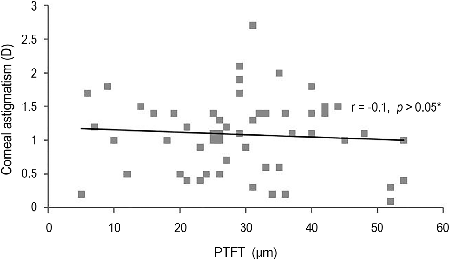 | Figure 2.The relationship between PTFT and corneal astig-matism. PTFT = precorneal tear film thickness; D = diopter; r = Pearson’s correlation coefficient. * p-value by pearson’s correlation analysis. |
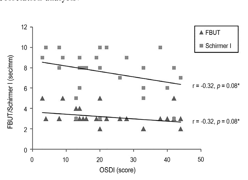 | Figure 3.The relationship among FBUT, Schirmer I test and OSDI. FBUT = fluorescein break-up time; OSDI = ocular surface disease index; r = Pearson’s correlation coefficient. * p-value by Pearson’s correlation analysis. |
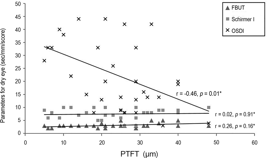 | Figure 4.The relationship between the PTFT and parameters for dry eye (FBUT, Schirmer I and OSDI). PTFT = pre-corneal tear film thickness; FBUT = fluorescein break-up time; OSDI = ocular surface disease index; r = Pearson’s cor-relation coefficient. * p-value by Pearson’s correlation analysis. |
Table 1.
The comparison of the corneal thickness and TFT between normal group and dry eye group
| Normal group (n = 31) | Dry eye group (n = 31) | p-value* | |
|---|---|---|---|
| Age (years) | 32.8 ± 9.3 | 49.5 ± 15.0 | <0.01 |
| Sex (No. of female, %) | 27 (87.1) | 28 (90.3) | 0.32‡ |
| Corneal spherical power (D) | 43.7 ± 1.0 | 43.8 ± 1.3 | 0.7 |
| Corneal astigmatism (D) | 1.0 ± 0.6 | 1.2 ± 0.5 | 0.3 |
| CCT (μ m) | 548.6 ± 4.4 | 549.4 ± 4.4 | 0.01† |
| Central TFT (μ m) | 37.6 ± 2.0 | 21.1 ± 2.0 | <0.01† |
Table 2.
The thickness of the localized tear film and the comparison of the distribution of the thickest area between normal and dry eye group
| Normal group | Dry eye group | p-value* | |
|---|---|---|---|
| Area | |||
| Superior (μ m) | 38.7 ± 18.3 | 28.1 ± 17.2 | 0.03 |
| Inferior (μ m) | 29.6 ± 18.4 | 20.3 ± 17.9 | 0.07 |
| Nasal (μ m) | 29.7 ± 13.1 | 25.2 ± 14.9 | 0.24 |
| Temporal (μ m) | 34.4 ± 16.5 | 21.1 ± 16.6 | <0.01 |
| Central (μ m) | 35.5 ± 10.0 | 23.2 ± 11.2 | <0.01 |
| Distribution of the thickest area | |||
| Superior (n, %) | 18 (58) | 13 (42) | |
| Inferior (n, %) | 9 (29) | 4 (13) | |
| Nasal (n, %) | 1 (3) | 8 (26) | 0.03† |
| Temporal (n, %) | 3 (10) | 3 (10) | |
| Central (n, %) | 0 (0) | 3 (10) | |
 XML Download