

 PDF
PDF ePub
ePub Citation
 Print
Print


Abstract
Purpose
To compare the choroidal thickness of unaffected and affected eyes in children with unilateral high myopia when measured using enhanced depth imaging and to analyze the relationship among choroidal thickness and axial length, spherical equivalent and best corrected visual acuity (BCVA).
Methods
Twenty children with high unilateral high myopia who received optical coherence tomography from December 2012 to May 2014 were retrospectively analyzed. Choroidal thickness was measured with a caliper at 500 μm apart from fovea superiorly and inferiorly, 2,500 μm apart nasally and at 2,500 μm apart temporally at 500 μm intervals. For statistical analyses, paired t-test was used for choroidal thickness and linear regression analysis for the relationship among choroidal thickness and axial length, spherical equivalent and BCVA.
Results
The patients were 10.4 ± 3.5 years of age and the average BCVA of myopic eye was 0.38 ± 0.2. Mean spherical equivalent was −9.8 ± 1.9D. The choroidal thickness was significantly thinner in the myopic eye (102.5 ± 16.9 μm) than the fellow eye (282.9 ± 14.0 μm). The thickest choroid in the myopic eye was the temporal and the thinnest was the nasal area (p = 0.008). Choroidal thickness was significantly associated with axial length and spherical equivalent but not BCVA.
Go to : 
References
1. Pang Y, Goodfellow GW, Allison C, et al. A prospective study of macular thickness in amblyopic children with unilateral high myopia. Invest Ophthalmol Vis Sci. 2011; 52:2444–9.

2. Kutschke PJ, Scott WE, Keech RV. Anisometropic amblyopia. Ophthalmology. 1991; 98:258–63.
3. Sen DK. Results of treatment in amblyopia associated with unilateral high myopia without strabismus. Br J Ophthalmol. 1984; 68:681–5.
4. Nucci P, Drack AV. Refractive surgery for unilateral high myopia in children. J AAPOS. 2001; 5:348–51.
5. Saw SM, Gazzard G, Shih-Yen EC, Chua WH. Myopia and associated pathological complications. Ophthalmic Physiol Opt. 2005; 25:381–91.
6. Ikuno Y, Fujimoto S, Jo Y, et al. Choroidal thinning in high myopia measured by optical coherence tomography. Clin Ophthalmol. 2013; 7:889–93.
7. Margolis R, Spaide RF. A pilot study of enhanced depth imaging optical coherence tomography of the choroid in normal eyes. Am J Ophthalmol. 2009; 147:811–5.
8. Kim JH, Kim JS, Lee KW, Lee JH. The posterior choroidal profiles measured by spectral domain optical coherence tomography in healthy Korean children. J Korean Ophthalmol Soc. 2013; 54:1708–14.
9. Kim EJ, Kim JH, Koo SH, et al. Choroidal thickness changes according to the refractive errors and axial length in Korean myopia patients. J Korean Ophthalmol Soc. 2012; 53:1814–22.
10. Flores-Moreno I, Lugo F, Duker JS, Ruiz-Moreno JM. The relationship between axial length and choroidal thickness in eyes with high myopia. Am J Ophthalmol. 2013; 155:314–9. e1.
11. Tanabe H, Ito Y, Iguchi Y, et al. Correlation between cross-sectional shape of choroidal veins and choroidal thickness. Jpn J Ophthalmol. 2011; 55:614–9.
12. Grossniklaus HE, Green WR. Pathologic findings in pathologic myopia. Retina. 1992; 12:127–33.
13. Abbott CJ, Grünert U, Pianta MJ, McBrien NA. Retinal thinning in tree shrews with induced high myopia: optical coherence tomography and histological assessment. Vision Res. 2011; 51:376–85.
14. Kim KH, Kim DG. The relationship among refractive power, axial length and choroidal thickness measured by SD-OCT in myopia. J Korean Ophthalmol Soc. 2012; 53:626–31.
15. Chui TY, Yap MK, Chan HH, Thibos LN. Retinal stretching limits peripheral visual acuity in myopia. Vision Res. 2005; 45:593–605.
16. Fujiwara T, Imamura Y, Margolis R, et al. Enhanced depth imaging optical coherence tomography of the choroid in highly myopic eyes. Am J Opthalmol. 2009; 148:445–50.
17. Agawa T, Miura M, Ikuno Y, et al. Choroidal thickness measurement in healthy Japanese sujects by three-dimensional high-penetration optical coherence tomography. Graefes Arch Clin Exp Ophthalmol. 2011; 249:1485–92.
18. Lam DS, Leung KS, Mohamed S, et al. Regional variations in the relationship between macular thickness measurements and myopia. Invest Ophthalmol Vis Sci. 2007; 48:376–82.
Go to :
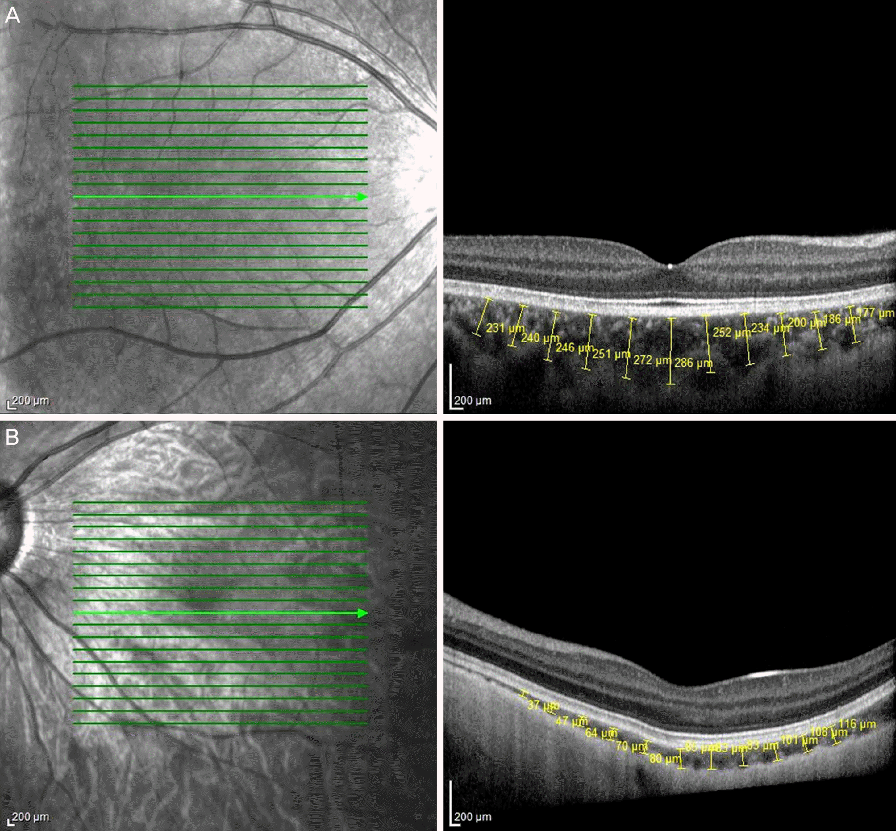 | Figure 1.Measurements of choroidal thickness in normal (A) and high myopic eye (B) using enhanced depth imaging. Choroidal thickness was measured by calipers at 500 μm apart from the fovea superiorly and inferiorly, from 2,500 μm apart nasally and to 2,500 μm apart temporally with 500 μm intervals. |
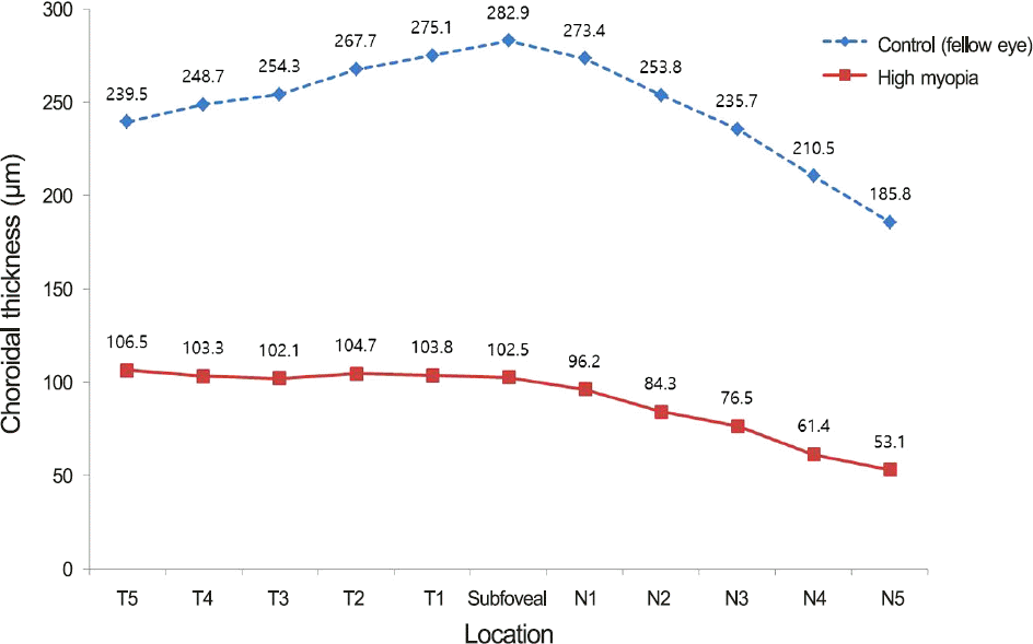 | Figure 2.Choroidal thickness of the myopic eye and the fellow eye graph shows the choroidal thickness in the myopic eye and the fellow eye. Mean thickness at each of the 11 locations measured by 500 μm intervals from temporal (T5) to the nasal (N5) including the subfoveal area. The thickest choroid in the fellow eye (top) is in the subfoveal area, and the thickest choroid in the myopic eye (bottom) is in the temporal area. T = temporal; N = nasal. |
Table 1.
Characteristics and baseline data for the children with unilateral high myopia
Table 2.
Optical characteristics of the myopic eyes and the fellow eyes
| Myopic eye | Fellow eye | p-value† | |
|---|---|---|---|
| Axial length (mm) | 28.4 ± 1.8 | 23.4 ± 1.0 | 0.028 |
| Spherical equivalent (diopters) | –9.8 ± 1.9 | –1.8 ± 1.4 | 0.005 |
| Best corrected visual acuity* (log MAR) | 0.38 ± 0.2 | 0.01 ± 0.16 | 0.015 |
| SFCT (μm) | 102.5 ± 16.9 | 282.9 ± 14.0 | 0.018 |
| Mean CT (μm) | 87.2 ± 28.1 | 243.7 ± 31.9 | 0.005 |
 XML Download