Abstract
Purpose
To evaluate the clinical outcomes of epiretinal membrane (ERM) surgery with minimal exposure to indocyanine green (ICG) dye-assisted internal limiting membrane (ILM) peeling compared with no ICG dye.
Methods
We divided 33 eyes with ERM treated by vitrectomy into 2 groups. ICG dye was used in the first group of 18 eyes (ICG group) but not in the second group of 15 eyes (no dye [ND] group). In the ICG group, 0.25% diluted ICG dye was injected into the fluid-filled eye and removed with a back-flushing needle after 3-5 seconds to peel ILM. Value changes in several parameters in-cluding visual acuity, central macular thickness, Humphrey automated kinetic perimetric analysis, and peripapillary retinal nerve fiber layer (RNFL) thickness were followed up and compared according to ICG dye use.
Results
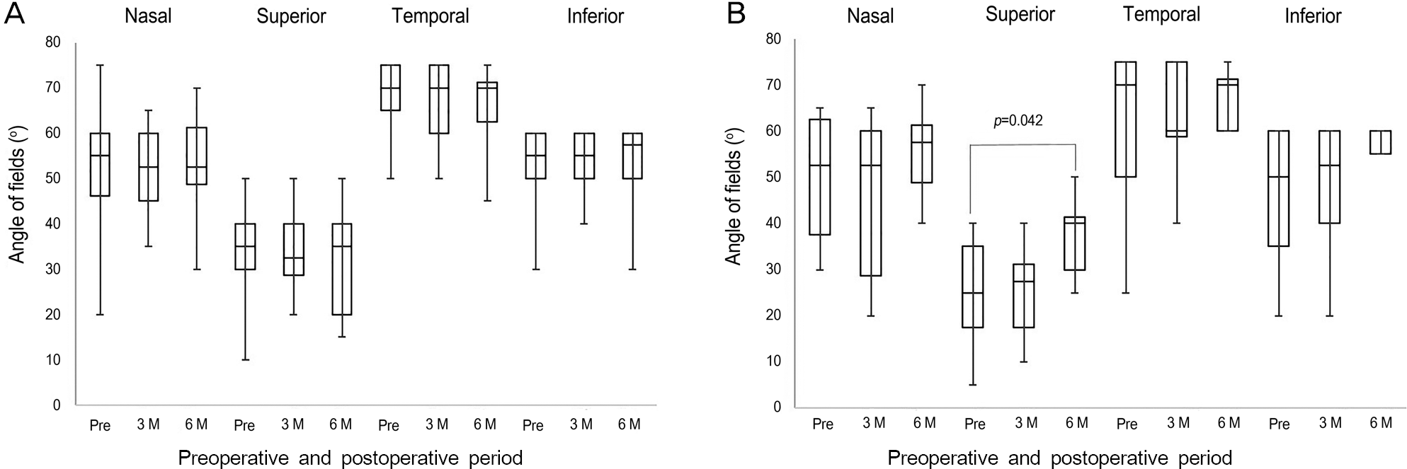
No differences were found between the 2 groups in terms of visual acuity, central macular thickness, and peripapillary RNFL thickness preoperatively and at 6 months postoperatively (p = 0.125 for visual acuity, p = 0.734 for central macular thick-ness, p = 0.615 for RNFL thickness). Six months after surgery, no significant increase was found in any region of visual field in the ICG group ( p = 0.392). The visual field was significantly increased in the superior region in the ND group ( p = 0.042). The RNFL thickness in the temporal quadrant was significantly reduced at 6 months postoperatively compared to baseline values in both groups ( p = 0.011 for ICG group, p = 0.042 for ND group).
References
1. Klein R, Klein BE, Wang Q, Moss SE. The epidemiology of epi-retinal membranes. Trans Am Ophthalmol Soc. 1994; 92:403–25. discussion 425-30.
2. de Bustros S, Thompson JT, Michels RG. . Vitrectomy for idio-pathic epiretinal membranes causing macular pucker. Br J Ophthalmol. 1988; 72:692–5.

3. Machemer R. The surgical removal of epiretinal macular mem-branes (macular puckers). Klin Monbl Augenheilkd. 1978; 173:36–42.
4. Grewing R, Mester U. Results of surgery for epiretinal membranes and their recurrences. Br J Ophthalmol. 1996; 80:323–6.

5. Shimada H, Nakashizuka H, Hattori T. . Double staining with brilliant blue G and double peeling for epiretinal membranes. Ophthalmology. 2009; 116:1370–6.

6. Lee JE, Yoon TJ, Oum BS. . Toxicity of indocyanine green in-jected into the subretinal space: subretinal toxicity of indocyanine green. Retina. 2003; 23:675–81.
7. Da Mata AP, Burk SE, Riemann CD. . Indocyanine green-as-sisted peeling of the retinal internal limiting membrane during vi-trectomy surgery for macular hole repair. Ophthalmology. 2001; 108:1187–92.

8. Ando F, Sasano K, Ohba N. . Anatomic and visual outcomes after indocyanine green-assisted peeling of the retinal internal lim-iting membrane in idiopathic macular hole surgery. Am J Ophthalmol. 2004; 137:609–14.

9. Gass CA, Haritoglou C, Schaumberger M, Kampik A. Functional outcome of macular hole surgery with and without indocyanine green-assisted peeling of the internal limiting membrane. Graefes Arch Clin Exp Ophthalmol. 2003; 241:716–20.

10. Uemura A, Kanda S, Sakamoto Y, Kita H. Visual field defects after uneventful vitrectomy for epiretinal membrane with indocyanine green-assisted internal limiting membrane peeling. Am J Ophthalmol. 2003; 136:252–7.

11. Kanda S, Uemura A, Yamashita T. . Visual field defects after intravitreous administration of indocyanine green in macular hole surgery. Arch Ophthalmol. 2004; 122:1447–51.
12. Nagai N, Ishida S, Shinoda K. . Surgical effects and complica-tions of indocyanine green-assisted internal limiting membrane peeling for idiopathic macular hole. Acta Ophthalmol Scand. 2007; 85:883–9.

13. Sekiryu T, Iida T. Long-term observation of fundus infrared fluo-rescence after indocyanine green-assisted vitrectomy. Retina. 2007; 27:190–7.

14. von Jagow B, Höing A, Gandorfer A. . Functional outcome of indocyanine green-assisted macular surgery: 7-year follow-up. Retina. 2009; 29:1249–56.
15. Gandorfer A, Haritoglou C, Gass CA. . Indocyanine green-as-sisted peeling of the internal limiting membrane may cause retinal damage. Am J Ophthalmol. 2001; 132:431–3.

16. Yam HF, Kwok AK, Chan KP. . Effect of indocyanine green and illumination on gene expression in human retinal pigment epi-thelial cells. Invest Ophthalmol Vis Sci. 2003; 44:370–7.

17. Murata M, Shimizu S, Horiuchi S, Sato S. The effect of in-docyanine green on cultured retinal glial cells. Retina. 2005; 25:75–80.

18. Gandorfer A, Haritoglou C, Gandorfer A, Kampik A. Retinal dam-age from indocyanine green in experimental macular surgery. Invest Ophthalmol Vis Sci. 2003; 44:316–23.

19. Haritoglou C, Priglinger S, Gandorfer A. . Histology of the vit-reoretinal interface after indocyanine green staining of the ILM, with illumination using a halogen and xenon light source. Invest Ophthalmol Vis Sci. 2005; 46:1468–72.

20. Song BY, Ahn KY, Seo MS. Internal limiting membrane peeling using indocyanine green in vitrectomy for idiopathic macular hole. J Korean Ophthalmol Soc. 2004; 45:444–50.
21. Lee JE, Oum BS. Macular hole surgery with or without in-docyanine green-assisted internal limiting membrane peeling. J Korean Ophthalmol Soc. 2003; 44:2553–9.
22. Nam DH, Hwang S, Huh K. Idiopathic macular hole surgery with or without indocyanine green-stained internal limiting membrane peeling. J Korean Ophthalmol Soc. 2004; 45:1086–91.
23. Hillenkamp J, Saikia P, Herrmann WA. . Surgical removal of idiopathic epiretinal membrane with or without the assistance of indocyanine green: a randomised controlled clinical trial. Graefes Arch Clin Exp Ophthalmol. 2007; 245:973–9.

24. Wu Y, Zhu W, Xu D. . Indocyanine green-assisted internal lim-iting membrane peeling in macular hole surgery: a meta-analysis. PLoS One. 2012; 7:e48405.

25. Schmid-Kubista KE, Lamar PD, Schenk A. . Comparison of macular function and visual fields after membrane blue or in-fracyanine green staining in vitreoretinal surgery. Graefes Arch Clin Exp Ophthalmol. 2010; 248:381–8.

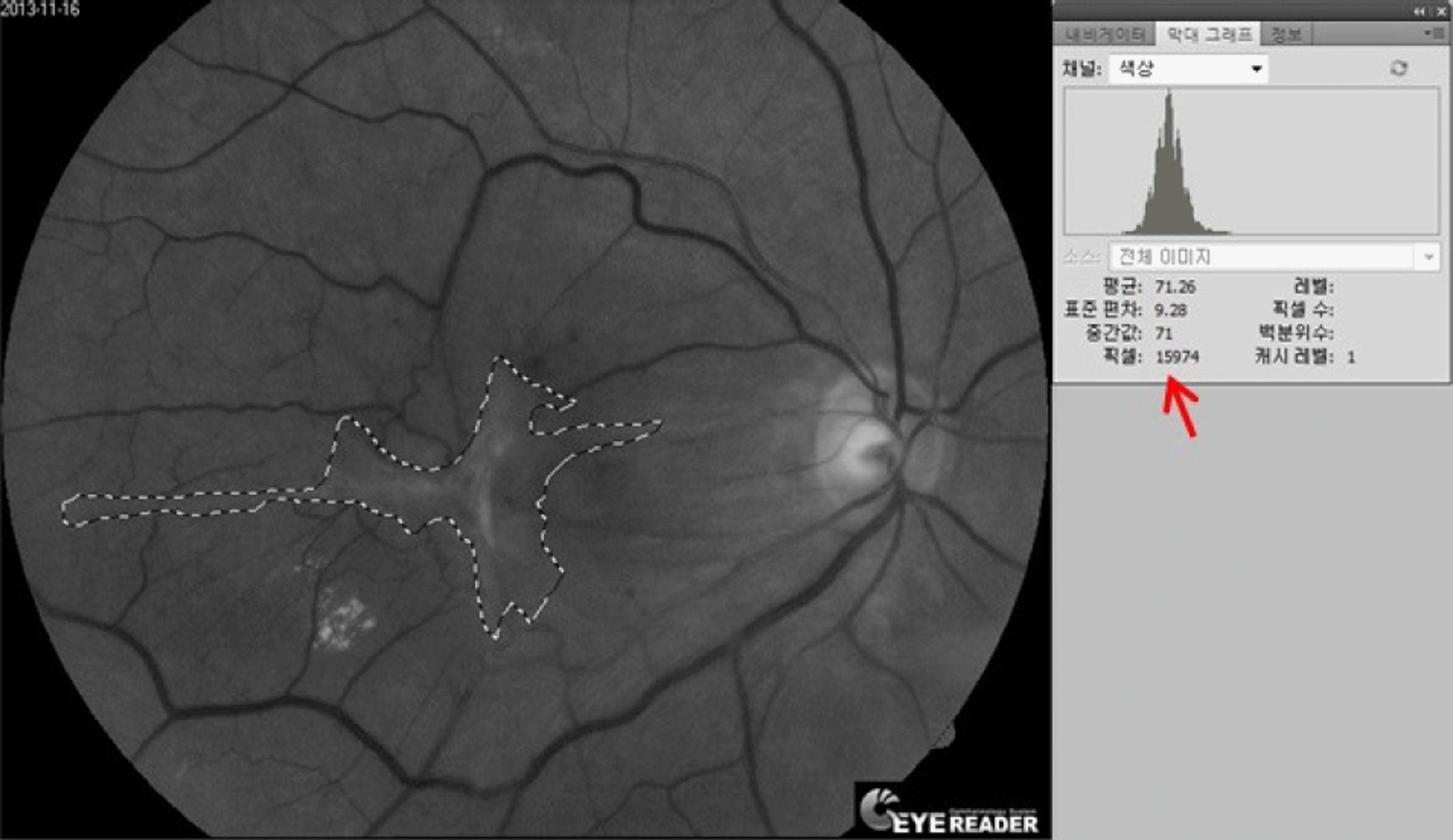
Figure 1.
Measuring the size of epiretinal membrane with red-free fundus photography and the Photoshop program (Photoshop 5.5, Adobe Systems Inc., San Jose, CA, USA). The selected area (dotted line) is expressed in pixels (red arrow) by the histogram option.

Figure 2.
Changes of visual fields according to regions and study points in the indocyanine green (ICG) group (A), and in the ND group (B). Superior visual fields significantly improved at 6 months after surgery in the ND group. ICG group is indocyanine green dye assisted internal limiting membrane peeling and ND group is internal limiting membrane peeling without staining. ND = no dye; Pre = preoperative period; M = months.

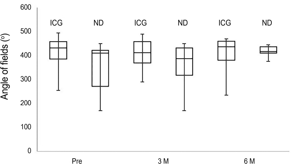
Figure 3.
Changes of preoperative and postoperative total visu-al fields. There was no significant difference between the groups. ICG group is indocyanine green dye assisted internal limiting membrane peeling and ND group is internal limiting membrane peeling without staining. ICG = indocyanine green. ND = no dye; Pre = preoperative period; M = months.

Figure 4.
Changes of nerve fiber layer thickness according to regions and study points in the ICG group (A), and in the ND group (B). It showed that temporal peripapillary RNFL thickness were significantly thinning at 3 months and 6 months in both groups. ICG group is indocyanine green dye assisted internal limiting membrane peeling and ND group is internal limiting membrane peeling without staining. ICG = indocyanine green; ND = no dye; RNFL = retinal nerve fiber layer; Pre = preoperative period; M = months.

Table 1.
Preoperative demographic findings
| Total (n = 33) | ICG group* (n = 18) | ND group† (n = 15) | p-value | |
|---|---|---|---|---|
| Age (years) | 63.9 (49-74) | 63.3 (49-74) | 64.5 (55-71) | 0.438 |
| Sex (M:F) | 10:23 | 6:12 | 4:11 | 0.541 |
| OD:OS | 19:14 | 13:5 | 6:9 | 0.094 |
| Preoperative visual acuity (log MAR) | 0.46 ± 0.29 | 0.40 ± 0.34 | 0.53 ± 0.34 | 0.263 |
| Preoperative IOP (mm Hg) | 14.6 ± 3.2 | 15.1 ± 2.8 | 13.9 ± 3.3 | 0.112 |
| Combined surgery (eyes) | 26 | 17 | 9 | 0.017 |
Table 2.
Visual acuity changes over time
|
Best corrected visual acuity (log MAR) |
|||
|---|---|---|---|
| Baseline | 3 months | 6 months | |
| ICG group* | 0.40 ± 0.34 | 0.20 ± 0.26 | 0.15 ± 0.50 |
| p-value† | - | 0.003 | 0.001 |
| ND group‡ | 0.53 ± 0.34 | 0.34 ± 0.26 | 0.29 ± 0.50 |
| p-value† | - | 0.045 | 0.038 |
| p-value§ | 0.278 | 0.235 | 0.125 |


 PDF
PDF ePub
ePub Citation
 Print
Print


 XML Download