Abstract
Purpose
To evaluate the efficacy of Tecnis® toric intraocular lens (IOL) implantation for the correction of astigmatism and rota-tional stability during cataract surgery in patients with cataract and astigmatism.
Methods
We prospectively analyzed 17 eyes of 14 patients with 1 to 4 diopters (D) of corneal astigmatism who underwent pha-coemulsification and Tecnis® toric IOL implantation at Seoul National University Hospital from June 2013 to May 2014. Informed consent was obtained from all participants before the clinical trial. We evaluated the changes in visual acuity, refraction, astigma-tism, IOL axis and higher order aberration for 3 months postoperatively. Power vector analysis was used to analyze astigmatism.
Results
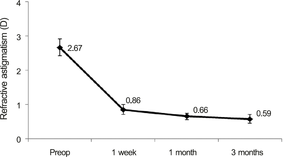
The mean uncorrected visual acuity (log MAR) significantly improved from 0.58 ± 0.34 to 0.26 ± 0.43 at 3 months postoperatively. The mean refractive astigmatism was significantly decreased by 77.9% from a mean value of -2.67 ± 0.89 D to -0.59 ± 0.48 D at 3 months postoperatively. According to power vector analysis, M, B, J0, and J45 were significantly reduced after the surgery. The mean difference between achieved and intended IOL axis was 3.26 degrees clockwise at postoperative 3 months, which was statistically insignificant. Most of the rotational changes were observed within a month after the surgery.
Go to : 
References
1. Rubenstein JB, Raciti M. Approaches to corneal astigmatism in cataract surgery. Curr Opin Ophthalmol. 2013; 24:30–4.

2. Kretz FT, Bastelica A, Carreras H. . Clinical outcomes and sur-geon assessment after implantation of a new diffractive multifocal toric intraocular lens. Br J Ophthalmol. 2015; 99:405–11.

3. Chen W, Zuo C, Chen C. . Prevalence of corneal astigmatism before cataract surgery in Chinese patients. J Cataract Refract Surg. 2013; 39:188–92.

4. Ferrer-Blasco T, Montés-Micó R, Peixoto-de-Matos SC. . Prevalence of corneal astigmatism before cataract surgery. J Cataract Refract Surg. 2009; 35:70–5.

5. Fouda S, Kamiya K, Aizawa D, Shimizu K. Limbal relaxing in-cision during cataract extraction versus photoastigmatic keratec-tomy after cataract extraction in controlling pre-existing corneal astigmatism. Graefes Arch Clin Exp Ophthalmol. 2010; 248:1029–35.

6. Kim DH, Wee WR, Lee JH, Kim MK. The short term effects of a single limbal relaxing incision combined with clear corneal incision. Korean J Ophthalmol. 2010; 24:78–82.

7. Kaufmann C, Peter J, Ooi K. . Limbal relaxing incisions versus on-axis incisions to reduce corneal astigmatism at the time of cata-ract surgery. J Cataract Refract Surg. 2005; 31:2261–5.

8. Lim R, Borasio E, Ilari L. Long-term stability of keratometric as-tigmatism after limbal relaxing incisions. J Cataract Refract Surg. 2014; 40:1676–81.

9. Hirnschall N, Gangwani V, Crnej A. . Correction of moderate corneal astigmatism during cataract surgery: toric intraocular lens versus peripheral corneal relaxing incisions. J Cataract Refract Surg. 2014; 40:354–61.

10. Jeon JH, Hyung Taek Tyler R, Seo KY. . Comparison of re-fractive stability after non-toric versus toric intraocular lens im-plantation during cataract surgery. Am J Ophthalmol. 2014; 157:658–65.e1.

11. Visser N, Beckers HJ, Bauer NJ. . Toric vs aspherical control intraocular lenses in patients with cataract and corneal astigma-tism: a randomized clinical trial. JAMA Ophthalmol. 2014; 132:1462–8.
12. Na JH, Lee HS, Joo CK. The clinical result of acrySof toric intra-ocular lens implantation. J Korean Ophthalmol Soc. 2009; 50:831–8.

13. Jeon HM, Lee KH. Analysis of miscorrection after implantation of the toric intraocular lens. J Korean Ophthalmol Soc. 2014; 55:1636–41.

14. Han JM, Choi HJ, Kim MK. . Comparative analysis of corneal refraction and astigmatism measured with autokeratometer, IOL Master, and topography. Korean Ophthalmol Soc. 2011; 52:1427–33.

15. Lee H, Kim TI, Kim EK. Corneal astigmatism analysis for toric in-traocular lens implantation: precise measurements for perfect correction. Curr Opin Ophthalmol. 2015; 26:34–8.
16. Hirnschall N, Maedel S, Weber M, Findl O. Rotational stability of a single-piece toric acrylic intraocular lens: a pilot study. Am J Ophthalmol. 2014; 157:405–11.e1.

17. Mencucci R, Favuzza E, Guerra F. . Clinical outcomes and ro-tational stability of a 4-haptic toric intraocular lens in myopic eyes. J Cataract Refract Surg. 2014; 40:1479–87.

18. Preussner PR, Hoffmann P, Wahl J. Impact of posterior corneal sur-face on toric intraocular lens (IOL) calculation. Curr Eye Res 2015; 40. 809–14.

19. Savini G, Hoffer KJ, Carbonelli M. . Influence of axial length and corneal power on the astigmatic power of toric intraocular lenses. J Cataract Refract Surg. 2013; 39:1900–3.

20. Waltz KL, Featherstone K, Tsai L, Trentacost D. Clinical outcomes of TECNIS toric intraocular lens implantation after cataract re-moval in patients with corneal astigmatism. Ophthalmology 2015; 122. 39–47.
21. Kaye SB, Campbell SH, Davey K, Patterson A. A method for as-sessing the accuracy of surgical technique in the correction of astigmatism. Br J Ophthalmol. 1992; 76:738–40.

22. Miyake T, Kamiya K, Amano R. . Long-term clinical outcomes of toric intraocular lens implantation in cataract cases with preex-isting astigmatism. J Cataract Refract Surg. 2014; 40:1654–60.

23. Ferreira TB, Almeida A. Comparison of the visual outcomes and OPD-scan results of AMO Tecnis toric and Alcon Acrysof IQ toric intraocular lenses. J Refract Surg. 2012; 28:551–5.

24. Hayashi K, Kondo H, Yoshida M. . Higher-order aberrations and visual function in pseudophakic eyes with a toric intraocular lens. J Cataract Refract Surg. 2012; 38:1156–65.

25. Eom Y, Kang SY, Song JS. . Effect of effective lens position on cylinder power of toric intraocular lenses. Can J Ophthalmol. 2015; 50:26–32.

26. Goggin M, Moore S, Esterman A. Outcome of toric intraocular lens implantation after adjusting for anterior chamber depth and in-traocular lens sphere equivalent power effects. Arch Ophthalmol. 2011; 129:998–1003.

27. Bascaran L, Mendicute J, Macias-Murelaga B. . Efficacy and stability of AT TORBI 709 M toric IOL. J Refract Surg. 2013; 29:194–9.

28. Ale Magar JB, Cunningham F, Brian G. Comparison of automated and partial coherence keratometry and resulting choice of toric IOL. Optom Vis Sci. 2013; 90:385–91.

29. Chang M, Kang SY, Kim HM. Which keratometer is most reliable for correcting astigmatism with toric intraocular lenses? Korean J Ophthalmol. 2012; 26:10–4.
30. Savini G, Næser K. An analysis of the factors influencing the re-sidual refractive astigmatism after cataract surgery with toric intra-ocular lenses. Invest Ophthalmol Vis Sci. 2015; 56:827–35.

31. Hasegawa Y, Okamoto F, Nakano S. . Effect of preoperative corneal astigmatism orientation on results with a toric intraocular lens. J Cataract Refract Surg. 2013; 39:1846–51.

32. Miyake T, Shimizu K, Kamiya K. Distribution of posterior corneal astigmatism according to axis orientation of anterior corneal astigmatism. PloS One. 2015; 10:e0117194.

33. Teichman JC, Baig K, Ahmed II. Simple technique to measure tor-ic intraocular lens alignment and stability using a smartphone. J Cataract Refract Surg. 2014; 40:1949–52.

34. Kasthurirangan S, Feuchter L, Smith P, Nixon D. Software-based evaluation of toric IOL orientation in a multicenter clinical study. J Refract Surg. 2014; 30:820–6.

Go to : 
 | Figure 1.Image analysis for the rotation of the cylindrical axis on the toric intraocular lens. |
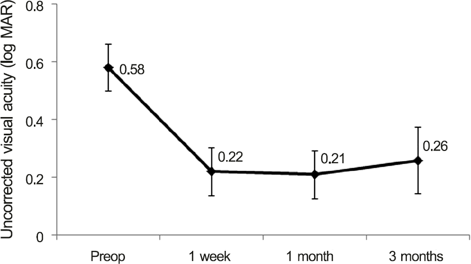 | Figure 2.Changes in visual acuity. There were statistically significant improvements in visual acuity at postoperative 1 week, 1 month, and 3 months compared with pre-operative values. log MAR = logarithm of the minimum angle of reso-lution; Preop = preoperative. |
 | Figure 3.Change in refractive astigmatism. There were stat-istically significant decreases in refractive astigmatism at post-operative 1 week, 1 month, and 3 months compared with pre- operative values. Preop = preoperative. |
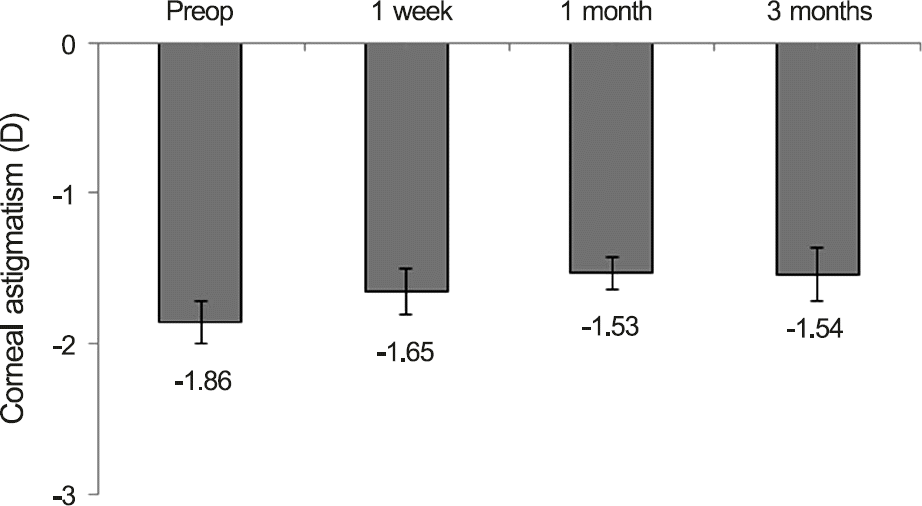
 | Figure 4.Changes in the corneal astigmatism between pre- and post- operation. There is no significant changes between the follow-ups. Preop = preoperation. |
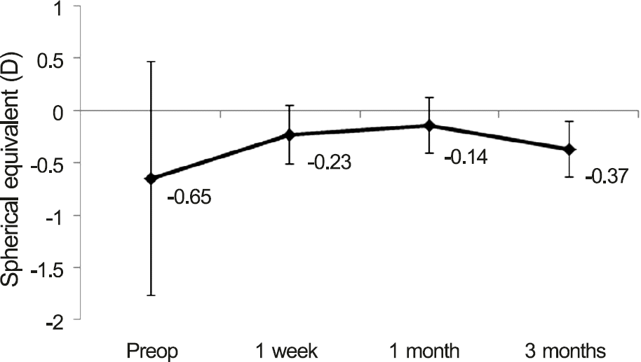 | Figure 5.Changes in spherical equivalents between pre- and post- operation. Preop = preoperation. |
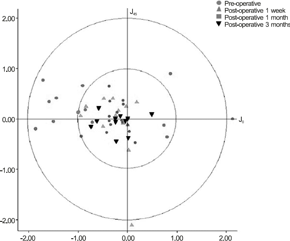 | Figure 6.Diagram showing refractive astigmatism in diopters measured before surgery and 1 week, 1 month, and 3 months after implanting a single-piece toric acrylic intraocular lens. Astigmatism is shown as vectors at J0 and J45. Each ring repre-sents 1 diopter. |
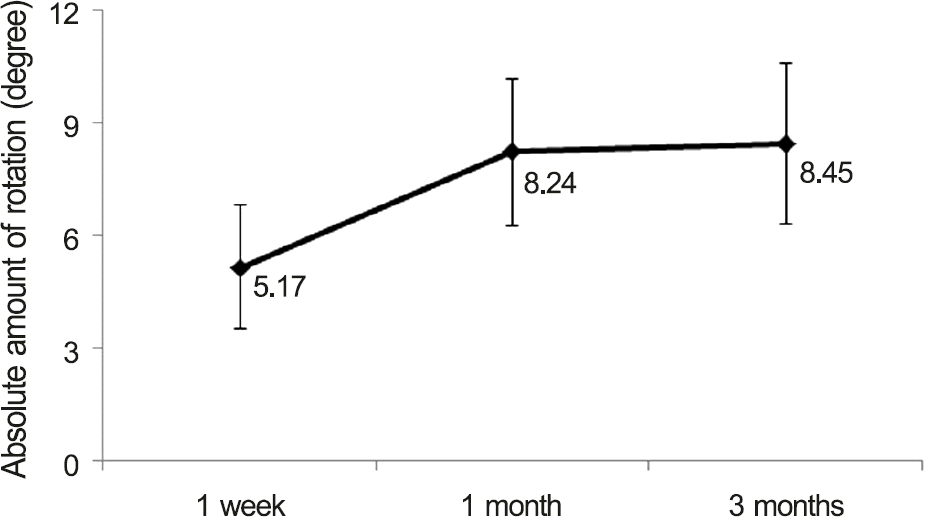 | Figure 7.Absolute amount of rotation of a single-piece toric acrylic intraocular lens (IOL) compared with preoperative IOL axis. There was significant rotation between 1 week and 1 month after surgery. However, axis was stable after 1 month. |
Table 1.
Demographics and preoperative status of the enrolled patients
Table 2.
Vector analysis of astigmatism
| Preop | Postop 1 week | Postop 1 month | Postop 3 months | |
|---|---|---|---|---|
| M | -3.43 ± 6.57 D | -0.66 ± 0.97 D | -0.47 ± 0.94 D | -0.66 ± 0.94 D |
| p-value | 0.038* | 0.060 | 0.305 | |
| J0 | -0.55 ± 1.26 D | -0.26 ± 0.34 D | -0.20 ± 0.19 D | -0.12 ± 0.28 D |
| p-value | 0.286 | 0.198 | 0.028* | |
| J45 | 0.18 ± 0.41 D | 0.05 ± 0.29 D | 0.001 ± 0.27 D | -0.09 ± 0.17 D |
| p-value | 0.021* | 0.034* | 0.131 | |
| B | 4.61 ± 5.92 D | 1.03 ± 0.74 D | 0.88 ± 0.67 D | 0.82 ± 0.59 D |
| p-value | 0.008* | 0.002* | 0.003* |
Table 3.
Post-operative high order aberration of the enrolled patients


 PDF
PDF ePub
ePub Citation
 Print
Print


 XML Download