Abstract
Purpose
To compare central corneal thickness (CCT) as measured using noncontact specular microscopy (NCSM), dual rotat-ing Scheimpflug camera (Galilei®), and ultrasound pachymetry (USP).
Methods
The measurements of CCT using NCSM, dual rotating Scheimpflug camera and USP in 70 eyes of 70 healthy subjects were compared.
Results
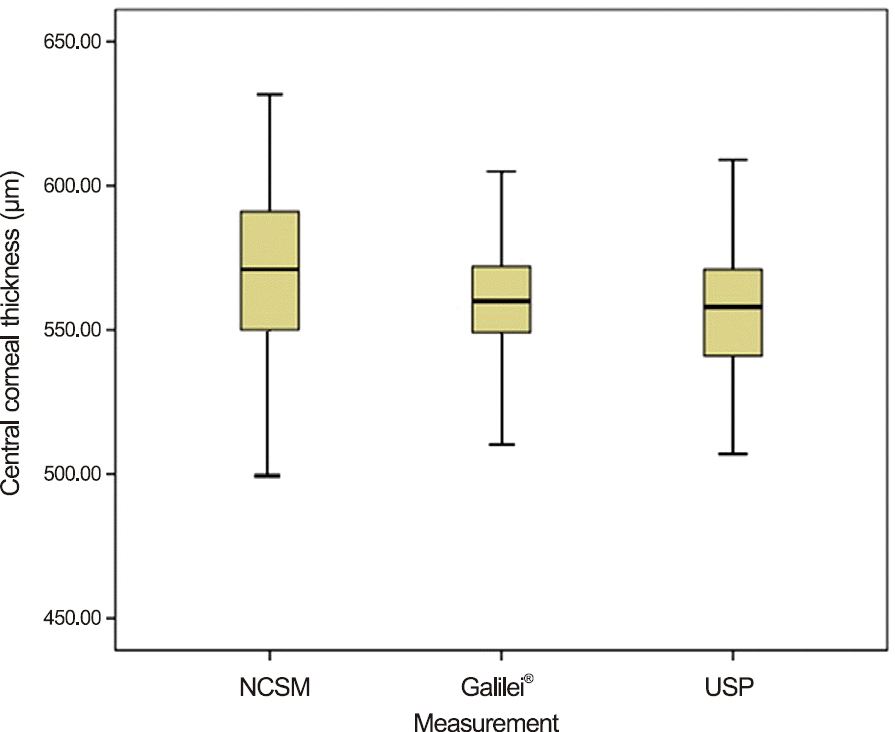
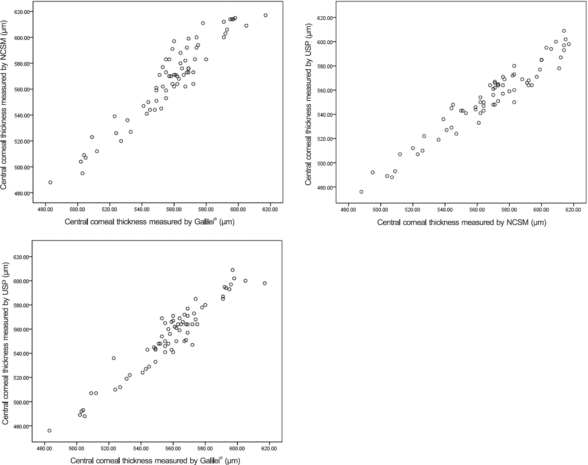
The average measurements of CCT using NCSM, dual rotating Scheimpflug camera, and USP were 567.70 ± 31.21 µm, 557.84 ± 26.29 µm, and 553.31 ± 29.69 µm, respectively. The CCT measurement using NCSM was statistically significantly thicker than when measured using USP ( p < 0.05). There was no significant difference between the NCSM and dual rotating Scheimpflug camera ( p = 0.138). Additionally, there was no significant difference between the dual rotating Scheimpflug camera and USP ( p = 0.656). A significant linear correlation was observed among the NCSM, dual rotating Scheimpflug camera, and USP (r > 0.900, p < 0.001).
Conclusions
The results of the 3 methods were significantly correlated but the measurement using NCSM was significantly thicker than when using USP. CCT measurements of healthy eyes using dual rotating Scheimpflug camera were more corre-lated with USP than NCSM. The CCT measurements using dual rotating Scheimpflug camera is a better alternative for USP than NCSM.
References
1. Ou RJ, Shaw EL, Glasgow BJ. Keratectasia after laser in situ kera-tomileusis (LASIK): evaluation of the calculated residual stromal bed thickness. Am J Ophthalmol. 2002; 134:771–3.

2. Wang Z, Chen J, Yang B. Posterior corneal surface topographic changes after laser in situ keratomileusis are related to resicdual corneal bed thickness. Ophthalmology. 1999; 106:406–9. discussion 409-10.
3. Yau CW, Cheng HC. Microkeratome blades and corneal flap thick-ness in LASIK. Ophthalmic Surg Lasers Imaging. 2008; 39:471–5.

4. Whitacre MM, Stein RA, Hassanein K. The effect of corneal thick-ness on applanation tonometry. Am J Ophthalmol. 1993; 115:592–6.

5. Kim DH, Kim MS, Kim JH. Early corneal-thickness changes after penetrating keratoplasty. J Korean Ophthalmol Soc. 1997; 38:1355–61.
6. Doughty MJ, Zaman ML. Human corneal thickness and its impact on intraocular pressure measures: a review and meta-analysis approach. Surv Ophthalmol. 2000; 44:367–408.
7. Jonas JB, Stroux A, Velten I. . Central corneal thickness corre-lated with glaucoma damage and rate of progression. Invest Ophthalmol Vis Sci. 2005; 46:1269–74.

8. Li EY, Mohamed S, Leung CK. . Agreement among 3 methods to measure corneal thickness: ultrasound pachymetry, Orbscan II, and Visante anterior segment optical coherence tomography. Ophthalmology. 2007; 114:1842–7.

9. Choi KS, Nam SM, Lee HK. . Comparison of central corneal thickness after the instillation of topical anesthetics: proparacaine versus oxybuprocaine. J Korean Ophthalmol Soc. 2005; 46:757–62.
10. Kim HS, Kim JH, Kim HM, Song JS. Comparison of corneal thick-ness measured by specular, US pachymetry, and Orbscan in post-PKP eyes. J Korean Ophthalmol Soc. 2007; 48:245–50.
11. Menassa N, Kaufmann C, Goggin M. . Comparison and re-producibility of corneal thickness and curvature readings obtained by the Galilei and the Orbscan II analysis systems. J Cataract Refract Surg. 2008; 34:1742–47.

12. Shim HS, Choi CY, Lee HG. . Utility of the anterior segment optical coherence tomography for measurements of central corneal thickness. J Korean Ophthalmol Soc. 2007; 48:1643–8.

13. Jung YG, Song JS, Kim HM, Jung HR. Comparison of corneal thickness measurements with noncontact specular microscope and ultrasonic pachymeter. J Korean Ophthalmol Soc. 2004; 45:1060–5.
14. Kim HY, Budenz DL, Lee PS. . Comparison of central corneal thickness using anterior segment optical coherence tomography vs ultrasound pachymetry. Am J Ophthalmol. 2008; 145:228–32.

15. Kim DW, Yi KY, Choi DG, Shin YJ. Corneal thickness measured by dual Scheimpflug, anterior segment optical coherence tomog-raphy, and ultrasound pachymetry. J Korean Ophthalmol Soc. 2012; 53:1412–8.

16. Thomas J, Wang J, Rollins AM, Sturm J. Comparison of corneal thickness measured with optical coherence tomography, ultrasonic pachymetry, and a scanning slit method. J Refract Surg 2006; 22. 671–8.

17. Ling T, Ho A, Holden BA. Method of evaluating ultrasonic pachometers. Am J Optom Physiol Opt. 1986; 63:462–6.

18. Copt RP, Thomas R, Mermoud A. Corneal thickness in ocular hy-pertension, primary open-angle glaucoma, and normal tension glaucoma. Arch Ophthalmol. 1999; 117:14–6.

19. Harper CL, Boulton ME, Bennett D. . Diurnal variations in hu-man corneal thickness. Br J Ophthalmol. 1996; 80:1068–72.

20. Ventura AC, Wälti R, Böhnke M. Corneal thickness and endothe-lial density before and after cataract surgery. Br J Ophthalmol. 2001; 85:18–20.

21. Savini G, Carbonelli M, Barboni P, Hoffer KJ. Repeatability of au-tomatic measurements performed by a dual Scheimpflug analyzer in unoperated and post-refractive surgery eyes. J Cataract Refract Surg. 2011; 37:302–9.

22. Bovelle R, Kaufman SC, Thompson HW, Hamano H. Corneal thickness measurements with the Topcon SP-2000P specular mi-croscope and an ultrasound pachymeter. Arch Ophthalmol 1999; 117. 868–70.

23. Módis L Jr, Langenbucher A, Seitz B. Corneal thickness measure-ments with contact and noncontact specular microscopic and ultra-sonic pachymetry. Am J Ophthalmol. 2001; 132:517–21.
24. Jung YG, Song JS, Kim HM, Jung HR. Comparison of corneal thickness measurements with noncontact specular microscope and ultrasonic pachymeter. J Korean Ophthalmol Soc. 2004; 45:1060–5.
25. Yang YS, Koh JW. Utility of the noncontact specular microscopy for measurements of central corneal thickness. J Korean Ophthalmol Soc. 2014; 55:59–65.

26. Yeter V, Sönmez B, Beden U. Comparison of central corneal thickness measurements by Galilei Dual-Scheimpflug analyzer(R) and ultrasound pachymeter in myopic eyes. Ophthalmic Surg Lasers Imaging. 2012; 43:128–34.
27. Ladi JS, Shah NA. Comparison of central corneal thickness meas-urements with the Galilei dual Scheimpflug analyzer and ultra-sound pachymetry. Indian J Ophthalmol. 2010; 58:385–8.

28. Prakash G, Agarwal A, Jacob S. . Comparison of fourier-domain and time-domain optical coherence tomography for assessment of corneal thickness and intersession repeatability. Am J Ophthalmol. 2009; 148:282–90.e2.

29. Feizi S, Jafarinasab MR, Karimian F. . Central and peripheral corneal thickness measurement in normal and keratoconic eyes us-ing three corneal pachymeters. J Ophthalmic Vis Res. 2014; 9:296–304.
Figure 1.
Mean value, 95% CI and range of CCT measured by NCSM, dual rotating Scheimpflug (Galilei®), and USP. CI = confidence interval; CCT = central corneal thickness; NCSM = noncontact specular microscopy; USP = ultrasound pachymetry.

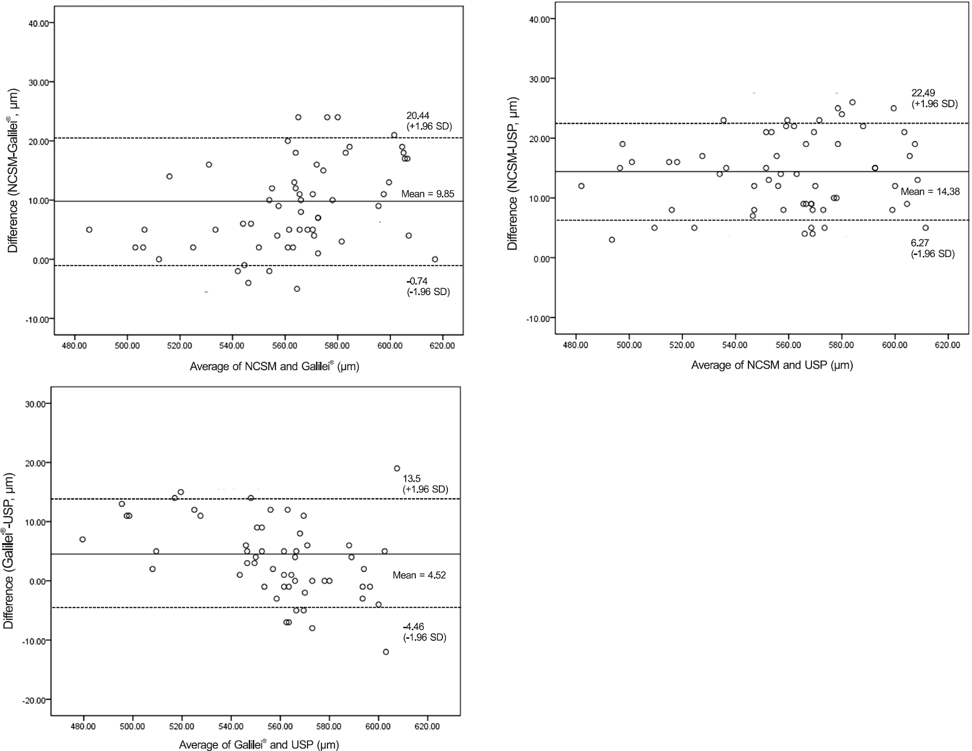
Figure 2.
Bland Altman plots between the 2 methods. The middle line is the mean and the lines on the side represent the upper and lower 95% LoA. (A) NCSM and dual rotating Scheimpflug (Galilei®). (B) NCSM and USP. (C) Galilei® and USP. LoA = limits of agreement; NCSM = noncontact specular microscopy; USP = ultrasound pachymetry.

Figure 3.
Scattergram showing the correlation of central corneal thickness measured by NCSM, dual rotating Scheimpflug (Galilei®), and USP. (A) Correlation between NCSM and Galilei® (r = 0.946, p < 0.001). (B) Correlation between NCSM and USP (r = 0.966, p < 0.001). (C) Correlation between Galilei® and USP (r = 0.956, p < 0.001). NCSM = noncontact specular microscopy; USP = ultrasound pachymetry.

Table 1.
Mean CCT measured by NCSM, dual rotating Scheimpflug (Galilei®), and USP
| NCSM | Galilei® | USP | p-value* | |
|---|---|---|---|---|
| CCT (μ m) | 567.70 ± 31.21 | 557.84 ± 26.29 | 553.31 ± 29.69 | 0.013* |
Table 2.
Pairwise comparison of central corneal thickness measurements
| Comparison | Mean difference ± SD (μ m) | p-value* |
|---|---|---|
| NCSM and Galilei® | 9.85 ± 4.92 | 0.138 |
| NCSM and USP | 14.38 ± 4.87 | 0.015 |
| Galilei® and USP | 4.52 ± 5.14 | 0.656 |


 PDF
PDF ePub
ePub Citation
 Print
Print


 XML Download