Abstract
Case summary
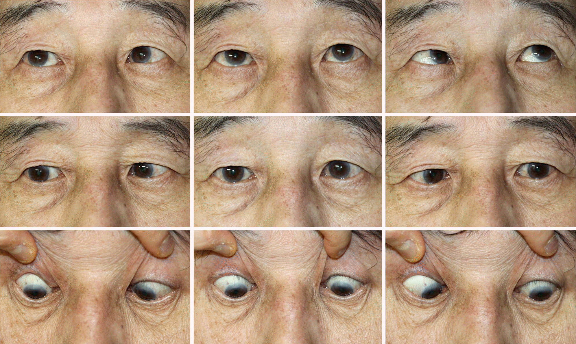
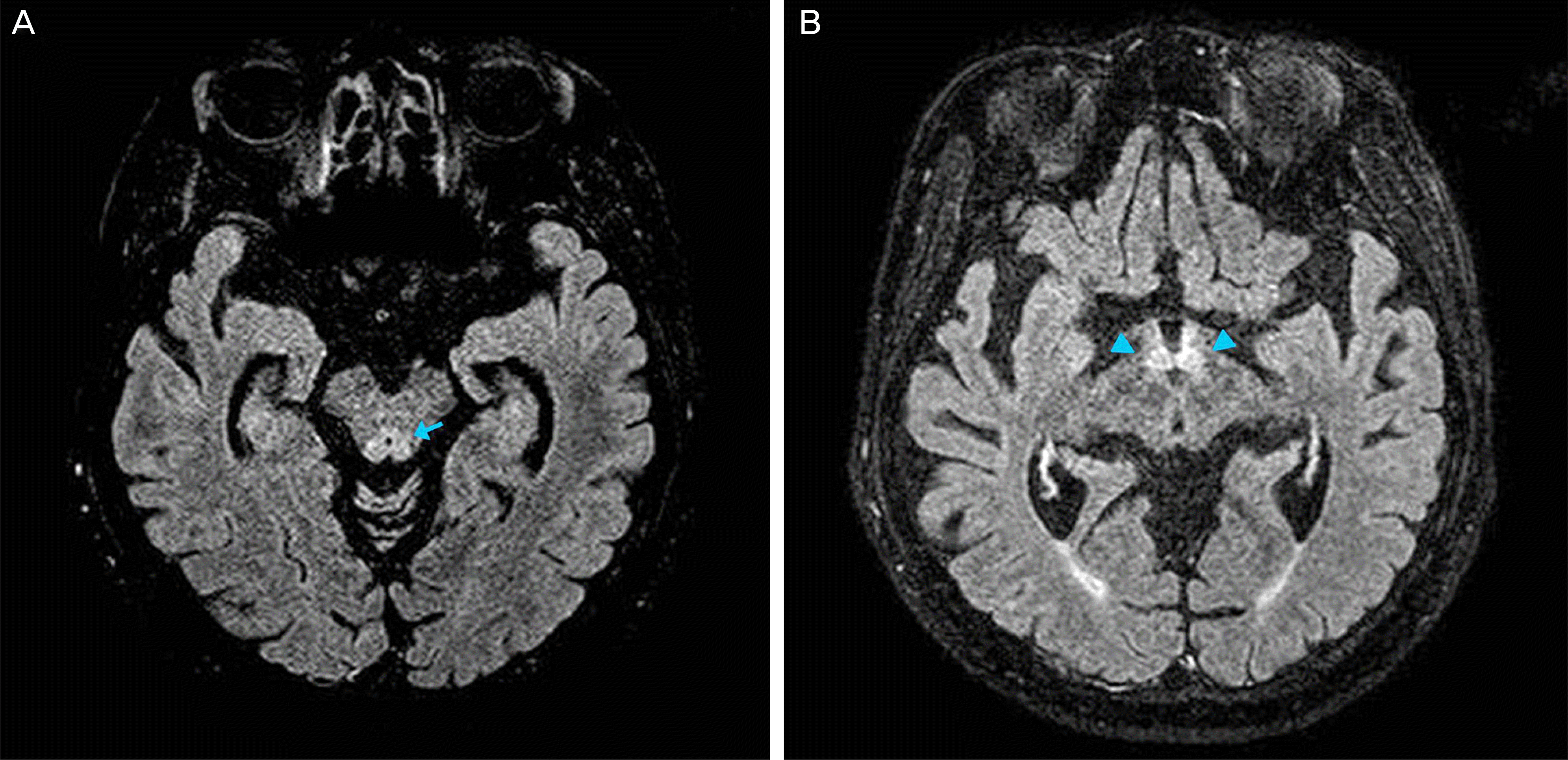
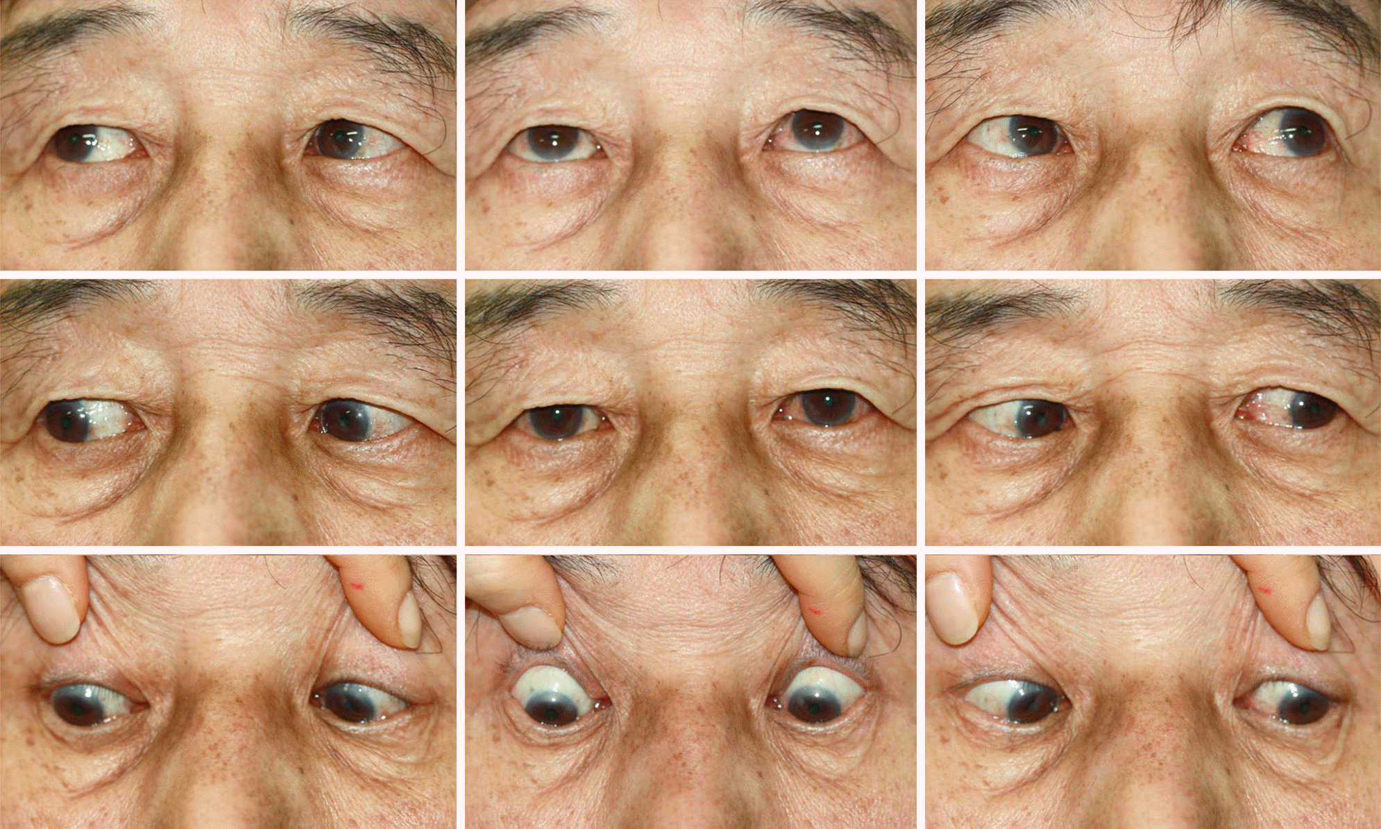
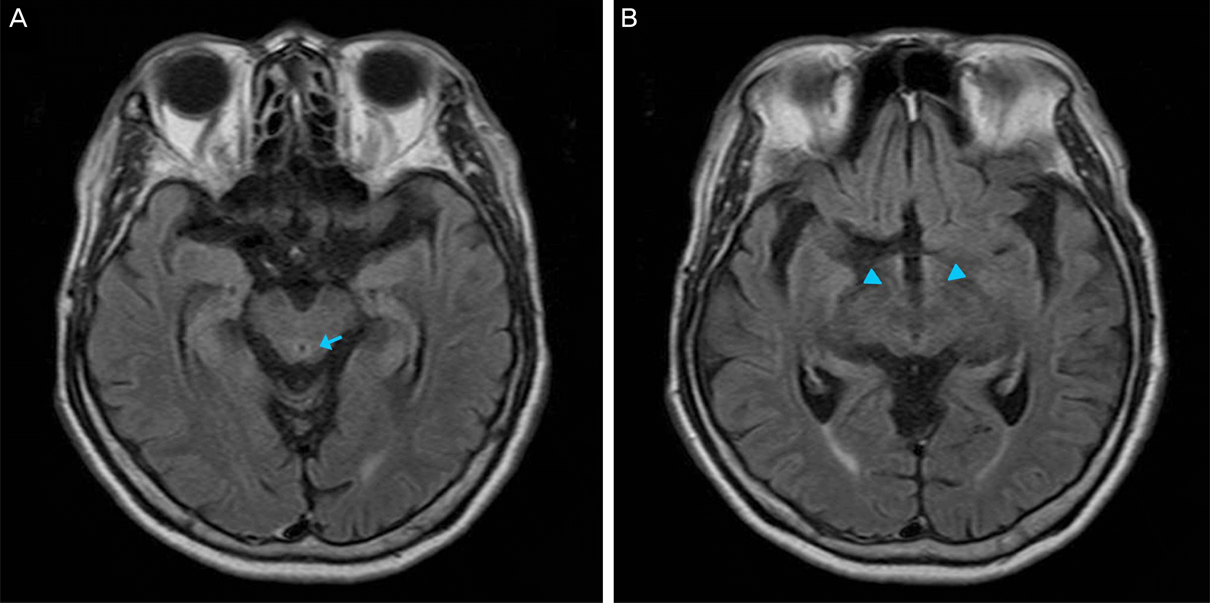
A 64-year-old male visited our hospital because of diplopia lasting a week. He was a chronic alcoholic drinking two bottles of makgeolli daily and eating little for a month. He showed -2 underaction of bilateral lateral rectus muscles and 45 prism diopters of esotropia at the primary position at the first visit. He had ataxia and mild cognitive impairment. There were high signal intensities in the periaqueductal area and mammillary bodies in the brain fluid attenuated inversion recovery magnetic resonance image. He was diagnosed with Wernike’s encephalopathy clinically and was immediately treated with intravenous thiamine. He showed -0.5 underaction of bilateral lateral muscles and 8 prism diopters of esotropia at the primary position 3 days after thiamine treatment.
Conclusions
Wernicke’s encephalopathy is a medical emergency. If diagnosis and treatment are delayed, patients may have neurological sequelae that can lead to death. Esotropia and diplopia can be the presenting manifestations in Wernike’s syndrome without other symptoms. In taking patient histories, physicians should ask about alcohol consumption and low food intake because of the possibility of acute incomitant esotropia associated with Wernicke’s encephalopathy.
Go to : 
References
2. Holub BJ. 1982 Borden Award lecture. Nutritional, biochemical, and clinical aspects of inositol and phosphatidylinositol metabolism. Can J Physiol Pharmacol. 1984; 62:1–8.

3. Tanphaichitr V. Thiamin. Shils M, Olson JA, Shike M, Ross AC, editors. Modern Nutrition in Health and Disease. 9th ed.Baltimore: Lippincott Williams & Wilkins;1999. p. 381.
4. Schenker S, Henderson GI, Hoyumpa AM Jr, McCandless DW. Hepatic and Wernicke's encephalopathies: current concepts of pathogenesis. Am J Clin Nutr. 1980; 33:2719–26.

6. Victor M, Adams RD, Collins GH. The Wernicke-Korsakoff syndrome. A clinical and pathological study of 245 patients, 82 with postmortem examinations. Contemp Neurol Ser. 1971; 7:1–206.
7. Eckardt MJ, Martin PR. Clinical assessment of cognition in alcoholism. Alcohol Clin Exp Res. 1986; 10:123–7.

8. Harper CG, Giles M, Finlay-Jones R. Clinical signs in the Wernicke- Korsakoff complex: a retrospective analysis of 131 cases diagnosed at necropsy. J Neurol Neurosurg Psychiatry. 1986; 49:341–5.
9. Torvik A, Lindboe CF, Rogde S. Brain lesions in alcoholics. A neu-ropathological study with clinical correlations. J Neurol Sci. 1982; 56:233–48.
10. Wallis WE, Willoughby E, Baker P. Coma in the Wernicke- Korsakoff syndrome. Lancet. 1978; 2:400–1.
11. Victor M. The Wernicke-Korsakoff syndrome. Vinken PJ, Bruyn GW, editors. Handbook of clinical neurology. Amsterdam: North-Holland Publishing Company;1976; v. 28:part II. 24370.
12. Reuler JB, Girard DE, Cooney TG. Current concepts. Wernicke's encephalopathy. N Engl J Med. 1985; 312:1035–9.
13. Kulkarni S, Lee AG, Holstein SA, Warner JE. You are what you eat. Surv Ophthalmol. 2005; 50:389–93.

14. Cooke CA, Hicks E, Page AB, Mc Kinstry S. An atypical pre-sentation of Wernicke's encephalopathy in an 11-year-old child. Eye (Lond). 2006; 20:1418–20.

15. Desai SD, Shah DS2. Atypical Wernicke's syndrome sans ence-phalopathy with acute bilateral vision loss due to post-chiasmatic optic tract edema. Ann Indian Acad Neurol. 2014; 17:103–5.

16. Tatsumoto M, Odaka M, Hirata K, Yuki N. Isolated abducens nerve palsy as a regional variant of Guillain-Barré syndrome. J Neurol Sci. 2006; 243:35–8.

17. Dwarakanath S, Ravichandra , Gopal S, Venkataramana NK. Posttraumatic bilateral abducens nerve palsy. Neurol India. 2006; 54:221–2.
18. Radhakrishnan K, Ahlskog JE, Garrity JA, Kurland LT. Idiopathic intracranial hypertension. Mayo Clin Proc. 1994; 69:169–80.

19. Anwar S, Nalla S, Fernando DJ. Abducens nerve palsy as a complication of lumbar puncture. Eur J Intern Med. 2008; 19:636–7.

20. Zuccoli G, Pipitone N. Neuroimaging findings in acute Wernicke's encephalopathy: review of the literature. AJR Am J Roentgenol. 2009; 192:501–8.

21. Antunez E, Estruch R, Cardenal C. . Usefulness of CT and MR imaging in the diagnosis of acute Wernicke's encephalopathy. AJR Am J Roentgenol. 1998; 171:1131–7.

22. Sechi G, Serra A. Wernicke's encephalopathy: new clinical settings and recent advances in diagnosis and management. Lancet Neurol. 2007; 6:442–55.

23. Mancinelli R, Ceccanti M, Guiducci MS. . Simultaneous liquid chromatographic assessment of thiamine, thiamine monophosphate and thiamine diphosphate in human erythrocytes: a study on alcoholics. J Chromatogr B Analyt Technol Biomed Life Sci. 2003; 789:355–63.

24. Lockman PR, Mumper RJ, Allen DD. Evaluation of blood-brain barrier thiamine efflux using the in situ rat brain perfusion method. J Neurochem. 2003; 86:627–34.

25. Thomson AD, Guerrini I, Marshall EJ. Wernicke's encephalop-athy: role of thiamine. Pract Gastroenterol. 2009; 23:21–30.
26. Cook CC, Hallwood PM, Thomson AD. B Vitamin deficiency and neuropsychiatric syndromes in alcohol misuse. Alcohol Alcohol. 1998; 33:317–36.

Go to : 
 | Figure 1.Nine cardinal gaze photographs at the first visit showing -2 underaction of bilateral lateral rectus muscles and 45-prism diopter esotropia at primary position. |
 | Figure 2.Brain fluid attenuated inversion recovery magnetic resonance image. There are high signal intensities in the periaqueductal area (arrow, A) and mammillary body (arrow heads, B). |


 PDF
PDF ePub
ePub Citation
 Print
Print


 XML Download