

 PDF
PDF ePub
ePub Citation
 Print
Print


Abstract
Purpose
To report multimodality diagnostic imaging in a case of unilateral acute idiopathic maculopathy.
Case summary
A 32-year-old woman with reduced vision in the right eye had experienced fatigue and flu-like symptoms, including sore throat and fever a few days before. Her best corrected visual acuity (BCVA) was 1.0 in the right eye. There were no cells in the anterior chamber and vitreous. Fundus photographs of the right eye on presentation showed gray-white thickening of the fovea and retinal hemorrhage next to the fovea. Fluorescein angiography demonstrated ring-shaped mottled hyperfluorescence in the early phase and dye pooling in the late phase. Spectral-domain optical coherence tomography (OCT) showed abnormal hyper-reflective thickening at the level of the outer retina and retinal pigment epithelium (RPE) and detachment of the neuro-sensory retina in the foveal lesion. The inner segment/outer segment junction and photoreceptor elevation/disruption was noted. Nineteen months after onset, the BCVA of the right eye was 1.0 and fundus photographs showed increased retinal pigment hy-perplasia, and residual RPE changes resembling a bull’s eye maculopathy. The OCT of the right macula showed that the inner segment/outer segment junction elevation/disruption almost completely regressed. The patient was diagnosed with unilateral acute idiopathic maculopathy.
Go to : 
References
1. Yannuzzi LA, Jampol LM, Rabb MF. . Unilateral acute idiopathic maculopathy. Arch Ophthalmol. 1991; 109:1411–6.

2. Beck AP, Jampol LM, Glaser DA, Pollack JS. Is coxsackievirus the cause of unilateral acute idiopathic maculopathy? Arch Ophthalmol. 2004; 122:121–3.
3. Kim IT, Jang SD. Unilateral idiopathic maculopathy. J Korean Ophthalmol Soc. 1999; 40:1260–8.
4. Choi KS, Kim JS. Bilateral acute idiopathic maculopathy. J Korean Ophthalmol Soc. 2000; 41:1626–30.
5. Lee SS, Kim EC, Kim YY, Kim SD. Unilateral acute idiopathic maculopathy: A case of atypical presentation. J Korean Ophthalmol Soc. 2000; 41:1796–800.
6. de la Fuente MA, Cuadrado R. Unilateral acute idiopathic macul-opathy: angiography, optical coherence tomography and micro-perimetry findings. J Ophthalmic Inflamm Infect. 2011; 1:125–7.
7. Jung CS, Payne JF, Bergstrom CS. . Multimodality diagnostic imaging in unilateral acute idiopathic maculopathy. Arch Ophthalmol. 2012; 130:50–6.
8. Aggio FB, Farah ME, Meirelles RL, de Souza EC. STRATUSOCT and multifocal ERG in unilateral acute idiopathic maculopathy. Graefes Arch Clin Exp Ophthalmol. 2006; 244:510–6.
9. Matsushita E, Fukuda K, Nakahira A. . Resolution of photo-receptor outer segment damage in a patient with unilateral acute idiopathic maculopathy observed using spectral-domain optical coherence tomography. Graefes Arch Clin Exp Ophthalmol. 2012; 250:765–8.
10. Milani P, Cacioppo V, Raimondi G, Scialdone A. Spectral domain OCT and autofluorescence imaging of unilateral acute idiopathic maculopathy. Eur J Ophthalmol. 2012; 22:499–502.
11. Ooto S, Hangai M, Yoshimura N. Photoreceptor restoration in unilateral acute idiopathic maculopathy on adaptive optics scanning laser ophthalmoscopy. Arch Ophthalmol. 2011; 129:1633–5.
12. Srour M, Querques G, Rostaqui O, Souied EH. Early spectral-do-main optical coherence tomography findings in unilateral acute idiopathic maculopathy. Retina. 2013; 33:2182–4.
Go to :
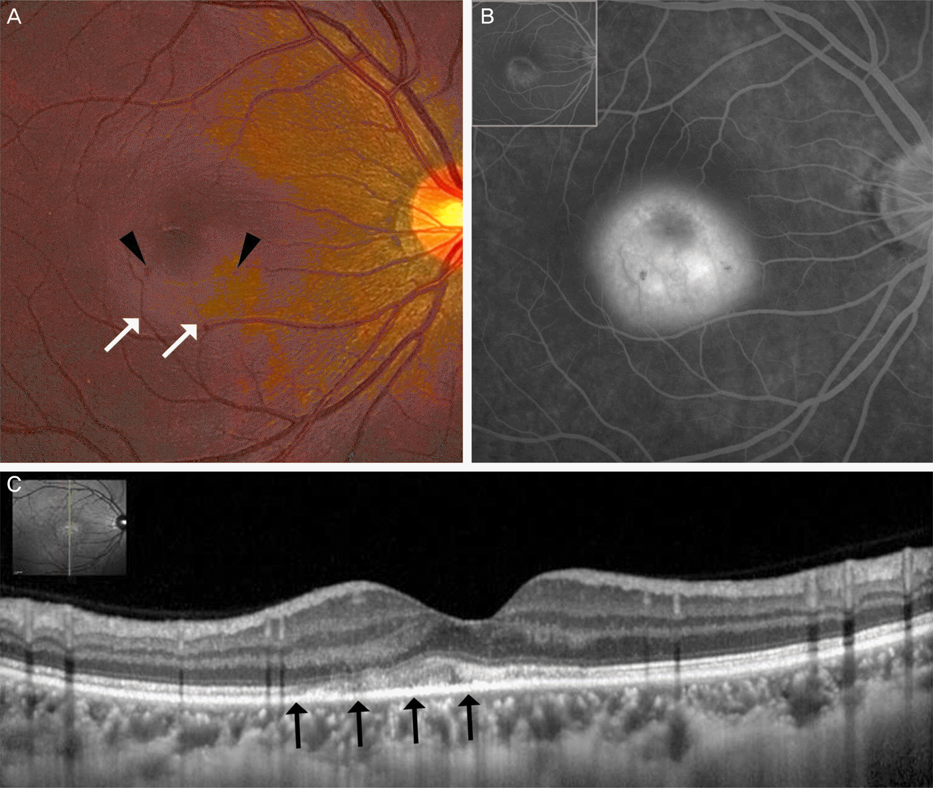 | Figure 1.(A) Fundus photographs of the right eye on presentation showed gray-white thickening of the fovea (white arrows) and retinal hemorrhage next to the fovea (black arrowheads). (B) Fluorescein angiography demonstrated ring shape mottled hyperfluorescence in the early phase and dye pooling in the late phase at the initial visit. (C) Spectral-domain optical coherence tomography showed abnormal hyperreflective thickening at the level of the outer retina and retinal pigment epithelium and detachment of the neurosensory retina at the foveal lesion. The outer plexiform layer swelling was noted. The inner seg-ment/outer segment junction and photoreceptor elevation/disruption was noted with preservation of the external limiting membrane and inner retina (black arrows). |
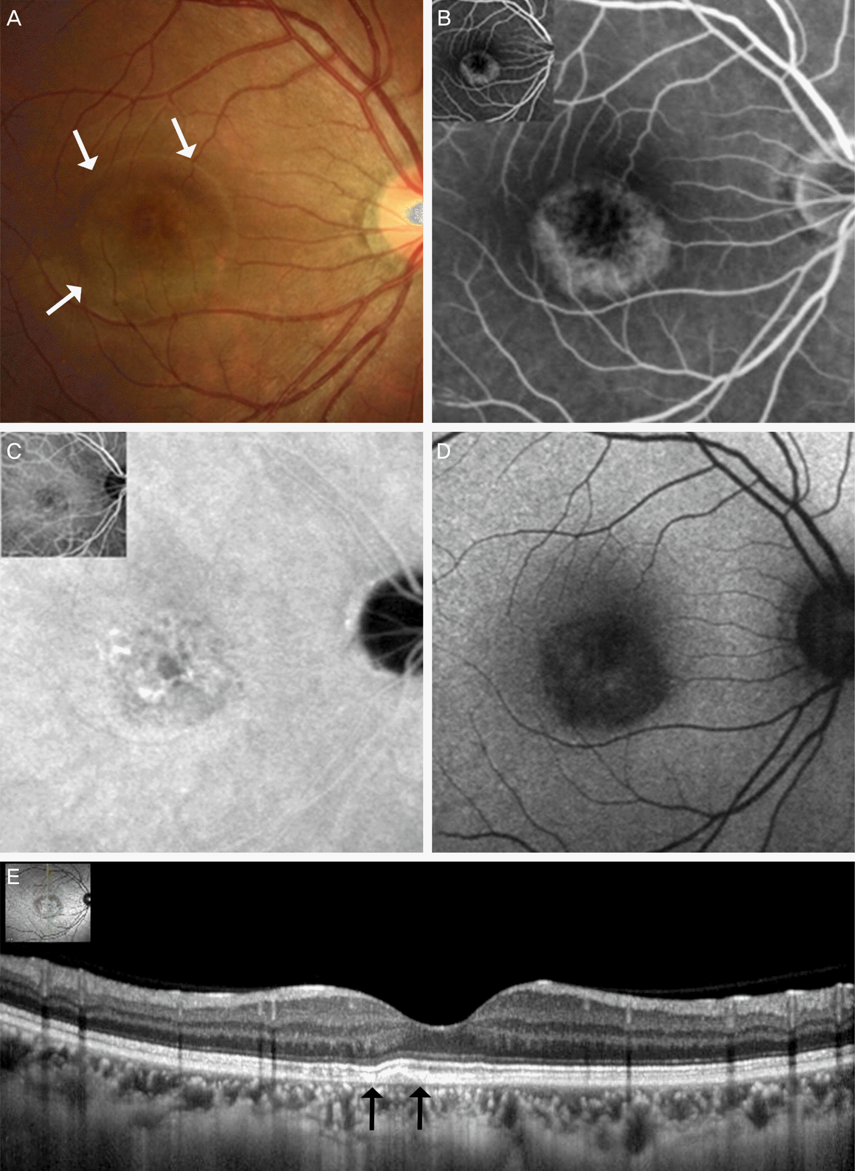 | Figure 2.(A) Nineteen months after onset, fundus photographs of the right eye showed increased retinal pigment hyperplasia and these residual retinal pigment epithelium (RPE) changes resembling bull’s eye maculopathy (white arrows). (B) Fluorescein angiography demonstrated mild hyperfluorescence staining of the central macula in the early phase (upper left framed) and the late phase. (C) Indocyanine green angiography showed hypofluor-escence in the macular area. (D) Autofluorescence imaging showed hypoautofluorescence corresponding to the clinically significant lesion on funduscopic examination. (E) The spectral-domain optical coherence tomography of the right macula showed the inner segment/outer segment junction elevation/disruption almost completely re-gressed, but slight elevation of RPE and outer retina was remained (black arrows). |
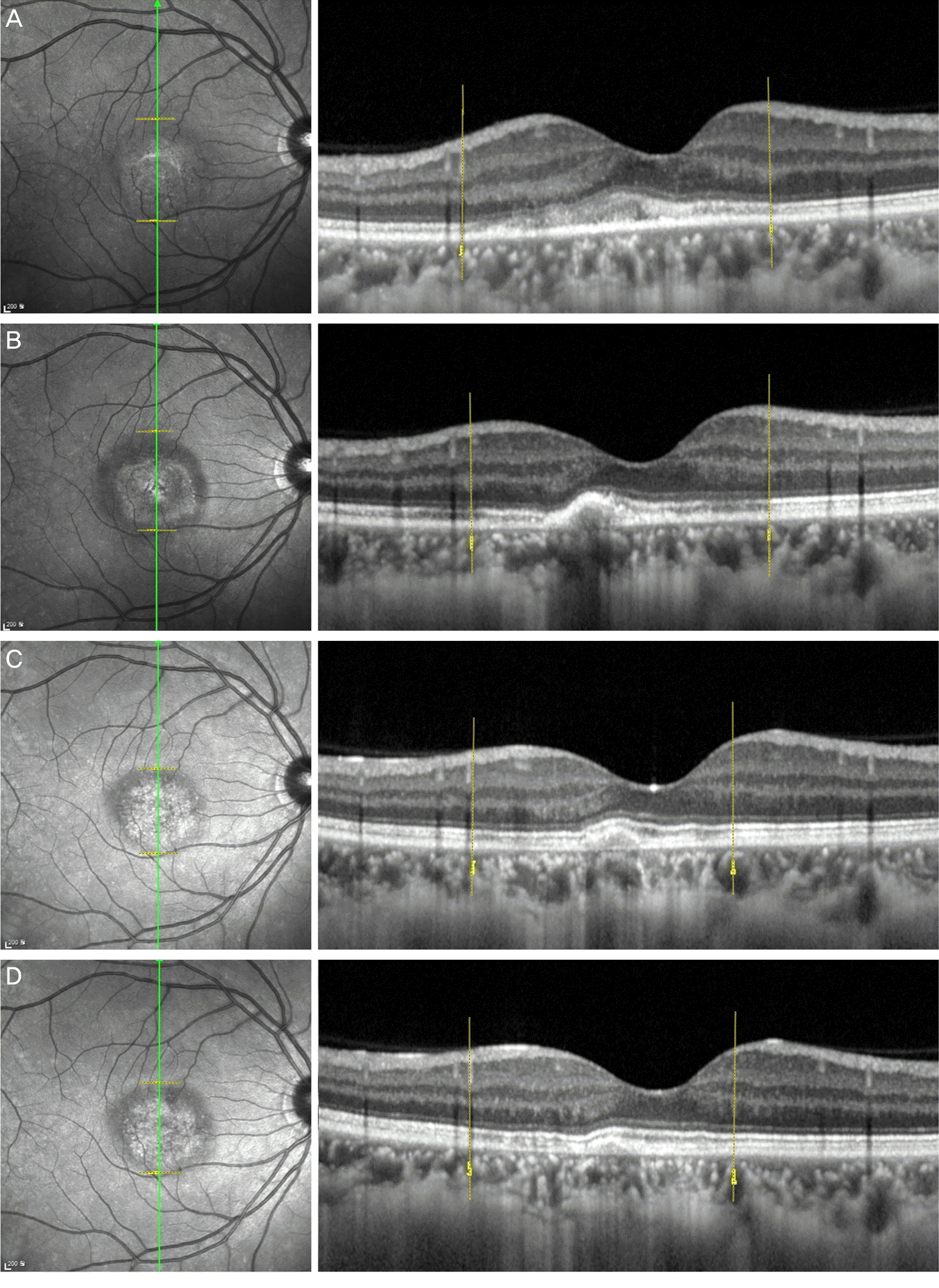 | Figure 3.Optical coherence tomography (OCT) evaluation of the macular region in the vertical plane of the right eye at initial presentation (A), and at one month (B), 19 months (C), 22 months (D) of follow-up. OCT showed abnormal hyperreflective thickening at the level of the outer retina and retinal pigment epithelium (RPE) and detachment of the neurosensory retina in the foveal lesion at presentation (A). One month follow-up presentation (B), it showed decrease in size of the lesion. 22 months after onset (D), RPE elevation was much decreased. Outer plexiform layer swelling was noted at presentation (A) and it decreased at one month after onset (B). The inner segment/outer segment junction and photoreceptor elevation/disruption was noted with preservation of the external limiting membrane and inner retina at presentation (A). Nineteen months after onset (C), the OCT showed the inner segment/outer segment junction elevation/disruption almost completely regressed, and 22 months after onset (D), the OCT showed the inner segment/outer segment junction elevation/disruption disappeared. Outer photoreceptor segment irregularity was still remains. |
 XML Download