

 PDF
PDF ePub
ePub Citation
 Print
Print


Abstract
Purpose
In this study we compared the intraocular pressures (IOPs) measured using dynamic contour tonometry (DCT) and Goldmann applanation tonometry (GAT) and investigated the correlation between central corneal thickness (CCT) and IOP.
Methods
In a prospective study, 178 eyes of 91 subjects with glaucoma and glaucoma suspect were enrolled. IOP was meas-ured using DCT and GAT and CCT was measured using ultrasound pachymetry. Each eye was classified into 1 of 3 groups ac-cording to their CCT: low CCT (Group A; CCT < 525 μ m), normal CCT (Group B; 525 ≤ CCT < 561 μ m), and high CCT (Group C; CCT ≥ 561 μ m). In each group, we investigated the correlation between CCT and IOP measurement using GAT and DCT.
Results
A significant correlation was found between CCT and IOP measured using GAT ( p < 0.001), but not between CCT and IOP measured using DCT ( p = 0.108) in all patients. Subgroup analysis showed that CCT affected IOP measured with GAT only in Group A ( p = 0.027) and IOP measured with DCT was not affected by CCT in all 3 groups.
Go to : 
References
1. Epstein DL, Krug JH Jr, Hertzmark E. . A long-term clinical trial of timolol therapy versus no treatment in the management of glaucoma suspects. Ophthalmology. 1989; 96:1460–7.

2. Kass MA, Gordon MO, Hoff MR. . Topical timolol admin-istration reduces the incidence of glaucomatous damage in ocular hypertensive individuals. A randomized, double-masked, long-term clinical trial. Arch Ophthalmol. 1989; 107:1590–8.
3. Schulzer M, Drance SM, Douglas GR. A comparison of treated and untreated glaucoma suspects. Ophthalmology. 1991; 98:301–7.
4. Goldmann H, Schmidt T. Applanation tonometry. Ophthalmologica. 1957; 134:221–42.
5. Ehlers N, Bramsen T, Sperling S. Applanation tonometry and cen-tral corneal thickness. Acta Ophthalmol (Copenh). 1975; 53:34–43.
6. Doughty MJ, Zaman ML. Human corneal thickness and its impact on intraocular pressure measures: a review and meta-analysis approach. Surv Ophthalmol. 2000; 44:367–408.
7. Doughty MJ, Jonuscheit S. Effect of central corneal thickness on Goldmann applanation tonometry measures-a different result with different pachymeters. Graefes Arch Clin Exp Ophthalmol. 2007; 245:1603–10.
8. Kanngiesser HE, Kniestedt C, Robert YC. Dynamic contour ton-ometry: presentation of a new tonometer. J Glaucoma. 2005; 14:344–50.
9. Siganos DS, Papastergiou GI, Moedas C. Assessment of the Pascal dynamic contour tonometer in monitoring intraocular pressure in unoperated eyes and eyes after LASIK. J Cataract Refract Surg. 2004; 30:746–51.
10. Kaufmann C, Bachmann LM, Thiel MA. Intraocular pressure measurements using dynamic contour tonometry after laser in situ keratomileusis. Invest Ophthalmol Vis Sci. 2003; 44:3790–4.
11. Choi HJ, Kim SW, Kim TI, Kim EK. Intraocular pressure measure-ments using dynamic contour tonometer after photorefractive keratectomy. J Korean Ophthalmol Soc. 2008; 49:577–82.
12. Han KE, Kim H, Kim NR. . Comparison of intraocular pres-sures after myopic laser-assisted subepithelial keratectomy: ton-ometry-pachymetry, Goldmann applanation tonometry, dynamic contour tonometry, and noncontact tonometry. J Cataract Refract Surg. 2013; 39:888–97.
13. Schneider E, Grehn F. Intraocular pressure measurement-comparison of dynamic contour tonometry and goldmann applanation tonometry. J Glaucoma. 2006; 15:2–6.
14. Carbonaro F, Andrew T, Mackey DA. . Comparison of three methods of intraocular pressure measurement and their relation to central corneal thickness. Eye (Lond). 2010; 24:1165–70.
15. Regev G, Harris A, Siesky B. . Goldmann applanation tonom-etry and dynamic contour tonometry are not correlated with central corneal thickness in primary open angle glaucoma. J Glaucoma. 2011; 20:282–6.
16. Grieshaber MC, Schoetzau A, Zawinka C. . Effect of central corneal thickness on dynamic contour tonometry and Goldmann applanation tonometry in primary open-angle glaucoma. Arch Ophthalmol. 2007; 125:740–4.
17. Francis BA, Hsieh A, Lai MY. . Effects of corneal thickness, corneal curvature, and intraocular pressure level on Goldmann ap-planation tonometry and dynamic contour tonometry. Ophthalmology. 2007; 114:20–6.
18. Doyle A, Lachkar Y. Comparison of dynamic contour tonometry with goldman applanation tonometry over a wide range of central corneal thickness. J Glaucoma. 2005; 14:288–92.
19. Seo JW, Shin DM, Rho SH. Comparison of dynamic contour ton-ometry and Goldmann applanation tonometry. J Korean Ophthalmol Soc. 2009; 50:242–6.
20. Lee J, Lee CH, Choi J. . Comparison between dynamic contour tonometry and Goldmann applanation tonometry. Korean J Ophthalmol. 2009; 23:27–31.
21. Kass MA. Standardizing the measurement of intraocular pressure for clinical research. Guidelines from the Eye Care Technology Forum. Ophthalmology. 1996; 103:183–5.
22. Altman DG, Bland JM. Measurement in medicine: the analysis of method comparison studies. The Statistician. 1983; 32:307–17.
23. Kwag JY, Choi SH. Comparison of ocular biometry measured by ultrasound and two kinds of partial coherence interferometers. J Korean Ophthalmol Soc. 2011; 52:169–74.
24. Allingham RR, Damji KF, Freedman SF. . Shields' textbook of glaucoma. 6th. Philadelphia: Lippincott Williams & Wilkins;2010. p. 30–2.
25. Shih CY, Graff Zivin JS, Trokel SL, Tsai JC. Clinical significance of central corneal thickness in the management of glaucoma. Arch Ophthalmol. 2004; 122:1270–5.
26. Faucher A, Grégoire J, Blondeau P. Accuracy of Goldmann tonom-etry after refractive surgery. J Cataract Refract Surg. 1997; 23:832–8.
27. Mardelli PG, Piebenga LW, Whitacre MM, Siegmund KD. The ef-fect of excimer laser photorefractive keratectomy on intraocular pressure measurements using the Goldmann applanation tonometer. Ophthalmology. 1997; 104:945–8. discussion 949.
28. Boehm AG, Weber A, Pillunat LE. . Dynamic contour tonom-etry in comparison to intracameral IOP measurements. Invest Ophthalmol Vis Sci. 2008; 49:2472–7.
29. Tzamalis A, Kynigopoulos M, Chalvatzis N. . Association of ocular hypotensive medication types with dynamic contour tonom-etry and Goldmann applanation tonometry measurements in a glaucoma and ocular hypertensive population. J Ocul Pharmacol Ther. 2013; 29:41–7.
30. Hwang YH, Kim HK, Sohn YH. Namil Study Group Korean Glaucoma Society Central corneal thickness in a Korean pop-ulation: the Namil Study. Invest Ophthalmol Vis Sci. 2012; 53:6851–5.
31. Hager A, Schroeder B, Sadeghi M. . The influence of corneal hysteresis and corneal resistance factor on the measurement of in-traocular pressure. Ophthalmologe. 2007; 104:484–9.
32. Oh JH, Yoo C, Kim YY. . The effect of contact lens-induced corneal edema on Goldmann applanation tonometry and dynamic contour tonometry. Graefes Arch Clin Exp Ophthalmol. 2009; 247:371–5.
33. Yoo C, Eom YS, Kim YY. Goldmann applanation tonometry and dynamic contour tonometry in eyes with elevated intraocular pres-sure (IOP): comparison in the same eyes after subsequent medical normalization of IOP. Graefes Arch Clin Exp Ophthalmol. 2010; 248:1611–6.
Go to :
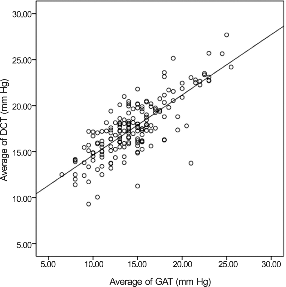 | Figure 1.Pearson correlation of intraocular pressure measure-ments obtained by Goldmann applanation tonometer (GAT) and Dynamic contour tonometer (DCT) (r = 0.791, p < 0.001). |
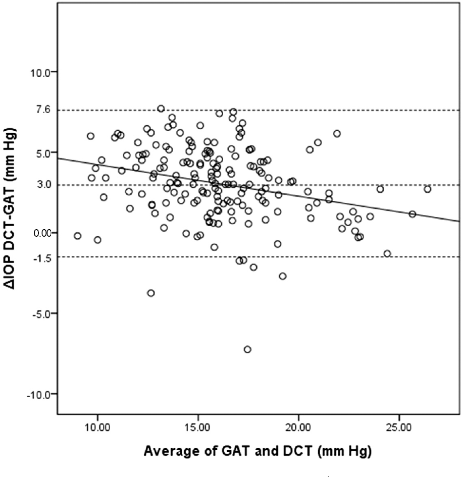 | Figure 2.Bland-Altman plot of the difference between Goldmann applanation tonometer (GAT) and Dynamic con-tour tonometry (DCT) against the mean of GAT and DCT. IOP = intraocular pressure. |
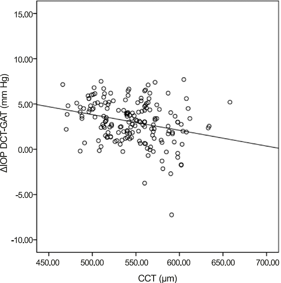 | Figure 3.Pearson correlation of the difference between Dynamic contour tonometry (DCT) and Goldmann applanation ton-ometer (GAT) with CCT (r = -0.272, p < 0.001). IOP = in-traocular pressure; CCT = central corneal thickness. |
Table 1.
Patient demographics
| Overall (n = 178) | Group A (n = 59) | Group B (n = 59) | Group C (n = 60) | p-value | |
|---|---|---|---|---|---|
| Sex | 0.353∗ | ||||
| Male | 80 (44.94) | 25 (42.37) | 23 (38.98) | 32 (53.33) | |
| Female | 98 (55.06) | 34 (57.63) | 36 (61.02) | 28 (46.67) | |
| Age (years) | 59.80 ± 14.49 | 59.41 ± 13.57 | 60.85 ± 14.28 | 59.15 ± 15.71 | 0.791† |
| Dx | 0.031∗ | ||||
| POAG | 86 (48.31) | 26 (44.07) | 22 (37.29) | 38 (63.33) | |
| NTG | 54 (30.34) | 22 (37.29) | 19 (32.20) | 13 (21.67) | |
| GS | 38 (21.35) | 11 (18.64) | 18 (30.51) | 9 (15.00) | |
| CCT (μ m) | 545.15 ± 36.36 | 504.54 ± 14.55 | 545.36 ± 9.61 | 584.88 ± 20.33 | <0.001† |
| GAT1 (mm Hg)‡ | 14.4 ± 3.8 | 12.7 ± 2.9 | 14.9 ± 3.8 | 15.7 ± 4.0 | <0.001† |
| GAT2 (mm Hg)‡ | 14.7 ± 3.8 | 12.9 ± 3.0 | 15.2 ± 3.7 | 16.0 ± 4.0 | <0.001† |
| DCT1 (mm Hg)‡ | 17.7 ± 3.2 | 16.6 ± 2.9 | 18.1 ± 3.3 | 18.2 ± 3.2 | 0.012† |
| DCT2 (mm Hg)‡ | 17.5 ± 3.1 | 16.7 ± 3.0 | 17.9 ± 3.2 | 18.1 ± 3.1 | 0.032† |
| OPA1 (mm Hg)§ | 2.5 ± 0.9 | 2.2 ± 0.8 | 2.4 ± 0.8 | 2.9 ± 1.0 | <0.001† |
| OPA2 (mm Hg)§ | 2.5 ± 1.0 | 2.1 ± 0.8 | 2.5 ± 1.1 | 2.8 ± 1.0 | 0.001† |
| AXL (mm) | 24.26 ± 1.84 | 24.52 ± 1.46 | 23.91 ± 1.41 | 24.31 ± 2.46 | 0.280† |
| MD (dB) | -6.43 ± 7.02 | -7.28 ± 7.23 | -5.46 ± 6.35 | -6.62 ± 7.47 | 0.371† |
| No. of Mx (n) | 1.0 ± 0.9 | 0.9 ± 0.6 | 0.9 ± 1.0 | 1.1 ± 1.1 | 0.383† |
Values are presented as mean ± SD unless otherwise indicated. Group A is CCT<525 μ m, Group B is 525 μ m ≤ CCT < 561 μ m and Group C is 561 μ m ≤ CCT. Dx = types of glaucoma; POAG = primary open-angle glaucoma; NTG = normotensive glaucoma; GS = glaucoma suspect; CCT = central corneal thickness; GAT = Goldmann applanation tonometer; DCT = Dynamic contour tonometry; OPA = ocular pulse amplitude; AXL = axial length; MD = mean deviation; No. of Mx = number of medications.
Table 2.
Reproducibility and repeatability of each intraocular measurement by Goldmann applanation tonometry and dynamic con-tour tonometry
| Mean ± SD (mm Hg) | ICC | CV (%) | CR (mm Hg) | RR (%) | |
|---|---|---|---|---|---|
| GAT | 14.6 ± 3.8 | 0.981 (95% CI; 0.975-0.986) | 3.78 | ±1.4 | 0.10 |
| DCT | 17.6 ± 3.2 | 0.958 (95% CI; 0.943-0.968) | 3.73 | ±1.8 | 0.10 |
Table 3.
Correlation of types of glaucoma, CCT, axial length, MD, and number of medications with the difference between DCT and GAT
| Dx | CCT | AXL | MD | No. of Mx | |
|---|---|---|---|---|---|
| △ IOP (DCT-GAT) | |||||
| p-value | 0.403∗ | <0.001† | 0.133† | 0.287† | 0.940† |
| r | - | -0.272 | -0.130 | -0.082 | 0.006 |
 XML Download