Abstract
Purpose
Central retinal vein occlusion (CRVO) as a complication of acute leukemia has rarely been reported. Here, we report a favorable outcome of radiation therapy for CRVO with severe macular edema in a patient with acute lymphocytic leukemia (ALL).
Case summary
A 21-year-old female presented with acute visual loss in the left eye and headache. Best-corrected visual acuity in the left eye was 0.3. Fundus examination showed some hemorrhagic spots in the right eye and flame-shaped retinal hemor-rhage, tortuous retinal vessels, and a retinal infiltrative lesion in the left eye. Fluorescein angiography revealed CRVO in the left eye and severe central macular edema was observed by optical coherence tomography. Hematologic study revealed ALL. Even after leukapheresis and commencement of systemic chemotherapy, fundus findings showed no remarkable change. She was given low dose (400 cGy) ocular external beam radiation therapy (EBRT). Three days after EBRT, macular edema, fundus in-filtration, and visual acuity improved dramatically. Visual acuity improved to 0.4 and to 0.8 at 1 month and 1 year after EBRT respectively.
References
1. Reddy SC, Jackson N, Menon BS. Ocular involvement in leuke-mia--a study of 288 cases. Ophthalmologica. 2003; 217:441–5.
2. Tseng MY, Chen YC, Lin YY. . Simultaneous bilateral central retinal vein occlusion as the initial presentation of acute myeloid leukemia. Am J Med Sci. 2010; 339:387–9.

3. Williamson TH. Central retinal vein occlusion: what's the story? Br J Ophthalmol. 1997; 81:698–704.

4. Boyd SR, Zachary I, Chakravarthy U. . Correlation of increased vascular endothelial growth factor with neovascularization and permeability in ischemic central vein occlusion. Arch Ophthalmol. 2002; 120:1644–50.

5. Finger PT, Pro MJ, Schneider S. . Visual recovery after radiation therapy for bilateral subfoveal acute myelogenous leukemia (AML). Am J Ophthalmol. 2004; 138:659–62.

6. Semeraro F, Morescalchi F, Duse S. . Systemic thromboem-bolic adverse events in patients treated with intravitreal anti-VEGF drugs for neovascular age-related macular degeneration: an overview. Expert Opin Drug Saf. 2014; 13:785–802.

7. Vertes D, Snyers B, De Potter P. Cytomegalovirus retinitis after low-dose intravitreous triamcinolone acetonide in an immuno- competent patient: a warning for the widespread use of intra-vitreous corticosteroids. Int Ophthalmol. 2010; 30:595–7.
Figure 1.
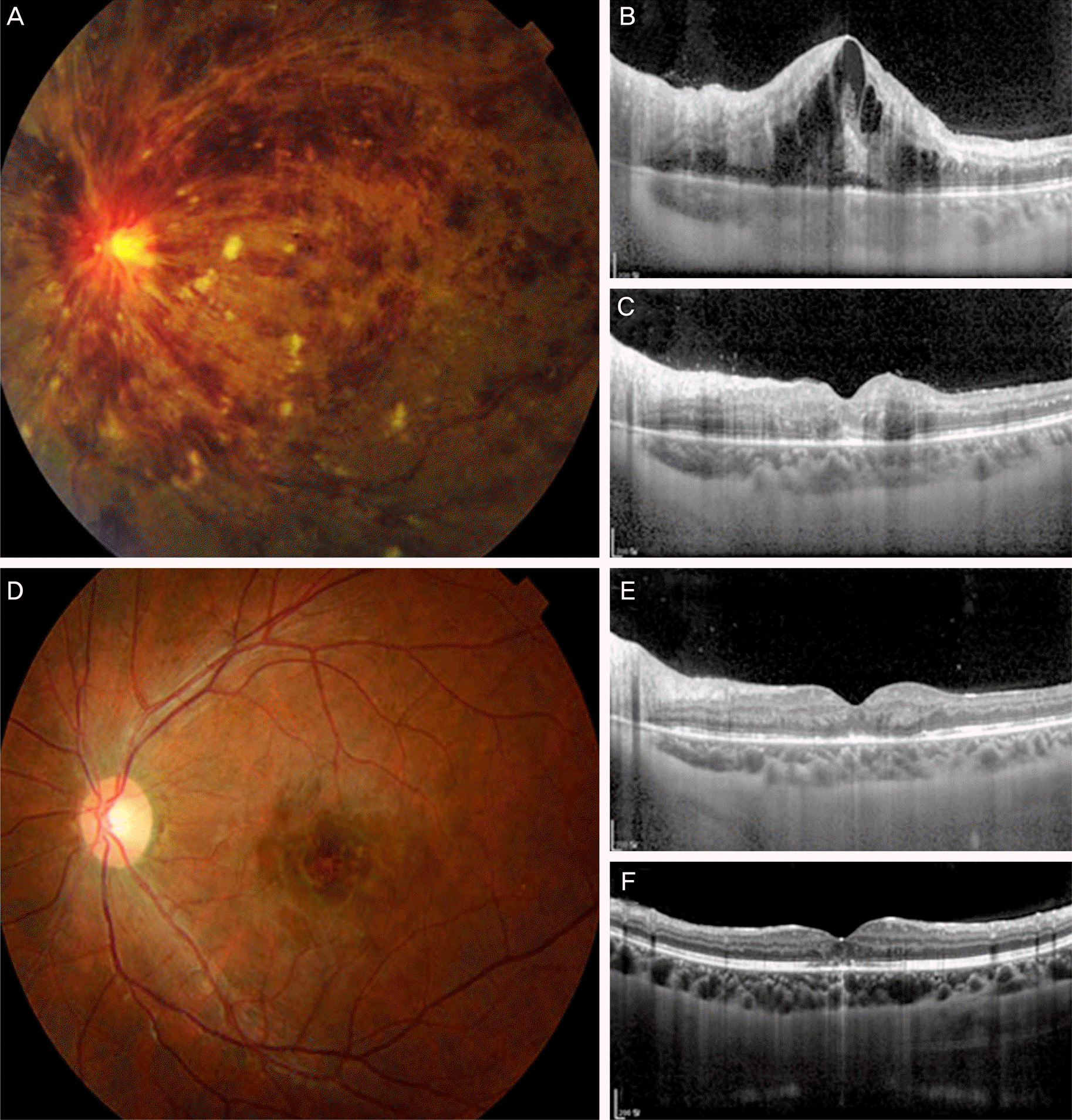
21-year-old female patient presented with acute visual loss of left eye. Best corrected visual acuity in the left eye was 0.3. (A) Fundus photo showed a few hemorrhagic spots in the right eye and flame-shaped retinal hemorrhage, dilated and tortuous retinal vessels, retinal infiltrative lesion in the left eye. (B) Central macular thickness was over 1000 μ m measured by optical coherence tomography (OCT). (C) Three days after ocular external beam radiation therapy (EBRT), prompt resolution of macular edema was observed on OCT. (E) One month after EBRT, nearly complete resolution of macular edema was observed on OCT. (F) At the last visit (one year after EBRT), macular edema was totally resolved except photoreceptor inner and outer segment defect. (D) At the last visit, fundus photo showed disappearance of retinal hemorrhage and retinal infiltrative lesion.


 PDF
PDF ePub
ePub Citation
 Print
Print


 XML Download