Abstract
Purpose
To evaluate the clinical effectiveness of topical diquafosol tetrasodium (DQS) after laser epithelial keratomileusis (LASEK).
Methods
This randomized prospective study included 97 eyes of 49 patients who were scheduled for LASEK. Patients in the DQS group used both 0.3% sodium hyaluronate and 3% DQS for 3 months after surgery while patients in the control group used only 0.3% sodium hyaluronate. Corneal staining score, tear film break-up time (TF-BUT), Schirmer test and ocular surface dis-ease index (OSDI) were evaluated before surgery and 2, 4, 8, 12 and 16 weeks after surgery.
Results
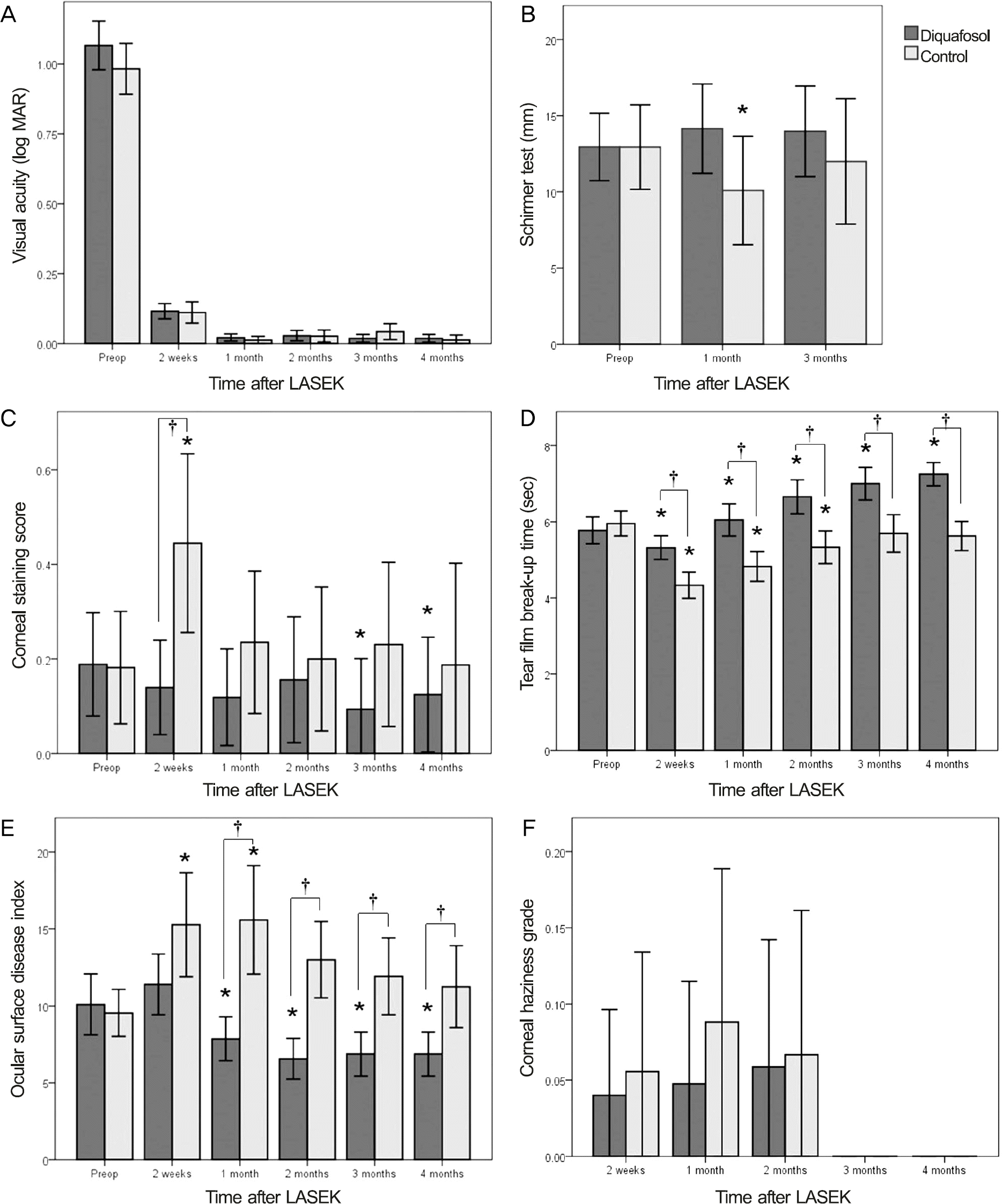
There was no significant difference in visual acuity, spherical equivalent and corneal haziness between the 2 groups af-ter surgery. Corneal staining score was significantly lower in the DQS group than in the control group 2 weeks after LASEK ( p < 0.01) and increased in the control group after LASEK compared with the preoperative value (2 weeks after LASEK, p < 0.01), but decreased in the DQS group (12 and 16 weeks after LASEK, p < 0.05). TF-BUT was significantly higher in the DQS group than in the control group 2 to 16 weeks after LASEK ( p < 0.01) and increased values were observed in the DQS group after LASEK compared with the preoperative value (4 to 16 weeks after LASEK, p < 0.05). The mean OSDI was significantly higher 4 to 16 weeks after LASEK in the control group than in the DQS group ( p < 0.01).
Go to : 
References
1. Camellin M. LASEK may offer the advantages of both LASIK and PRK. Ocular Surgery News. 1999; 28.
2. Lee SJ, Kim JK, Seo KY. . Comparison of corneal nerve re-generation and sensitivity between LASIK and laser epithelial ker-atomileusis (LASEK). Am J Ophthalmol. 2006; 141:1009–15.

3. Herrmann WA, Shah CP, von Mohrenfels CW. . Tear film func-tion and corneal sensation in the early postoperative period after LASEK for the correction of myopia. Graefes Arch Clin Exp Ophthalmol. 2005; 243:911–6.

4. Dooley I, D'Arcy F, O'Keefe M. Comparison of dry-eye disease se-verity after laser in situ keratomileusis and laser-assisted sub-epithelial keratectomy. J Cataract Refract Surg. 2012; 38:1058–64.

5. Hovanesian JA, Shah SS, Maloney RK. Symptoms of dry eye and recurrent erosion syndrome after refractive surgery. J Cataract Refract Surg. 2001; 27:577–84.

6. Pflugfelder SC, Tseng SC, Sanabria O. . Evaluation of sub-jective assessments and objective diagnostic tests for diagnosing tear-film disorders known to cause ocular irritation. Cornea. 1998; 17:38–56.

7. Gündüz K, Ozdemir O. Topical cyclosporin treatment of kerato-conjunctivitis sicca in secondary Sjögren's syndrome. Acta Ophthalmol (Copenh). 1994; 72:438–42.

8. Sall K, Stevenson OD, Mundorf TK, Reis BL. Two multicenter, randomized studies of the efficacy and safety of cyclosporine oph-thalmic emulsion in moderate to severe dry eye disease. CsA Phase 3 Study Group. Ophthalmology. 2000; 107:631–9.
9. Stevenson D, Tauber J, Reis BL. Efficacy and safety of cyclosporin A ophthalmic emulsion in the treatment of moderate-to-severe dry eye disease: a dose-ranging, randomized trial. The Cyclosporin A Phase 2 Study Group. Ophthalmology. 2000; 107:967–74.
10. Lee JS, Yoon TJ, Kim KH. Cinical effect of Restasis(R) eye drops in mild dry eye syndrome. J Korean Ophthalmol Soc. 2009; 50:1489–94.
11. Kang KW, Kim HK. Efficacy of topical cyclosporine in mild dry eye patients having refractive surgery. J Korean Ophthalmol Soc. 2014; 55:1752–7.

12. Nichols KK, Yerxa B, Kellerman DJ. Diquafosol tetrasodium: a novel dry eye therapy. Expert Opin Investig Drugs. 2004; 13:47–54.

13. Tauber J, Davitt WF, Bokosky JE. . Double-masked, place-bo-controlled safety and efficacy trial of diquafosol tetrasodium (INS365) ophthalmic solution for the treatment of dry eye. Cornea. 2004; 23:784–92.

14. Miyata K, Amano S, Sawa M, Nishida T. A novel grading method for superficial punctate keratopathy magnitude and its correlation with corneal epithelial permeability. Arch Ophthalmol. 2003; 121:1537–9.

15. Fantes FE, Hanna KD, Waring GO. 3rd. . Wound healing after excimer laser keratomileusis (photorefractive keratectomy) in monkeys. Arch Ophthalmol. 1990; 108:665–75.

16. Schiffman RM, Christianson MD, Jacobsen G. . Reliability and validity of the ocular surface disease index. Arch Ophthalmol. 2000; 118:615–21.

17. Benitez-del-Castillo JM, del Rio T, Iradier T. . Decrease in tear secretion and corneal sensitivity after laser in situ keratomileusis. Cornea. 2001; 20:30–2.

18. Yu EY, Leung A, Rao S, Lam DS. Effect of laser in situ keratomi-leusis on tear stability. Ophthalmology. 2000; 107:2131–5.

19. Aras C, Ozdamar A, Bahcecioglu H. . Decreased tear secretion after laser in situ keratomileusis for high myopia. J Refract Surg. 2000; 16:362–4.

20. Siganos DS, Popescu CN, Siganos CS, Pistola G. Tear secretion following spherical and astigmatic excimer laser photorefractive keratectomy. J Cataract Refract Surg. 2000; 26:1585–9.

21. Ozdamar A, Aras C, Karakas N. . Changes in tear flow and tear film stability after photorefractive keratectomy. Cornea. 1999; 18:437–9.

22. Lee JB, Ryu CH, Kim J. . Comparison of tear secretion and tear film instability after photorefractive keratectomy and laser in situ keratomileusis. J Cataract Refract Surg. 2000; 26:1326–31.

23. Mori Y, Nejima R, Masuda A. . Effect of diquafosol tetraso-dium eye drop for persistent dry eye after laser in situ keratomileusis. Cornea. 2014; 33:659–62.

24. Toda I, Ide T, Fukumoto T. . Combination therapy with diqua-fosol tetrasodium and sodium hyaluronate in patients with dry eye after laser in situ keratomileusis. Am J Ophthalmol. 2014; 157:616–22.e1.

Go to : 
 | Figure 1.Changes in visual acuity (A), Schirmer test (B), corneal staining score (C), tear film break-up time (D), ocular surface dis-ease index (E), and corneal haziness grade (F) in the 3% diquafosol tetrasodium and the control groups before and after LASEK. LASEK = laser-assisted sub-epithelial keratectomy; Preop = preoperation. * p < 0.05 compared with preoperative value; † p < 0.05 between Diquafosol and control group. |
Table 1.
Characteristics of patients who underwent LASEK and were treated with or without topical 3% diquafosol tetrasodium
| Diquafosol group | Control group | p-value | |
|---|---|---|---|
| Sex (male:female) | 13:14 | 10:12 | 0.10∗ |
| Age (years) | 24.55 ± 7.98 | 25.68 ± 5.90 | 0.44† |
| UCVA (log MAR) | 1.07 ± 0.32 | 0.98 ± 0.29 | 0.99† |
| Spherical equivalent (diopters) | -5.26 ± 1.92 | -5.56 ± 2.09 | 0.93† |
| Central corneal thickness (μ m) | 536.04 ± 26.51 | 545.54 ± 31.79 | 0.46† |
| Schirmer test (mm) | 12.9 ± 8.0 | 12.9 ± 9.1 | 0.46† |
| Corneal staining score | 0.2 ± 0.4 | 0.2 ± 0.4 | 0.26† |
| Tear film break-up time (sec) | 5.8 ± 1.3 | 6.0 ± 1.1 | 0.11† |
| Ocular surface disease index | 10.1 ± 7.2 | 9.5 ± 5.0 | 0.47† |
| Ablation depth (μ m) | 97.17 ± 25.92 | 103.36 ± 27.24 | 0.66† |


 PDF
PDF ePub
ePub Citation
 Print
Print


 XML Download