Abstract
Purpose
To compare the clinical outcomes between AQUA ICL® (V4C) and conventional implantable collamer lens (ICL, V4B) in patients with high myopia.
Methods
We compared preoperative and postoperative visual acuities, spherical equivalent, intraocular pressure, postoperative vault and visual quality assessed using optical quality analyzing system (OQAS®) between 20 eyes implanted with ICL (V4B) and 22 eyes implanted with AQUA ICL® (V4C).
Results
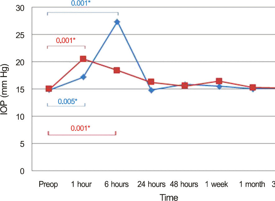
Visual acuity (log MAR) and spherical equivalent at postoperative 3 months were 0.06 ± 0.09 and -0.26 ± 0.17 D in the V4B group and 0.03 ± 0.03 and -0.23 ± 0.19 D in the V4C group, respectively. There was no statistical difference in visual acuity and spherical equivalent between the 2 groups ( p > 0.05). Postoperative intraocular pressure (IOP) elevated significantly in both groups until 6 hours after the operation ( p < 0.05) compared with preoperative IOP. No significant IOP elevation was observed from the first postoperative day to postoperative 3 months ( p > 0.05). V4C resulted in lower IOP than V4B 6 hours postoperatively. In ICL (V4B) and AQUA ICL® (V4C) groups, the objective scattering index ( OSI) was 1.29 ± 0.90 and 1.36 ± 0.68, modulation transfer function (MTF) cut off value was 29.62 ± 11.31 c/deg and 29.61 ± 9.56 c/deg and Sterhl ratio was 0.18 ± 0.07 and 0.17 ± 0.04, respectively, 3 months postoperatively. None of these values were significantly different between the 2 groups.
Go to : 
References
1. Heitzmann J, Binder PS, Kassar BS, Nordan LT. The correction of high myopia using the excimer laser. Arch Ophthalmol. 1993; 111:1627–34.

2. Holladay JT, Dudeja DR, Chang J. Functional vision and corneal changes after laser in situ keratomileusis determined by contrast sensitivity, glare testing, and corneal topography. J Cataract Refract Surg. 1999; 25:663–9.

3. Lee SY, Cheon HJ, Baek TM, Lee KH. Implantable contact lens to correct high myopia (Clinical Study with 24 months follow-up). J Korean Ophthalmol Soc. 2000; 41:1515–22.
4. Dejaco-Ruhswurm I, Scholz U, Pieh S. . Long-term endothe-lial changes in phakic eyes with posterior chamber intraocular lenses. J Cataract Refract Surg. 2002; 28:1589–93.

5. Edelhauser HF, Sanders DR, Azar R. . Corneal endothelial as-sessment after ICL implantation. J Cataract Refract Surg. 2004; 30:576–83.

6. Sanders DR. Anterior subcapsular opacities and cataracts 5 years after surgery in the visian implantable collamer lens FDA trial. J Refract Surg. 2008; 24:566–70.

7. Sánchez-Galeana CA, Zadok D, Montes M. . Refractory intra-ocular pressure increase after phakic posterior chamber intraocular lens implantation. Am J Ophthalmol. 2002; 134:121–3.

8. Smallman DS, Probst L, Rafuse PE. Pupillary block glaucoma sec-ondary to posterior chamber phakic intraocular lens implantation for high myopia. J Cataract Refract Surg. 2004; 30:905–7.

9. Murphy PH, Trope GE. Monocular blurring. A complication of YAG laser iridotomy. Ophthalmology. 1991; 98:1539–42.
10. Kumar N, Feyi-Waboso A. Intractable secondary glaucoma from hyphema following YAG iridotomy. Can J Ophthalmol. 2005; 40:85–6.

11. Siam GA, de Barros DS, Gheith ME. . Post-peripheral iridot-omy inflammation in patients with dark pigmentation. Ophthalmic Surg Lasers Imaging. 2008; 39:49–53.

12. Spaeth GL. The normal development of the human anterior cham-ber angle: a new system of descriptive grading. Trans Ophthalmol Soc U K. 1971; 91:709–39.
13. Alfonso JF, Lisa C, Fernández-Vega Cueto L. . Clinical out-comes after implantation of a posterior chamber collagen copoly-mer phakic intraocular lens with a central hole for myopic correction. J Cataract Refract Surg. 2013; 39:915–21.

14. Higueras-Esteban A, Ortiz-Gomariz A, Gutiérrez-Ortega R. . Intraocular pressure after implantation of the visian implantable collamer lens with CentraFLOW without iridotomy. Am J Ophthalmol. 2013; 156:800–5.

15. Uozato H, Shimizu K, Kawamorita T, Ohmoto F. Modulation transfer function of intraocular collamer lens with a central artifi-cial hole. Graefes Arch Clin Exp Ophthalmol. 2011; 249:1081–5.

16. Park CW, Lee YE, Joo CK. Changes in optical quality of cataract patients' corrected visual acuity before and after phacoemulsification. J Korean Ophthalmol Soc. 2013; 54:1208–12.

17. Shin KH, Kim KH, Kwon JW. Two cases of glare after Iridotomy for phakic intraocular lens implantation. J Korean Ophthalmol Soc. 2011; 52:1537–40.

18. Zadok D, Chayet A. Lens opacity after neodymium: YAG laser iri-dectomy for phakic intraocular lens implantation. J Cataract Refract Surg. 1999; 25:592–3.
19. Dhawahir-Scala FE, Clark D. Neodymium: YAG laser peripheral iridotomy: cause of a visually incapacitating cataract? Ophthalmic Surg Lasers Imaging. 2006; 37:330–2.
20. Jeng S, Lee JS, Huang SC. Corneal decompensation after argon la-ser iridectomy-a delayed complication. Ophthalmic Surg. 1991; 22:565–9.

21. Wu SC, Jeng S, Huang S, Lin SM. Corneal endothelial damage af-ter neodymium: YAG laser iridotomy. Ophthalmic Surg Lasers. 2000; 31:411–6.
22. Anderson JE, Gentile RC, Sidoti PA, Rosen RB. Stage 1 macular hole as a complication of laser iridotomy. Arch Ophthalmol. 2006; 124:1658–60.

23. Kawamorita T, Uozato H, Shimizu K. Fluid dynamics simulation of aqueous humour in a posterior-chamber phakic intraocular lens with a central perforation. Graefes Arch Clin Exp Ophthalmol. 2012; 250:935–9.

24. Martinez-de-la-Casa JM, Garcia-Feijoo J, Castillo A, Garcia-Sanchez J. Reproducibility and clinical evaluation of rebound tonometry. Invest Ophthalmol Vis Sci. 2005; 46:4578–80.

25. Abraham LM, Epasinghe NC, Selva D, Casson R. Comparison of the ICare rebound tonometer with the Goldmann applanation ton-ometer by experienced and inexperienced tonometrists. Eye (Lond). 2008; 22:503–6.

26. Allan BD, Argeles-Sabate I, Mamalis N. Endophthalmitis rates af-ter implantation of the intraocular Collamer lens: survey of users between 1998 and 2006. J Cataract Refract Surg. 2009; 35:766–9.

27. Scuderi GL, Cascone NC, Regine F. . Validity and limits of the rebound tonometer (ICare[R]): clinical study. Eur J Ophthalmol. 2011; 21:251–7.
Go to : 
 | Figure 1.Intraocular pressure (IOP) changes in two groups postoperatively. Both of conventional implantable collamer lens (ICL) (V4B) group and AQUA ICL® (V4C) group presented temporary elevation of IOP postoperatively, which was stabilized thereafter. At postoperative 6 hours, IOP of V4B group showed higher value (27.36 ± 9.25) than that of V4C group (18.44 ± 4.21) significantly ( p = 0.001). Other val-ues at different instances showed no statistically significant difference. Preop = preoperative period. * p-value within each group was acquired from Wilcoxon signed rank test; † p-values between 2 groups were acquired from Mann-Whitney U-test. |
Table 1.
Demographic data of the patients
| Parameter | V4B (20 eyes, 10 patients) | V4C (22 eyes, 11 patients) | p-value∗ |
|---|---|---|---|
| Age (years) | 27.3 ± 7.73 | 30.1 ± 4.21 | 0.621 |
| Spherical equivalent (D) | -10.44 ± 3.53 | -10.43 ± 2.84 | 0.223 |
| Spherical R.E. (D) | -9.92 ± 3.39 | -9.74 ± 2.95 | 0.854 |
| Cylinderical R.E. (D) | -1.04 ± 1.20 | -1.39 ± 0.77 | 0.208 |
| CDVA (log MAR) | 0.03 ± 0.04 | 0.06 ± 0.05 | 0.092 |
| ACD (mm) | 3.17 ± 0.18 | 3.13 ± 0.24 | 0.520 |
| White to white (mm) | 11.26 ± 0.46 | 11.1 ± 0.22 | 0.833 |
| Endothelial cell density (cells/mm2) | 2,541.93 ± 191.67 | 2,595.75 ± 344.76 | 0.609 |
Table 2.
Postoperative UCDVA (log MAR) and spherical equivalent (diopter) at postoperative 1 week, 1 month and 3 months
| UCDVA (log MAR) | Spherical equivalent (diopter) | |||||
|---|---|---|---|---|---|---|
| V4B | V4C | p-value∗ | V4B | V4C | p-value∗ | |
| 1 week | 0.05 ± 0.07 | 0.07 ± 0.04 | 0.316 | -0.15 ± 0.28 | -0.11 ± 0.44 | 0.513 |
| 1 month | 0.10 ± 0.05 | 0.08 ± 0.08 | 0.627 | -0.22 ± 0.36 | -0.15 ± 0.12 | 0.854 |
| 3 months | 0.06 ± 0.09 | 0.03 ± 0.03 | 0.256 | -0.26 ± 0.17 | -0.23 ± 0.19 | 0.127 |
Table 3.
CDVA changes in two groups at postoperative three months
| Final CDVA change (in Snellen chart) | V4B | V4C |
|---|---|---|
| Loss ≥ 2 lines | None | None |
| Loss 1 line | None | None |
| Unchanged | 19 eyes | 20 eyes |
| Gain 1 line | 1 eye | 2 eyes |
| Gain ≥ 2 lines | None | None |
Table 4.
Central vaulting values measured at three months after the operation
| Parameters | V4B | V4C | p-value∗ |
|---|---|---|---|
| Vaulting value† (μ m) | 501.52 ± 130.88 | 485.24 ± 117.21 | 0.331 |
Table 5.
OQAS® values measured at three months after the operation
| Parameters | V4B | V4C | p-value∗ |
|---|---|---|---|
| OSI | 1.29 ± 0.90 | 1.36 ± 0.68 | 0.632 |
| MTF cut off (c/deg) | 29.62 ± 11.31 | 29.61 ± 9.56 | 0.934 |
| Sterhl ratio | 0.18 ± 0.07 | 0.17 ± 0.04 | 0.724 |


 PDF
PDF ePub
ePub Citation
 Print
Print


 XML Download