

 PDF
PDF ePub
ePub Citation
 Print
Print


Abstract
Purpose
To evaluate the effect of posterior sub-Tenon triamcinolone acetonide injection combined with vitrectomy for idiopathic epiretinal membrane (ERM).
Methods
This study included 40 eyes of 40 patients who underwent vitrectomy and membrane peeling for idiopathic ERM. Triamcinolone acetonide (40 mg) was injected into the posterior sub-Tenon space following vitrectomy in 20 eyes of the injected group. The other 20 eyes that did not receive the injection were selected as the control group to match preoperative visual acuity and central macular thickness with the injected group. Pre- and postoperative best-corrected visual acuity, central macular thick-ness, intraocular pressure, and complications were compared between the 2 groups.
Results
The average visual acuity in the injected group improved from 0.56 ± 0.23 (log MAR) at baseline to 0.52 ± 0.36 at 1 month, 0.44 ± 0.39 at 3 months, and 0.38 ± 0.41 at 6 months postoperatively. Central macular thickness decreased from 456.2 ± 86.2 to 399.0 ± 60.1 at 1 month, 377.1 ± 71.5 at 3 months, and 353.1 ± 57.4 at 6 months postoperatively. In the control group, average vis-ual acuity improved from 0.56 ± 0.23 at baseline to 0.53 ± 0.25 at 1 month, 0.41 ± 0.20 at 3 months, and 0.37 ± 0.24 at 6 months postoperatively. Central macular thickness decreased from 456.4 ± 74.8 to 394.9 ± 63.5 at 1 month, 377.2 ± 56.8 at 3 months, and 358.0 ± 57.2 at 6 months postoperatively. Significant differences in visual acuity and central macular thickness were not observed between the 2 groups before surgery and during the follow-up period. Intraocular pressure and complications were similar.
Go to : 
References
1. Mitchell P, Smith W, Chey T. . Prevalence and associations of epiretinal membranes. The Blue Mountains Eye Study, Australia. Ophthalmology. 1997; 104:1033–40.
2. de Bustros S, Rice TA, Michels RG. . Vitrectomy for macular pucker. Use after treatment of retinal tears or retinal detachment. Arch Ophthalmol. 1988; 106:758–60.
3. Niwa T, Terasaki H, Kondo M. . Function and morphology of macula before and after removal of idiopathic epiretinal membrane. Invest Ophthalmol Vis Sci. 2003; 44:1652–6.

4. Wise GN. Clinical features of idiopathic preretinal macular fibrosis. Am J Ophthalmol. 1975; 79:349–7.
5. Poliner LS, Olk RJ, Grand MG. . Surgical management of pre-macular fibroplasia. Arch Ophthalmol. 1988; 106:761–4.
6. de Bustros S, Thompson JT, Michels RG. . Vitrectomy for idio-pathic epiretinal membranes causing macular pucker. Br J Ophthalmol. 1988; 72:692–5.
7. Pesin SR, Olk RJ, Grand MG. . Vitrectomy for premacular fi-broplasia: prognostic factors, long-term follow-up, and time course of visual improvement. Ophthalmology. 1991; 98:1109–14.
8. Machemer R. The surgical removal of epiretinal macular mem-branes (macular puckers) (author's transl). Klin Monbl Augenheilkd. 1978; 173:36–42.
9. Massin P, Allouch C, Haouchine B. . Optical coherence tomog-raphy of idiopathic macular epiretinal membranes before and after surgery. Am J Ophthalmol. 2000; 130:732–9.
10. McDonald HR, Verre WP, Aaberg TM. Surgical management of idiopathic epiretinal membranes. Ophthalmology. 1986; 93:978–83.
11. Margherio RR, Cox MS Jr, Trese MT. . Removal of epimacular membranes. Ophthalmology. 1985; 92:1075–83.
12. Pesin SR, Olk RJ, Grand MG. . Vitrectomy for premacular fibroplasia. Prognostic factors, long-term follow-up, and time course of visual improvement. Ophthalmology. 1991; 98:1109–14.
13. Michalewski J, Michalewska Z, Cisiecki S, Nawrocki J. Morphologically functional correlations of macular pathology connected with epiretinal membrane formation in spectral optical coherence tomography (SOCT). Graefes Arch Clin Exp Ophthalmol. 2007; 245:1623–31.
14. Lee P, Lee TG, Kim MS. . Prognostic factors in vitrectomy for macular epiretinal membrane. J Korean Ophthalmol Soc. 2011; 52:1302–7.
15. Kadonosono K, Itoh N, Nomura E, Ohno S. Capillary blood flow velocity in patients with idiopathic epiretinal membranes. Retina. 1999; 19:536–9.
16. Danis RP, Ciulla TA, Pratt LM, Anliker W. Intravitreal triamcinolone acetonide in exudative age-related macular degeneration. Retina. 2000; 20:244–50.
17. Jonas JB, Söfker A. Intraocular injection of crystalline cortisone as adjunctive treatment of diabetic macular edema. Am J Ophthalmol. 2001; 132:425–7.
18. Antcliff RJ, Spalton DJ, Stanford MR. . Intravitreal tri-amcinolone for uveitic cystoid macular edema: an optical coher-ence tomography study. Ophthalmology. 2001; 108:765–72.
19. Kim JY, Kim JM, Lew YJ. . Effect of intravitreal injection as a primary treatment in cystoid macular edema after cataract surgery. J Korean Ophthalmol Soc. 2012; 53:428–33.
20. Ahn JH, Park HJ, Lee JE, Oum BS. Effect of intravitreal tri-amcinolone injection during vitrectomy for idiopathic epiretinal membrane. Retina. 2012; 32:892–6.
21. Chin HS, Park TS, Moon YS, Oh JH. Difference in clearance of in-travitreal triamcinolone acetonide between vitrectomized and non-vitrectomized eyes. Retina. 2005; 25:556–60.
22. Park HJ, Lee JE, Kim SI. . Intravitreal pharmacokinetics after posterior subtenon triamcinolone acetonide injection in vitrectom-ized rabbit eyes. Retina. 2014; 34:801–6.
23. Crafood S, Jemt M, Carlsson JO. . Long term results of mac-ular pucker surgery. Acta Ophthalmol Scand. 1997; 75:85–8.
24. Okamoto F, Okamoto Y, Hiraoka T, Oshika T. Effect of vitrectomy for epiretinal membrane on visual function and vision-related qual-ity of life. Am J Ophthalmol. 2009; 147:869–874. 874.e1.
25. Trese MT, Chandler DB, Machemer R. Macular pucker. I. Prognostic criteria. Graefes Arch Clin Exp Ophthalmol. 1983; 221:12–5.
26. Ishida M, Takeuchi S, Nakamura M. . The surgical outcome of vitrectomy for idiopathic epiretinal membranes and foveal thick-ness before and after surgery. Nihon Ganka Gakkai Zasshi. 2004; 108:18–22.
27. Smiddy WE, Maguire AM, Green WR. . Idiopathic epiretinal membranes. Ultrastructural characteristics and clinicopathologic correlation. Ophthalmology. 1989; 96:811–20.
28. Brasil OF, Smith SD, Galor A. . Predictive factors for short-term visual outcome after intravitreal triamcinolone aceto-nide injection for diabetic macular oedema: an optical coherence tomography study. Br J Ophthalmol. 2007; 91:761–5.
29. Toda J, Fukushima H, Kato S. Injection of triamcinolone acetonide into the posterior sub-tenon capsule for treatment of diabetic mac-ular edema. Retina. 2007; 27:764–9.
30. Gurram MM. Effect of posterior sub-tenon triamcinolone in mac-ular edema due to non-ischemic vein occlusions. J Clin Diagn Res. 2013; 7:2821–4.
31. Lin JM, Chiu YT, Hung PT, Tsai YY. Early treatment of severe cystoid macular edema in central retinal vein occlusion with posterior sub-tenon triamcinolone acetonide. Retina. 2007; 27:180–9.
32. Yoshikawa K, Kotake S, Ichiishi A. . Posterior sub-tenon in-jections of repository corticosteroids in uveitis patients with cys-toid macular edema. Jpn J Ophthalmol. 1995; 39:71–6.
33. Wilson CA, Berkowitz BA, Sato Y. . Treatment with intra-vitreal steroid reduces blood-retinal barrier breakdown due to reti-nal photocoagulation. Arch Ophthalmol. 1992; 110:1155–9.
34. Yoo WS, Seo SW, Park JM. . Effect of triamcinolone on retinal vessel-related factors in oxygen-induced retinopathy rats. J Korean Ophthalmol Soc. 2012; 53:1864–9.
35. Schindler RH, Chandler D, Thresher R, Machemer R. The clear-ance of intravitreal triamcinolone acetonide. Am J Ophthalmol. 1982; 93:415–7.
36. Brar M, Yuson R, Kozak I. . Correlation between morphologic features on spectral-domain optical coherence tomography and an-giographic leakage patterns in macular edema. Retina. 2010; 30:383–9.
37. Li Z, Zhang G, Su K. . Membrane peeling combined with intra-vitreal injection of bevacizumab for treatment of macular epiretinal membrane: analysis of 33 cases. Nan Fang Yi Ke Da Xue Xue Bao. 2014; 34:1207–9.
38. Suzuki T, Hayakawa K, Nakagawa Y. . Topical dorzolamide for macular edema in the early phase after vitrectomy and epi-retinal membrane removal. Clin Ophthalmol. 2013; 7:549–53.
Go to :
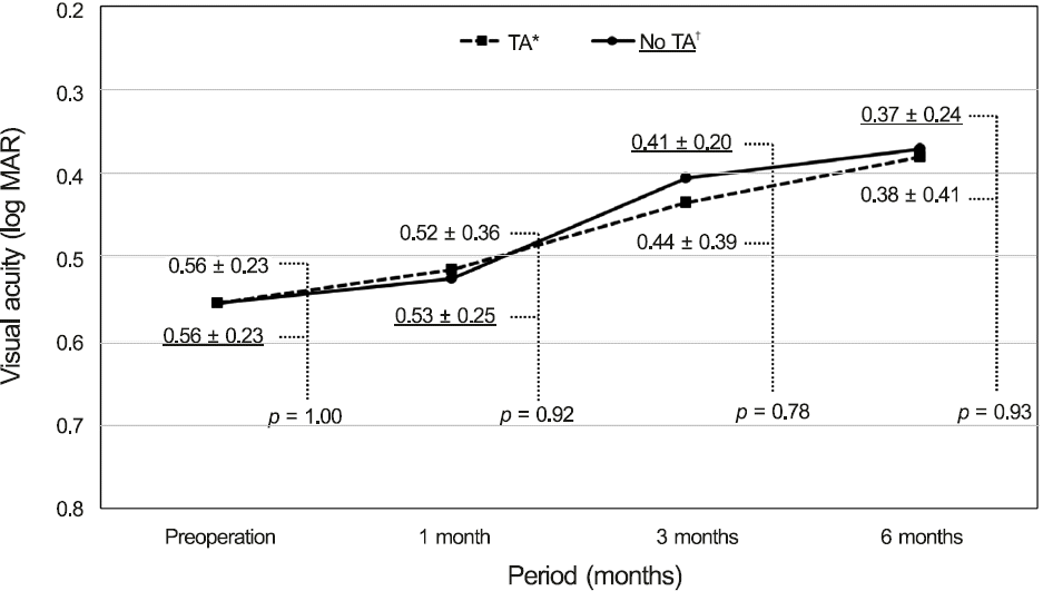 | Figure 1.Preoperative and postoperative best-corrected visual acuity after surgical removal of the epiretinal membrane with and without posterior sub-tenon triamcinolone acetonide injection. TA =triamcinolone acetonide. *Posterior sub-tenon triamcinolone acetonide was injected during vitrectomy; † Posterior sub-tenon triamcinolone acetonide was not injected during vitrectomy. |
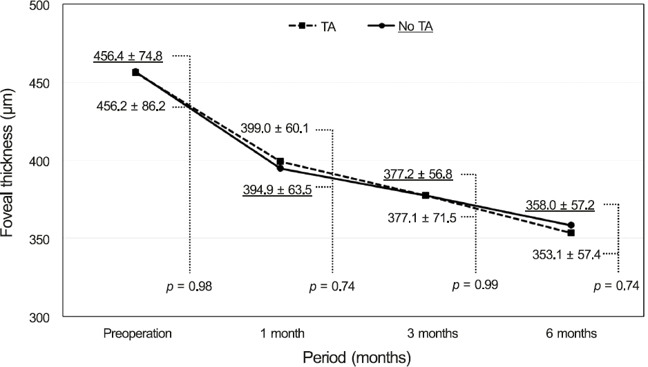 | Figure 2.Preoperative and postoperative foveal thickness (micrometers) after the surgical removal of the epiretinal membrane with and without posterior sub-tenon triamcinolone acetonide injection. TA = triamcinolone acetonide. |
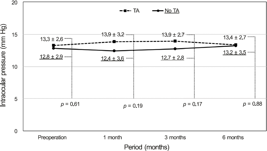 | Figure 3.Preoperative and postoperative intraocular pressure (mm Hg) after the surgical removal of the epiretinal membrane with and without posterior sub-tenon triamcinolone acetonide injection. TA = triamcinolone acetonide. |
Table 1.
Baseline characteristics
 XML Download