

 PDF
PDF ePub
ePub Citation
 Print
Print


Abstract
Methods
This study included 185 eyes of 112 patients treated with beveled, full thickness astigmatic keratotomy. Treated eyes were divided into 3 groups: beveled, full thickness astigmatic keratotomy after implantable collamer lens (ICL) implantation (group A), beveled, full thickness astigmatic keratotomy after cataract surgery (group B) and beveled, full thickness astigmatic keratotomy alone (group C). Follow-up visits were at 1 week, 1 month, 3 months and 6 months. The outcome measures included uncorrected distance visual acuity, astigmatism, efficacy, safety and predictability.
Results
At 6 months postoperatively, astigmatism was significantly reduced: 68.9 ± 18.24% in total, 69.24 ± 20.76%, in the group A, 67.84 ± 17.56% in the group B and 67.82 ± 13.97% in the group C. The proportion of eyes with astigmatism 1.0 or less was 88.65% in total, 91.49% in the group A, 87.5% in the group B and 70.0% in the group C. Mean improvement in corrected dis-tance visual acuity (CDVA) was 0.56 lines; no eyes lost 2 lines of CDVA after 6 months postoperatively. Postoperative complica-tions were not observed.
Go to : 
References
1. Pineda R, Jain V. Arcuate keratotomy: an option for astigmatism correction after laser in situ keratomileusis. Cornea. 2009; 28:1178–80.

2. Güell JL, Vazquez M. Correction of high astigmatism with astig-matic keratotomy combined with laser in situ keratomileusis. J Cataract Refract Surg. 2000; 26:960–6.
3. Song HB, Choi HJ, Kim MK, Wee WR. The short-term effect of limbal relaxing incision and compression suture on post-penetrat-ing keratoplasty astigmatism. J Korean Ophthalmol Soc. 2011; 52:1142–9.
4. Nubile M, Carpineto P, Lanzini M. . Femtosecond laser arcuate keratotomy for the correction of high astigmatism after keratoplasty. Ophthalmology. 2009; 116:1083–92.
5. Rosecan LR. Endophthalmitis and cystoid macular edema after as-tigmatic keratotomy. Ophthalmic Surg. 1994; 25:481–2.
6. Yoon JM, Moon SJ, Lee KH. Clinical outcomes of toric implant-able collamer lens implantation. J Korean Ophthalmol Soc. 2009; 50:839–51.
7. Ivarsen A, Næser K, Hjortdal J. Laser in situ keratomileusis for high astigmatism in myopic and hyperopic eyes. J Cataract Refract Surg. 2013; 39:74–80.
8. Binder PS. Analysis of ectasia after laser in situ keratomileusis: risk factors. J Cataract Refract Surg. 2007; 33:1530–8.
9. Nagy ZZ, Munkácsy G, Popper M. Photorefractive keratectomy using the meditec MEL 70 G-scan laser for hyperopia and hyper-opic astigmatism. J Refract Surg. 2002; 18:542–50.
10. Holladay JT, Dudeja DR, Chang J. Functional vision and corneal changes after laser in situ keratomileusis determined by contrast sensitivity, glare testing, and corneal topography. J Cataract Refract Surg. 1999; 25:663–9.
11. Jiménez-Alfaro I, Benítez del Castillo JM, García-Feijoó J. . Safety of posterior chamber phakic intraocular lenses for the cor-rection of high myopia: anterior segment changes after posterior chamber phakic intraocular lens implantation. Ophthalmology. 2001; 108:90–9.
12. Akura J, Matsuura K, Hatta S. . Experimental study using pig eyes for realizing ideal astigmatic keratotomy. Cornea. 2001; 20:325–8.
13. Duffey RJ, Jain VN, Tchah H. . Paired arcuate keratotomy. A surgical approach to mixed and myopic astigmatism. Arch Ophthalmol. 1988; 106:1130–5.
14. Akura J, Matsuura K, Hatta S. . Clinical application of full-arc, depth-dependent, astigmatic keratotomy. Cornea. 2001; 20:839–43.
15. Hoffart L, Touzeau O, Borderie V, Laroche L. Mechanized astig-matic arcuate keratotomy with the Hanna arcitome for astigmatism after keratoplasty. J Cataract Refract Surg. 2007; 33:862–8.
16. Poole TR, Ficker LA. Astigmatic keratotomy for post-keratoplasty astigmatism. J Cataract Refract Surg. 2006; 32:1175–9.
17. Cleary C, Tang M, Ahmed H. . Beveled femtosecond laser as-tigmatic keratotomy for the treatment of high astigmatism post- penetrating keratoplasty. Cornea. 2013; 32:54–62.
18. Buzzonetti L, Petrocelli G, Laborante A. . Arcuate keratotomy for high postoperative keratoplasty astigmatism performed with the intralase femtosecond laser. J Refract Surg. 2009; 25:709–14.
19. Kumar NL, Kaiserman I, Shehadeh-Mashor R. . IntraLase-en-abled astigmatic keratotomy for post-keratoplasty astigmatism: on-axis vector analysis. Ophthalmology. 2010; 117:1228–35.
20. Tsioulias G, Droutsas D, Moschos M. . Arcuate relaxing in-cisions with a 5.00-mm optical zone for the correction of high post-cataract astigmatism. Ophthalmologica. 2000; 214:385–9.
21. Guyton DL. Prescribing cylinders: the problem of distortion. Surv Ophthalmol. 1977; 22:177–88.
22. Lindquist TD, Rubenstein JB, Rice SW. . Trapezoidal astig-matic keratotomy. Quantification in human cadaver eyes. Arch Ophthalmol. 1986; 104:1534–9.
23. Akura J, Matsuura K, Hatta S. . Clinical application of full-arc, depth-dependent, astigmatic keratotomy. Cornea. 2001; 20:839–43.
24. Feldman RM, Crapotta JA, Feldman ST, Goldbaum MH. Retinal detachment following radial and astigmatic keratotomy. Refract Corneal Surg. 1991; 7:252–3.
25. Adrean SD, Cochrane R, Reilly CD, Mannis MJ. Infectious kerati-tis after astigmatic keratotomy in penetrating keratoplasty: review of three cases. Cornea. 2005; 24:626–8.
Go to :
 | Figure 1.(A) Corneal marking with a ring marker with cross wires (7.5 mm). (B) Making at 3, 9, 12 o’clock direction with marking pen. (C) Beveled, full thickness cornea incision with 2.8 mm blade. (D) Extension of corneal incision with wider blade. (E) Checking the wound with a Weck-Cel sponge if there is any leakage. (F) Subconjunctival injection with mixture of antibiotics, ste-roid, and lidocaine. |
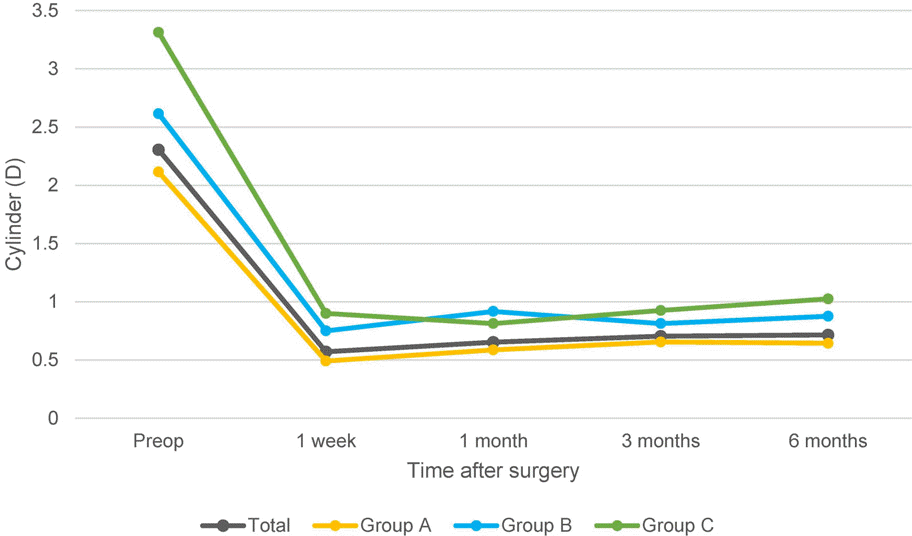 | Figure 2.Changes in cylinder after the beveled, full thickness astigmatic keratotomy. Preop = Preoperation. |
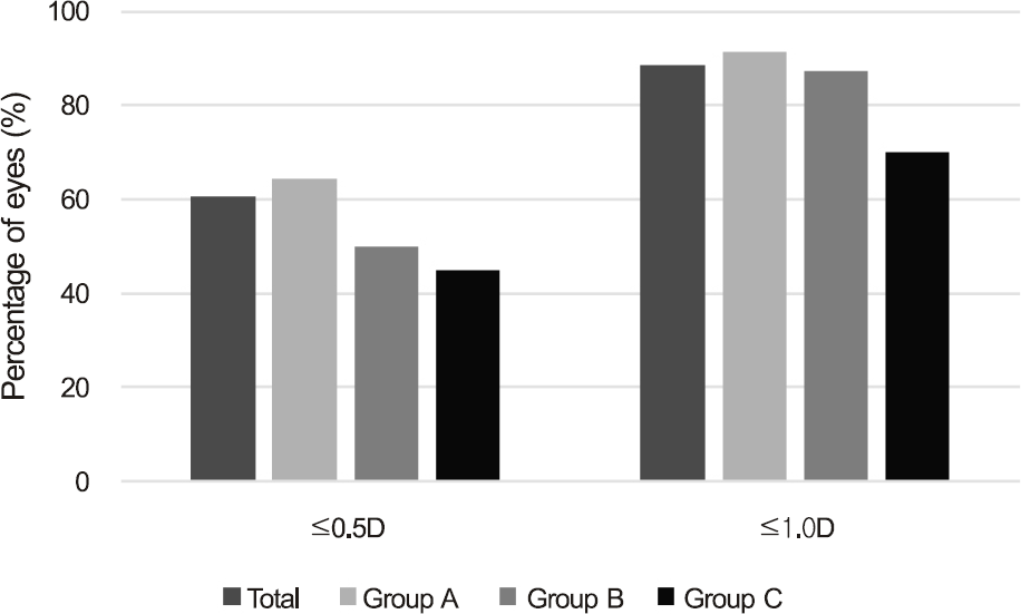 | Figure 4.Predictability. Bar graph represents the percentage of eyes within 0.5D and 1.0D of intended correction at 6 months postoperatively. D = diopters. |
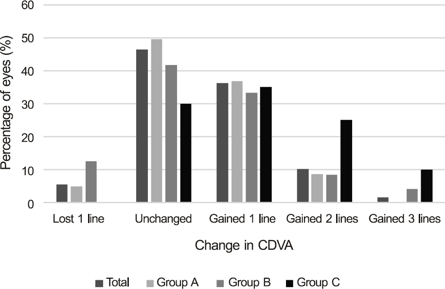 | Figure 6.Safety. Gain and loss of CDVA 6 months postoperatively. CDVA = corrected distance visual acuity. |
 | Figure 7.Case example. Beveled full thickness astigmatic keratotomy was performed on the left eye at 12:30 o’clock direction, 5.7-mm width in a patient after ICL implantation. (A) The incision is faintly seen on slit-lamp photography at 1 month post-operatively (dotted line). (B) Kmax was 44.6 D at steep axis on preoperative topography. UCVA was 20/30, CDVA was 20/20, re-fraction was +1.5 -2.75 × 165° preoperatively. (C) At the 1 month postoperative visit, UCVA and CDVA were 20/16, refraction was +0.25 -0.25 × 170°. Kmax was 42.5 D at steep axis. (D) Anterior OCT image of the incision site for ICL implantation. Perpendicular incision can be seen. (E) Anterior OCT image of the incision site for astigmatic keratotomy. Beveled, full thickness incision can be seen. ICL = implantable collamer lens; K = keratometry; UCVA = uncorrected visual acuity; CDVA = corrected distance visual acuity; OCT = optical coherence tomography. |
Table 1.
Demographics of patients
Table 2.
Changes in UDVA and cylinder after the beveled, full thickness astigmatic keratotomy
Table 3.
Amount of corrected astigmatism at 6 months after as the beveled, full thickness astigmatic keratotomy
Table 4.
Changes in refraction after the beveled, full thickness astigmatic keratotomy
Table 5.
Changes in irregularity index checked by ORB® scan
Table 6.
Changes in CDVA after the beveled, full thickness astigmatic keratotomy and safety index
 XML Download