Abstract
Purpose:
To report a case of nonparaneoplastic autoimmune retinopathy diagnosed using serum anti-retinal autoantibodies.
Case summary:
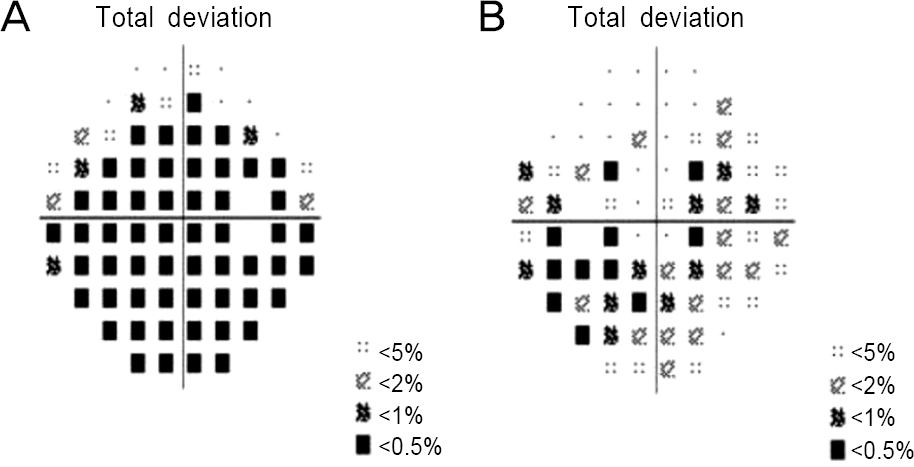
A 60-year-old female complained of progressive visual loss in both eyes over 3 months. Her best corrected visu-al acuity was hand motion in the right eye, 0.9 (decimal) in the left eye, and no definite abnormal findings were identified on fun-dus examinations. Automated visual field test revealed severely depressed visual fields in both eyes. Standard full-field electro-retinogram (ERG) revealed nearly extinguished scotopic b-waves and spectral-domain optical coherence tomography (SD-OCT) showed subtle obscuration and interruption of the inner segment/outer segment (IS/OS) junction of the photoreceptors. Using Western blotting with human retinal proteins and the patient’s serum, we diagnosed nonparaneoplastic autoimmune retinopathy and performed posterior subtenon steroid injection in the right eye, systemic corticosteroids, and oral mycophenolate mofetil. Full-field ERG after treatment showed slightly increased amplitude but there was no subjective visual improvement. OCT after treatment did not reveal significant changes in the photoreceptor layer.
Go to : 
References
1. Comlekoglu DU, Thompson IA, Sen HN. Autoimmune retinopathy. Curr Opin Ophthalmol. 2013; 24:598–605.

3. Grange L, Dalal M, Nussenblatt RB, Sen HN. Autoimmune retinopathy. Am J Ophthalmol. 2014; 157:266–72.e1.

4. Weleber RG, Watzke RC, Shults WT, et al. Clinical and electro-physiologic characterization of paraneoplastic and autoimmune retinopathies associated with antienolase antibodies. Am J Ophthalmol. 2005; 139:780–94.

5. Grewal DS, Fishman GA, Jampol LM. Autoimmune retinopathy and antiretinal antibodies: a review. Retina. 2014; 34:1023–41.
6. Lima LH, Greenberg JP, Greenstein VC, et al. Hyperautofluo-rescent ring in autoimmune retinopathy. Retina. 2012; 32:1385–94.

7. Abazari A, Allam SS, Adamus G, Ghazi NG. Optical coherence to-mography findings in autoimmune retinopathy. Am J Ophthalmol. 2012; 153:750–6,756.e1.

8. Faez S, Loewenstein J, Sobrin L. Concordance of antiretinal anti-body testing results between laboratories in autoimmune retinopathy. JAMA Ophthalmol. 2013; 131:113–5.

9. Ko AC, Hernandez J, Brinton JP, et al. Anti-gamma-enolase auto-immune retinopathy manifesting in early childhood. Arch Ophthalmol. 2010; 128:1590–5.
10. Pepple KL, Cusick M, Jaffe GJ, Mruthyunjaya P. SD-OCT and au-tofluorescence characteristics of autoimmune retinopathy. Br J Ophthalmol. 2013; 97:139–44.

11. Mohamed Q, Harper CA. Acute optical coherence tomographic findings in cancer-associated retinopathy. Arch Ophthalmol. 2007; 125:1132–3.

12. Shimazaki K, Jirawuthiworavong GV, Heckenlively JR, Gordon LK. Frequency of anti-retinal antibodies in normal human serum. J Neuroophthalmol. 2008; 28:5–11.

13. Adamus G. Antirecoverin antibodies and autoimmune retinopathy. Arch Ophthalmol. 2000; 118:1577–8.

14. Adamus G, Ren G, Weleber RG. Autoantibodies against retinal proteins in paraneoplastic and autoimmune retinopathy. BMC Ophthalmol. 2004; 4:5.

15. Adamus G. Autoantibody-induced apoptosis as a possible mecha-nism of autoimmune retinopathy. Autoimmun Rev. 2003; 2:63–8.

16. Mizener JB, Kimura AE, Adamus G, et al. Autoimmune retinop-athy in the absence of cancer. Am J Ophthalmol. 1997; 123:607–18.

17. Heckenlively JR, Ferreyra HA. Autoimmune retinopathy: a review and summary. Semin Immunopathol. 2008; 30:127–34.

18. Heckenlively JR, Fawzi AA, Oversier J, et al. Autoimmune retin-opathy: patients with antirecoverin immunoreactivity and pan-retinal degeneration. Arch Ophthalmol. 2000; 118:1525–33.
19. Heckenlively JR, Aptsiauri N, Nusinowitz S, et al. Investigations of antiretinal antibodies in pigmentary retinopathy and other reti-nal degenerations. Trans Am Ophthalmol Soc. 1996; 94:179–200. discussion. 200–6.
20. Mrejen S, Khan S, Gallego-Pinazo R, et al. Acute zonal occult out-er retinopathy: a classification based on multimodal imaging. JAMA Ophthalmol. 2014; 132:1089–98.
21. Or C, Collins DR, Merkur AB, et al. Intravenous rituximab for the treatment of cancer-associated retinopathy. Can J Ophthalmol. 2013; 48:e35–8.

22. Dy I, Chintapatla R, Preeshagul I, Becker D. Treatment of can-cer-associated retinopathy with rituximab. J Natl Compr Canc Netw. 2013; 11:1320–4.

23. Audemard A, de Raucourt S, Miocque S, et al. Melanoma-asso-ciated retinopathy treated with ipilimumab therapy. Dermatology. 2013; 227:146–9.

24. Rahimy E, Sarraf D. Paraneoplastic and non-paraneoplastic retin-opathy and optic neuropathy: evaluation and management. Surv Ophthalmol. 2013; 58:430–58.

25. Chan JW. Paraneoplastic retinopathies and optic neuropathies. Survey of ophthalmology. 2003; 48:12–38.

Go to : 
 | Figure 1.(A, B) Normal fundus photographs. (C) Fundus autofluorescence reveals a ring of outer hyperautofluorescence (between round dots and arrows) in the right eye. (D) Fundus autofluorescence showed no remarkable findings in the left eye. |
 | Figure 2.Automated visual field test revealed severely de-pressed visual fields in the (A) right eye and (B) left eye (more in the right eye than in the left eye). |
 | Figure 3.Standard full field ERG reveals nearly extinguished scotopic a and b waves, negative waves of combined responses, and severely de-creased amplitude of photopic response in both eyes. ERG = electroretinogram. |
 | Figure 4.(A) Multifocal electroretinogram shows decreased P1 amplitude and increased implicit time which is more prominent in the right eye. (B) Normal multifocal electroretinogram in the left eye. T = temporal; I = inferior; N =nasal; S = superior. |
 | Figure 5.Spectral-domain coherence tomography (SD-OCT) shows subtle obscuration and interruption of the inner seg-ment/outer segment (IS/OS) junction of the photoreceptors in the right eye (A) and in the left eye (B) (arrows). |
 | Figure 6.Western blot analysis-Bands at 50 kDa were constantly seen in all lanes and reflect immunoglobulin G (IgG) heavy chains present in the soluble protein extracts from human tissue. Arrows indicate proteins that react with serum from pa-tient, not with serum from control. Band A near 46 kDa and band B near 39 kDa. |


 PDF
PDF ePub
ePub Citation
 Print
Print


 XML Download