Abstract
Purpose:
To determine the incidence of steroid-induced ocular hypertension following myopic vision correction.
Methods:
This study retrospectively reviewed the medical records of 6,087 patients (12,164 eyes) who underwent myopic re-fractive surgery (laser-assisted in-situ keratomileusis [LASIK]/ photorefractive keratectomy [PRK]/phakic intraocular lens [IOL] implantation) at Eyereum Eye Clinic between July 2011 and February 2013. Ocular hypertension was defined when post-operative intraocular pressure (IOP) was increased more than 30% compared to predicted IOP adjusted according to corneal thickness. All preoperative IOPs were measured using Goldmann applanation tonometer (GAT). Postoperative IOPs were measured using non-contact tonometer first and with GAT when the IOP was suspiciously increased.
Results:
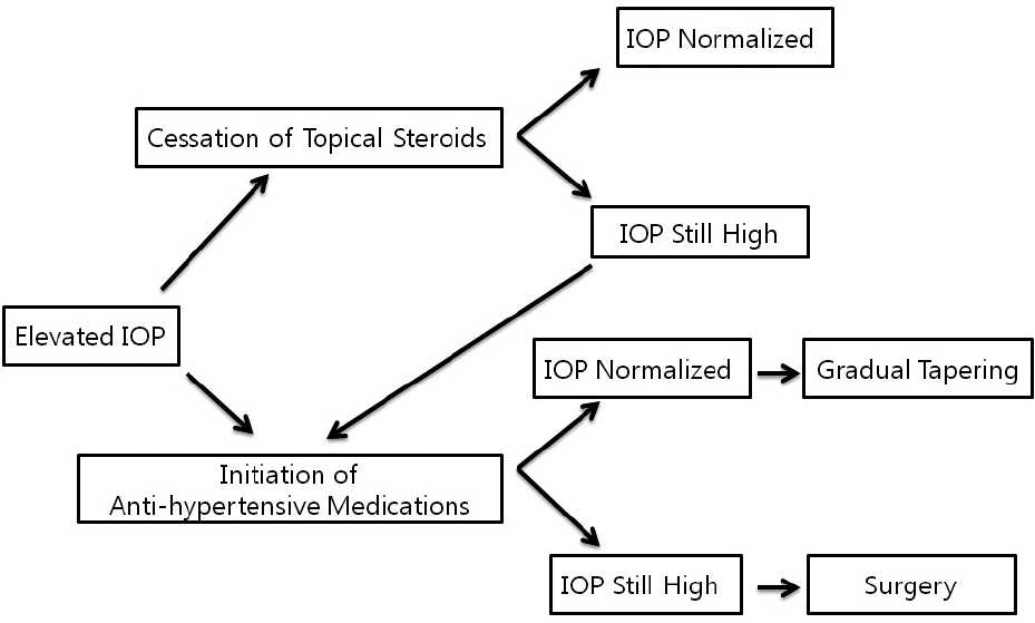
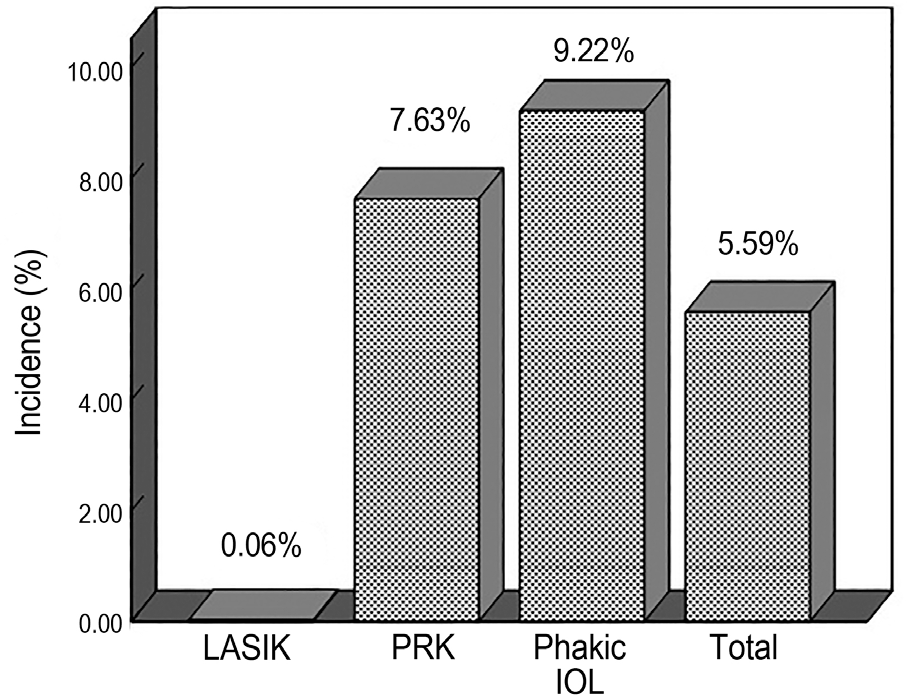
Steroid-induced ocular hypertension after a myopic refractive surgery occurred in 680 eyes (5.58%) of 404 patients (6.64%). The incidence based on surgery was LASIK (0.06%, 2/3, 514 eyes) followed by PRK (7.63%, 575/7,533 eyes) and phakic IOL implantation (9.2%, 103/1,117 eyes). The average increased IOP level in patients with steroid-induced ocular hyper-tension was 5.62 ± 3.73 mm Hg after PRK and 9.35 ± 4.95 mm Hg after phakic IOL implantation. A statistically significantly higher change in IOP was observed in the phakic IOL group ( p < 0.001). However, the PRK group had a longer treatment period for ocu-lar hypertension and used more antiglaucoma medications than the phakic IOL group ( p < 0.05). Most patients with ocular hyper-tension were successfully treated with cessation of topical steroid or use of antiglaucoma medications. Only 2 eyes required glaucoma surgery because IOP was not controlled.
Conclusions:
IOP measurements should be initiated no later than 1 week after surgery because steroid-induced ocular hyper-tension following myopic refractive surgery can occur in approximately 5.58% of patients and most cases of ocular hypertension can be controlled with careful follow-up and use of antiglaucoma medications.
Go to : 
References
1. Smith RJ, Maloney RK. Diffuse lamellar keratitis. A new syn-drome in lamellar refractive surgery. Ophthalmology. 1998; 105:1721–6.

2. Peters NT, Lingua RW, Kim CH. Topical intrastromal steroid dur-ing laser in situ keratomileusis to retard interface keratitis. J Cataract Refract Surg. 1999; 25:1437–40.

3. Nagy ZZ, Szabó A, Krueger RR, Süveges I. Treatment of intra-ocular pressure elevation after photorefractive keratectomy. J Cataract Refract Surg. 2001; 27:1018–24.

4. Tengroth B, Epstein D, Fagerholm P, et al. Excimer laser photo-refractive keratectomy for myopia: clinical results in sighted eyes. Ophthalmology. 1993; 100:739–45.
5. McPherson R, Hanna K, Agro A, et al. Cerivastatin versus branded pravastatin in the treatment of primary hypercholesterolemia in primary care practice in Canada: a one-year, open-label, random-ized, comparative study of efficacy, safety, and cost-effectiveness. Clin Ther. 2001; 23:1492–507.

6. Gaston H, Absolon MJ, Thurtle OA, Sattar MA. Steroid re-sponsiveness in connective tissue diseases. Br J Ophthalmol. 1983; 67:487–90.

7. Munjal VP, Dhir SP, Jain IS. Steroid induced glaucoma. Indian J Ophthalmol. 1982; 30:379–82.
8. Seiler T, Holschbach A, Derse M, et al. Complications of myopic photorefractive keratectomy with the excimer laser. Ophthalmology. 1994; 101:153–60.

9. Vetrugno M, Maino A, Quaranta G, Cardia L. A randomized, com-parative open-label study on the efficacy of latanoprost and timolol in steroid induced ocular hypertension after photorefractive keratectomy. Eur J Ophthalmol. 2000; 10:205–11.

10. Leibowitz HM, Ryan WJ Jr, Kupferman A. Comparative anti-in-flammatory efficacy of topical corticosteroids with low glauco-ma-inducing potential. Arch Ophthalmol. 1992; 110:118–20.

11. Sher NA, Chen V, Bowers RA, et al. The use of the 193-nm ex-cimer laser for myopic photorefractive keratectomy in sighted eyes: a multicenter study. Arch Ophthal. 1991; 109:1525–30.
12. Johnson M, Kass MA, Moses RA, Grodzki WJ. Increased corneal thickness simulating elevated intraocular pressure. Arch Ophthalmol. 1978; 96:664–5.

13. Ehlers N, Bramsen T, Sperling S. Applanation tonometry and cen-tral corneal thickness. Acta Ophthalmol (Copenh). 1975; 53:34–43.

14. Shimizu K, Amano S, Tanaka S. Photorefractive keratectomy for myopia: one-year follow-up in 97 eyes. J Refract Corneal Surg. 1994; 10((2 Suppl)):S178–87.

15. Ishibashi T, Takagi Y, Mori K, et al. cDNA microarray analysis of gene expression changes induced by dexamethasone in cultured human trabecular meshwork cells. Invest Ophthalmol Vis Sci. 2002; 43:3691–7.
16. Dickerson JE Jr, Steely HT Jr, English-Wright SL, Clark AF. The effect of dexamethasone on integrin and laminin expression in cul-tured human trabecular meshwork cells. Exp Eye Res. 1998; 66:731–8.
17. Alward WL. The genetics of open-angle glaucoma: the story of GLC1A and myocilin. Eye (Lond). 2000; 14((Pt 3B)):429–36.

18. Alward WL, Fingert JH, Coote MA, et al. Clinical features asso-ciated with mutations in the chromosome 1 open-angle glaucoma gene (GLC1A). N Engl J Med. 1998; 338:1022–7.

19. Tang WC, Yip SP, Lo KK, et al. Linkage and association of my-ocilin (MYOC) polymorphisms with high myopia in a Chinese population. Mol Vis. 2007; 13:534–44.
20. Zayats T, Yanovitch T, Creer RC, et al. Myocilin polymorphisms and high myopia in subjects of European origin. Mol Vis. 2009; 15:213–22.
21. Machat JJ, Tayfour F. Photorefractive keratectomy for myopia: preliminary results in 147 eyes. Refract Corneal Surg. 1993; 9((2 Suppl)):S16–9.

22. Gartry DS, Kerr Muir MG, Marshall J. Excimer laser photo-refractive keratectomy. 18-month follow-up. Ophthalmology. 1992; 99:1209–19.
23. Javadi MA, Mirbabaei-Ghafghazi F, Mirzade M, et al. Steroid in-duced ocular hypertension following myopic photorefractive keratectomy. J Ophthalmic Vis Res. 2008; 3:42–6.
Go to : 
 | Figure 1.Management of the steroid induced ocular hyper-tension after myopic refractive surgery. IOP = intraocular pressure. |
 | Figure 2.The incidence of the steroid-induced ocular hypertension in each group after myopic refractive surgery between July 2011 and February 2013. LASIK = laser-assisted in-situ keratomileusis; PRK = photorefractive keratectomy; IOL = intraocular lens. |
Table 1.
Baseline characteristics in non-steroid responder vs. steroid responder after myopic refractive surgery
| Total (n = 12,164) | Non responder (n = 11,485) | Responder (n = 680) | p-value |
|---|---|---|---|
| Age (years) | 26.43 ± 4.79 | 27.88 ± 4.91 | 0.713∗ |
| Gender (female, %) | 6,202 (54.0) | 316 (46.5) | 0.641† |
| SE (diopter) | -4.98 ± 2.40 | -9.12 ± 2.12 | <0.001∗ |
| Sim K1 (diopter) | 43.01 ± 1.74 | 42.87 ± 1.80 | 0.825∗ |
| Sim K2 (diopter) | 44.21 ± 1.50 | 44.19 ± 1.76 | 0.863∗ |
| CCT (μ m) | 542.67 ± 29 | 541.76 ± 32 | 0.742∗ |
Table 2.
Baseline characteristics in non-steroid responder vs. steroid responder after photorefractive keratectomy or phakic intra-ocular lens implantation
| PRK (n = 7,533) | Phakic IOL (n = 1,117) | |||||
|---|---|---|---|---|---|---|
| Non responder | Responder | p-value | Non responder | Responder | p-value | |
| No. of eyes | 6,958 | 575 | 1,014 | 103 | ||
| Age (years) | 26.25 ± 4.73 | 26.87 ± 4.97 | 0.832∗ | 27.72 ± 5.11 | 27.88 ± 5.69 | 0.741∗ |
| Gender (female, %) | 3,068 (44.1) | 259 (45.0) | 0.662† | 766 (75.5) | 57 (55.3) | <0.001† |
| SE (diopter) | -4.40 ± 3.40 | -4.88 ± 2.12 | 0.622∗ | -8.06 ± 4.12 | -9.12 ± 5.40 | 0.036∗ |
| CCT (μ m) | 545.67 ± 29 | 537.76 ± 32 | 0.742∗ | 521.30 ± 39 | 522.44 ± 36 | 0.981∗ |
Table 3.
Duration and number of medications required for IOP control and average IOP rise in steroid responders for each surgical groups
| PRK (n = 575) | Phakic IOL (n = 103) | p-value | |
|---|---|---|---|
| Duration of glaucoma medications (days) | 70.40 ± 49.06 | 31.35 ± 21.11 | <0.001∗ |
| No. of glaucoma medications | 1.90 ± 0.81 | 1.70 ± 0.73 | 0.020∗ |
| Average IOP rise (mm Hg) | 5.62 ± 3.73 | 9.35 ± 4.95 | <0.001∗ |
Table 4.
Characteristics of patient with steroid induced ocular hypertension undergoing femtosecond LASIK
| Pt | Gender | Eyes | Preop SE (D) | Preop CCT (μ m) | Postop CCT (μ m) | IOP change∗(mm Hg) | Meds (n) | Duration of Meds (days) |
|---|---|---|---|---|---|---|---|---|
| 1 | female | OD | -4.12 | 563 | 410 | 7.46 | 1 | 25 |
| OS | -4.19 | 571 | 442 | 8.03 | 1 | 25 |


 PDF
PDF ePub
ePub Citation
 Print
Print


 XML Download