

 PDF
PDF ePub
ePub Citation
 Print
Print


Abstract
Purpose:
To compare the surgical outcomes of triple procedure in patients with open-angle glaucoma and angle-closure glaucoma.
Methods:
The patients who underwent triple procedures for open-angle glaucoma and angle-closure glaucoma and were fol-lowed up for more than 1 year postoperatively were retrospectively reviewed. Preoperative and postoperative visual acuity, intra-ocular pressure (IOP), visual field mean deviation, refractive error, number of medications, and complications were analyzed. The effect of surgery on IOP reduction and refractive error correction was compared.
Results:
The IOP at 1 year postoperatively was 13.39 ± 2.25 mm Hg, 13.41 ± 2.79 mm Hg ( p = 0.981) and IOP reduction was 4.51 ± 6.35 mm Hg, 9.11 ± 8.27 mm Hg ( p = 0.042) in the open angle glaucoma group and angle closure glaucoma group, respectively. No patient in either group required reoperation due to uncontrolled IOP. The percentage of patients showing post-operative IOP reduction of at least 10% and 20% from baseline IOP was statistically higher in the angle-closure glaucoma group than in the open-angle glaucoma group. Prediction errors were -0.84 ± 0.88 D and -0.13 ± 0.65 D in the open-angle glaucoma group and angle-closure glaucoma group, respectively.
Go to : 
References
1. Acton J, Salmon JF, Scholtz R. Extracapsular cataract extraction with posterior chamber lens implantation in primary angle-closure glaucoma. J Cataract Refract Surg. 1997; 23:930–4.

2. Ming Zhi Z, Lim AS, Yin Wong T. A pilot study of lens extraction in the management of acute primary angle-closure glaucoma. Am J Ophthalmol. 2003; 135:534–6.
3. Strenk SA, Strenk LM, Guo S. Magnetic resonance imaging of the anteroposterior position and thickness of the aging, accommodat-ing, phakic, and pseudophakic ciliary muscle. J Cataract Refract Surg. 2010; 36:235–41.
4. Kook MS, Kim HB, Lee SU. Short-term effect of mitomycin-C augmented trabeculectomy on axial length and corneal astigmatism. J Cataract Refract Surg. 2001; 27:518–23.
5. Tan HY, Wu SC. Refractive error with optimum intraocular lens power calculation after glaucoma filtering surgery. J Cataract Refract Surg. 2004; 30:2595–7.
6. Lowe RF. Aetiology of the anatomical basis for primary angle-clo-sure glaucoma. Biometrical comparisons between normal eyes and eyes with primary angle-closure glaucoma. Br J Ophthalmol. 1970; 54:161–9.
7. George R, Paul PG, Baskaran M, et al. Ocular biometry in occlud-able angles and angle closure glaucoma: a population based survey. Br J Ophthalmol. 2003; 87:399–402.
8. Go GB, Kim DW, Baek NH. Intraocular pressure change following cataract surgery in patient with high intraocular pressure. J Korean Ophthalmol Soc. 1993; 34:1128–34.
9. Martinez-Bello C, Rodriguez-Ares T, Pazos B, et al. Changes in anterior chamber depth and angle width after filtration surgery: a quantitative study using ultrasound biomicroscopy. J Glaucoma. 2000; 9:51–5.
10. Karasheva G, Goebel W, Klink T, et al. Changes in macular thick-ness and depth of anterior chamber in patients after filtration surgery. Graefes Arch Clin Exp Ophthalmol. 2003; 241:170–5.
11. Francis BA, Wang M, Lei H, et al. Changes in axial length follow-ing trabeculectomy and glaucoma drainage device surgery. Br J Ophthalmol. 2005; 89:17–20.
12. Uretmen O, Ateş H, Andaç K, Deli B. Axial length changes accom-panying successful nonpenetrating glaucoma filtration surgery. Ophthalmologica. 2003; 217:199–203.
13. Chan JC, Lai JS, Tham CC. Comparison of postoperative re-fractive outcome in phacotrabeculectomy and phacoemulsification with posterior chamber intraocular lens implantation. J Glaucoma. 2006; 15:26–9.
14. Husain R, Li W, Gazzard G, et al. Longitudinal changes in anterior chamber depth and axial length in Asian subjects after trabeculec-tomy surgery. Br J Ophthalmol. 2013; 97:852–6.
15. Caporossi A, Casprini F, Tosi GM, Balestrazzi A. Long-term results of combined 1-way phacoemulsification, intraocular lens im-plantation, and trabeculectomy. J Cataract Refract Surg. 1999; 25:1641–5.
16. Rockwood EJ, Larive B, Hahn J. Outcomes of combined cataract extraction, lens implantation, and trabeculectomy surgeries. Am J Ophthalmol. 2000; 130:704–11.
17. Tow SL, Aung T, Oen FT, Seah SK. Combined phacoemulsifica-tion, intraocular lens implantation and trabeculectomy for chronic angle closure glaucoma. Int Ophthalmol. 2001; 24:283–9.
Go to :
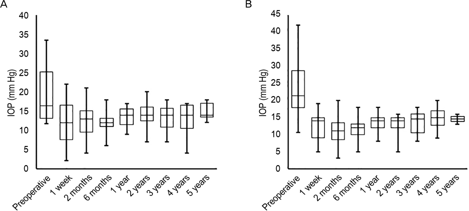 | Figure 1.The changes of IOP after triple procedure are shown at each time point. (A) OAG group. (B) AGC group. IOP = intra-ocular pressure; OAG = open angle glaucoma; ACG = angle closure glaucoma. |
Table 1.
Characteristics of subjects who underwent triple procedure for OAG and ACG
| OAG | ACG | p-value | |
|---|---|---|---|
| No. of subjects | 23 | 23 | |
| Sex (M:F) | 18:5 | 10:13 | 0.033∗ |
| Mean age ± SD (years) | 69.00 ± 9.13 | 68.09 ± 8.47 | 0.727† |
| Mean IOP (mm Hg) | 17.90 ± 5.96 | 22.91 ± 8.15 | 0.022† |
| Number of medications | 2.43 ± 0.66 | 2.57 ± 0.73 | 0.528† |
| MD of visual field test (dB) | -18.55 ± 9.52 | -17.82 ± 9.42 | 0.849† |
| Mean follow-up period (years) | 3.39 ± 1.69 | 2.92 ± 1.34 | 0.298† |
Table 2.
Postoperative 1 year outcome in OAG and ACG
| OAG | ACG | p-value | |
|---|---|---|---|
| No. of subjects | 23 | 23 | |
| IOP (mm Hg) | 13.39 ± 2.25 | 13.41 ± 2.79 | 0.981‡ |
| △ IOP∗ (mm Hg) | 4.51 ± 6.35 | 9.11 ± 8.27 | 0.042‡ |
| Number of medications | 0.65 ± 0.83 | 0.41 ± 0.67 | 0.287‡ |
| Postoperative management (n, %) | 13 (56.5) | 10 (43.5) | 0.556§ |
| Reoperation (n, %) | 0 (0) | 0 (0) | 1.000§ |
| Prediction error† (D) | -0.84 ± 0.88 | -0.13 ± 0.65 | 0.008‡ |
 XML Download