Abstract
Purpose:
To evaluate 0.025% brilliant blue G (BBG) for staining the internal limiting membrane (ILM) during vitrectomy.
Methods:
In a retrospective, non-comparative clinical case series, we analyzed consecutive 111 patients who underwent pars pla-na vitrectomy and removal of the ILM after staining using BBG solution. BBG was dissolved and diluted with balanced salt solution at a concentration of 0.025% and then sterilized by filtering through a 0.22 μ m millipore filter. The prepared BBG solution was in-jected into the vitreous cavity over the macula after removal of the vitreous and excessive solution was removed immediately.
Results:
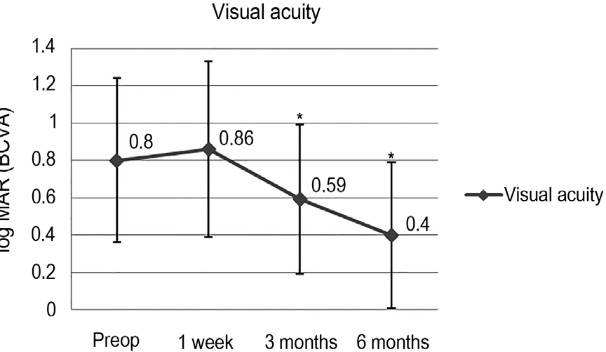
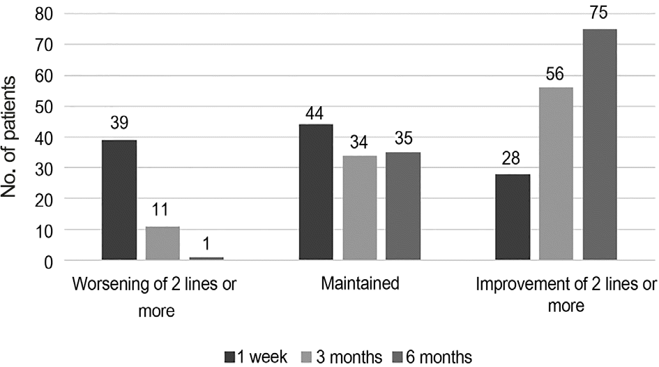
The ILM was successfully removed without use of additional adjuvant in all cases. Mean best corrected visual acuity (log MAR) was significantly improved from 0.80 ± 0.44 at baseline to 0.40 ± 0.39 at 6 months postoperatively ( p < 0.001). One case each of endophthalmitis and diabetic papillopathy developed. The relationship when using BBG solution was not identified as complications were not observed in the other patients who underwent vitrectomy using the same BBG solution on the same day. One idiopathic epiretinal membrane patient had visual acuity loss more than 2 lines. During the follow-up period, other com-plications suspected to be associated with the use of BBG solution were not observed.
References
1. Eckardt C, Eckardt U, Groos S, et al. Removal of the internal limit-ing membrane in macular holes. Clinical and morphological findings. Ophthalmologe. 1997; 94:545–51.
2. Brooks HL Jr. Macular hole surgery with and without internal lim-iting membrane peeling. Ophthalmology. 2000; 107:1939–48.

3. Kifuku K, Hata Y, Kohno RI, et al. Residual internal limiting mem-brane in epiretinal membrane surgery. Br J Ophthalmol. 2009; 93:1016–9.

4. Munuera JM, García-Layana A, Maldonado MJ, et al. Optical co-herence tomography in successful surgery of vitreomacular trac-tion syndrome. Arch Ophthalmol. 1998; 116:1388–9.
5. Ando F, Yasui O, Hirose H, Ohba N. Optic nerve atrophy after vi-trectomy with indocyanine green-assisted internal limiting mem-brane peeling in diffuse diabetic macular edema. Graefes Arch Clin Exp Ophthalmol. 2004; 242:995–9.

6. Taniuchi S, Hirakata A, Itoh Y, et al. Vitrectomy with or without in-ternal limiting membrane peeling for each stage of myopic traction maculopathy. Retina. 2013; 33:2018–25.

7. Burk SE, Da Mata AP, Snyder ME, et al. Indocyanine green-as-sisted peeling of the retinal internal limiting membrane. Ophthalmology. 2000; 107:2010–4.
8. Gandorfer A, Messmer EM, Ulbig MW, Kampik A. Indocyanine green selectively stains the internal limiting membrane. Am J Ophthalmol. 2001; 131:387–8.

9. Kimura H, Kuroda S, Nagata M. Triamcinolone acetonide-assisted peeling of the internal limiting membrane. Am J Ophthalmol. 2004; 137:172–3.

10. Yip H, Lai T, So K, Kwok A. Retinal ganglion cells toxicity caused by photosensitising effects of intravitreal indocyanine green with illumination in rat eyes. Br J Ophthalmol. 2006; 90:99–102.

11. Engelbrecht NE, Freeman J, Sternberg P Jr, et al. Retinal pigment epithelial changes after macular hole surgery with indocyanine green-assisted internal limiting membrane peeling. Am J Ophthalmol. 2002; 133:89–94.

12. Sippy BD, Engelbrecht NE, Hubbard GB, et al. Indocyanine green effect on cultured human retinal pigment epithelial cells: im-plication for macular hole surgery. Am J Ophthalmol. 2001; 132:433–5.

13. Narayanan R, Mungcal JK, Kenney MC, et al. Toxicity of tri-amcinolone acetonide on retinal neurosensory and pigment epi-thelial cells. Invest Ophthalmol Vis Sci. 2006; 47:722–8.

14. Horio N, Horiguchi M, Yamamoto N. Triamcinolone-assisted in-ternal limiting membrane peeling during idiopathic macular hole surgery. Arch Ophthalmol. 2005; 123:96–9.

15. Enaida H, Hisatomi T, Hata Y, et al. Brilliant blue G selectively stains the internal limiting membrane/brilliant blue G-assisted membrane peeling. Retina. 2006; 26:631–6.

16. Henrich PB, Haritoglou C, Meyer P, et al. Anatomical and func-tional outcome in brilliant blue G assisted chromovitrectomy. Acta Ophthalmol. 2010; 88:588–93.

17. Haritoglou C, Gass CA, Schaumberger M, et al. Long-term fol-low-up after macular hole surgery with internal limiting membrane peeling. Am J Ophthalmol. 2002; 134:661–6.

18. Haritoglou C, Gass CA, Schaumberger M, et al. Macular changes after peeling of the internal limiting membrane in macular hole surgery. Am J Ophthalmol. 2001; 132:363–8.

19. Horio N, Horiguchi M. Effect on visual outcome after macular hole surgery when staining the internal limiting membrane with in-docyanine green dye. Arch Ophthalmol. 2004; 122:992–6.
20. Da Mata AP, Burk SE, Riemann CD, et al. Indocyanine green-as-sisted peeling of the retinal internal limiting membrane during vi-trectomy surgery for macular hole repair. Ophthalmology. 2001; 108:1187–92.

21. Kwok AK, Lai TY, Man-Chan W, Woo DC. Indocyanine green as-sisted retinal internal limiting membrane removal in stage 3 or 4 macular hole surgery. Br J Ophthalmol. 2003; 87:71–4.

22. Song BY, Ahn KY, Seo MS. Internal limiting membrane peeling using indocyanine green in vitrectomy for idiopathic macular hole. J Korean Ophthalmol Soc. 2004; 45:444–50.
23. Yuen D, Gonder J, Proulx A, et al. Comparison of the in vitro safety of intraocular dyes using two retinal cell lines: a focus on brilliant blue G and indocyanine green. Am J Ophthalmol. 2009; 147:251–9.e252.

24. Hisatomi T, Enaida H, Matsumoto H, et al. Staining ability and bio-compatibility of brilliant blue G: preclinical study of brilliant blue G as an adjunct for capsular staining. Arch Ophthalmol. 2006; 124:514–9.
25. Chung CF, Liang CC, Lai JS, et al. Safety of trypan blue 1% and in-docyanine green 0.5% in assisting visualization of anterior capsule during phacoemulsification in mature cataract. J Cataract Refract Surg. 2005; 31:938–42.

26. Gandorfer A, Haritoglou C, Gandorfer A, Kampik A. Retinal dam-age from indocyanine green in experimental macular surgery. Invest Ophthalmol Vis Sci. 2003; 44:316–23.

27. Lee JE, Yoon TJ, Oum BS, et al. Toxicity of indocyanine green in-jected into the subretinal space: subretinal toxicity of indocyanine green. Retina. 2003; 23:675–81.
28. Rodrigues EB, Meyer CH. Meta-analysis of chromovitrectomy with indocyanine green in macular hole surgery. Ophthalmologica. 2008; 222:123–9.

29. Jonas J, Kreissig I, Degenring R. Intraocular pressure after intra-vitreal injection of triamcinolone acetonide. Br J Ophthalmol. 2003; 87:24–7.

30. Nomoto H, Shiraga F, Yamaji H, et al. Macular hole surgery with triamcinolone acetonide-assisted internal limiting membrane peel-ing: one-year results. Retina. 2008; 28:427–32.
31. Park UC, Park KH, Yu YS, Chung H. Comparison of indocyanine green and triamcinolone acetonide for internal limiting membrane peeling in macular hole. J Korean Ophthalmol Soc. 2005; 46:1995–2003.
32. Pierce J, Suelter CH. An evaluation of the Coomassie brilliant blue G-250 dye-binding method for quantitative protein determination. Anal Biochem. 1977; 81:478–80.

33. Enaida H, Hisatomi T, Goto Y, et al. Preclinical investigation of in-ternal limiting membrane staining and peeling using intravitreal brilliant blue G. Retina. 2006; 26:623–30.

34. Ejstrup R, la Cour M, Heegaard S, Kiilgaard JF. Toxicity profiles of subretinal indocyanine green, Brilliant Blue G, and triamcinolone acetonide: a comparative study. Graefes Arch Clin Exp Ophthal-mol. 2012; 250:669–77.

35. Baba T, Hagiwara A, Sato E, et al. Comparison of vitrectomy with brilliant blue G or indocyanine green on retinal microstructure and function of eyes with macular hole. Ophthalmology. 2012; 119:2609–15.

36. Thompson JT. Epiretinal membrane removal in eyes with good vis-ual acuities. Retina. 2005; 25:875–82.

Figure 1.
Intraoperative view of using 0.025% Brilliant Blue G (BBG). (A) During injection of 0.025% BBG for internal limiting membrane (ILM) stain. (B) ILM was stained well enough to remove safely using intraocular forcep.

Figure 2.
Mean best corrected visual acuity (log MAR) after vitrectomy and removal of the internal limiting membrane using brilliant blue G solution. Visual acuity improved significantly at 3 and 6 months. log MAR = logarithm of the minimum angle of resolution; BCVA = best corrected visual acuity; Preop = preoperation. *p < 0.05, wilcoxon signed rank test.

Table 1.
Baseline characteristics
| Value | |
|---|---|
| No. of eyes (patients) | 111 (111) |
| Age (years) | 64.7 ± 7.5 |
| Sex (male/female) | 33/78 |
| Preoperative visual acuity (log MAR) | 0.80 ± 0.44 |
| Pathology | |
| ERM | 58 (52.3) |
| MH | 37 (33.3) |
| DME | 12 (10.8) |
| VMTS | 4 (3.6) |
Table 2.
Mean best-corrected visual acuity (log MAR) changes of each disease after the operation combined with removal of the in-ternal limiting membrane using 0.025% brilliant blue G
| Preop | 1 week | p-value∗ | 3 months | p-value∗ | 6 months | p-value∗ | |
|---|---|---|---|---|---|---|---|
| ERM | 0.59 ± 0.36 | 0.61 ± 0.35 | 0.512 | 0.38 ± 0.28 | 0.001< | 0.31 ± 0.27 | 0.001< |
| MH | 1.06 ± 0.44 | 1.15 ± 0.44 | 0.213 | 0.78 ± 0.37 | 0.001< | 0.70 ± 0.42 | 0.001< |
| DME | 0.84 ± 0.40 | 1.04 ± 0.40 | 0.221 | 0.81 ± 0.37 | 0.878 | 0.75 ± 0.35 | 0.929 |
| VMTS | 1.21 ± 0.56 | 1.36 ± 0.43 | 0.593 | 1.03 ± 0.47 | 0.655 | 0.45 ± 0.19 | 0.068 |


 PDF
PDF ePub
ePub Citation
 Print
Print


 XML Download