

 PDF
PDF ePub
ePub Citation
 Print
Print


Abstract
Purpose:
To compare the clinical outcomes during phacoemulsification when using recently improved longitudinal (Stellaris®, Bausch & Lomb, Rochester, NY, USA) and torsional (Infiniti Ozil®, Alcon, Fort Worth, TX, USA) ultrasound.
Methods:
The present study included 74 eyes of 59 patients undergoing cataract surgery. Operated eyes with mild cataract (nuclear sclerosis grade 1 and 2), moderate cataract (nuclear sclerosis grade 3) and hard cataract (nuclear sclerosis grade 4 and 5) were compared in terms of the total phacoemulsification (phaco) time, average phaco power, total phaco energy and amount of fluid used during cataract surgery between the 2 modalities. Endothelial cell density, corneal edema, central corneal thickness (CCT), surgically induced astigmatism (SIA) and best-corrected visual acuity (BCVA) were also evaluated preoperatively and up to 3 month postoperatively.
Results:
In mild cataracts, the operative parameters and corneal changes were similar between the 2 modalities. In moderate cataracts, the total phaco time was significantly higher in the torsional group than the longitudinal group, but the average phaco power, total phaco energy, and amount of fluid were not significantly different. In patients with hard cataract, the torsional group showed higher total phaco time (27.50 ± 17.77 sec vs. 97.08 ± 12.67 sec), average phaco power, total phaco energy, persistent corneal edema at 1 week postoperatively and more endothelial cell loss at 3 month postoperatively (16.33 ± 15.50% vs. 38.71 ±26.49%). Postoperative CCT, SIA and BCVA were not significantly different in hard cataracts between the 2 modalities.
Go to : 
References
1. Hayashi K, Hayashi H, Nakao F, Hayashi F. Risk factors for cor-neal endothelial injury during phacoemulsification. J Cataract Refract Surg. 1996; 22:1079–84.

2. Cameron MD, Poyer JF, Aust SD. Identification of free radicals produced during phacoemulsification. J Cataract Refract Surg. 2001; 27:463–70.
3. Miyoshi T, Yoshida H. Emulsification action of longitudinal and torsional ultrasound tips and the effect on treatment of the nucleus during phacoemulsification. J Cataract Refract Surg. 2010; 36:1201–6.
4. Zacharias J, Ohl CD. Fluid dynamics, cavitation, and tip-to-tissue interaction of longitudinal and torsional ultrasound modes during phacoemulsification. J Cataract Refract Surg. 2013; 39:611–6.
5. Zeng M, Liu X, Liu Y. . Torsional ultrasound modality for hard nucleus phacoemulsification cataract extraction. Br J Ophthalmol. 2008; 92:1092–6.
6. Liu Y, Zeng M, Liu X. . Torsional mode versus conventional ultrasound mode phacoemulsification: randomized comparative clinical study. J Cataract Refract Surg. 2007; 33:287–92.
7. Reuschel A, Bogatsch H, Barth T, Wiedemann R. Comparison of endothelial changes and power settings between torsional and lon-gitudinal phacoemulsification. J Cataract Refract Surg. 2010; 36:1855–61.
8. Packer M, Fine IH, Hoffman RS. MICS with different platforms: stellaris vision enhancement system. Alió JL, Fine IH, editors. Minimizing Incisions and Maximizing Outcomes in Cataract Surgery. 1st. Heidelberg: Springer;2010. p. 89–94.
9. Holladay JT, Cravy TV, Koch DD. Calculating the surgically in-duced refractive change following ocular surgery. J Cataract Refract Surg. 1992; 18:429–43.
10. Aust SD, Hebdon T, Humbert J, Dimalanta R. Hydroxyl free radi-cal production during torsional phacoemulsification. J Cataract Refract Surg. 2010; 36:2146–9.
11. Tognetto D, Cecchini P, Leon P. . Stroke dynamics and fre-quency of 3 phacoemulsification machines. J Cataract Refract Surg. 2012; 38:333–42.
12. Christakis PG, Braga-Mele RM. Intraoperative performance and postoperative outcome comparison of longitudinal, torsional, and transversal phacoemulsification machines. J Cataract Refract Surg. 2012; 38:234–41.
13. Ryoo NK, Kwon JW, Wee WR. . Thermal imaging comparison of Signature, Infiniti, and Stellaris phacoemulsification systems. BMC Ophthalmol. 2013; 13:53–7.
14. Rekas M, Montés-Micó R, Krix-Jachym K. . Comparison of torsional and longitudinal modes using phacoemulsification parameters. J Cataract Refract Surg. 2009; 35:1719–24.
15. Gonen T, Sever O, Horozoglu F. . Endothelial cell loss: biaxial small-incision torsional phacoemulsification versus biaxial small- incision longitudinal phacoemulsification. J Cataract Refract Surg. 2012; 38:1918–24.
16. Kim DH, Wee WR, Lee JH, Kim MK. The comparison between torsional and conventional mode phacoemulsification in moderate and hard cataracts. Korean J Ophthalmol. 2010; 24:336–40.
17. Bozkurt E, Bayraktar S, Yazgan S. . Comparison of conven-tional and torsional mode (OZil) phacoemulsification: randomized prospective clinical study. Eur J Ophthalmol. 2009; 19:984–9.
18. Ratnarajan G, Packard R, Ward M. Combined occlusion-triggered longitudinal and torsional phacoemulsification during coaxial mi-croincision cataract surgery: effect on 30-degree mini-flared tip behavior. J Cataract Refract Surg. 2011; 37:825–9.
19. Vasavada AR, Praveen MR, Vasavada VA. . Impact of high and low aspiration parameters on postoperative outcomes of phacoe-mulsification: randomized clinical trial. J Cataract Refract Surg. 2010; 36:588–93.
20. Wong T, Hingorani M, Lee V. Phacoemulsification time and power requirements in phaco chop and divide and conquer nucleofractis techniques. J Cataract Refract Surg. 2000; 26:1374–8.
21. Baradaran-Rafii A, Rahmati-Kamel M, Eslani M. . Effect of hydrodynamic parameters on corneal endothelial cell loss after phacoemulsification. J Cataract Refract Surg. 2009; 35:732–7.
Go to :
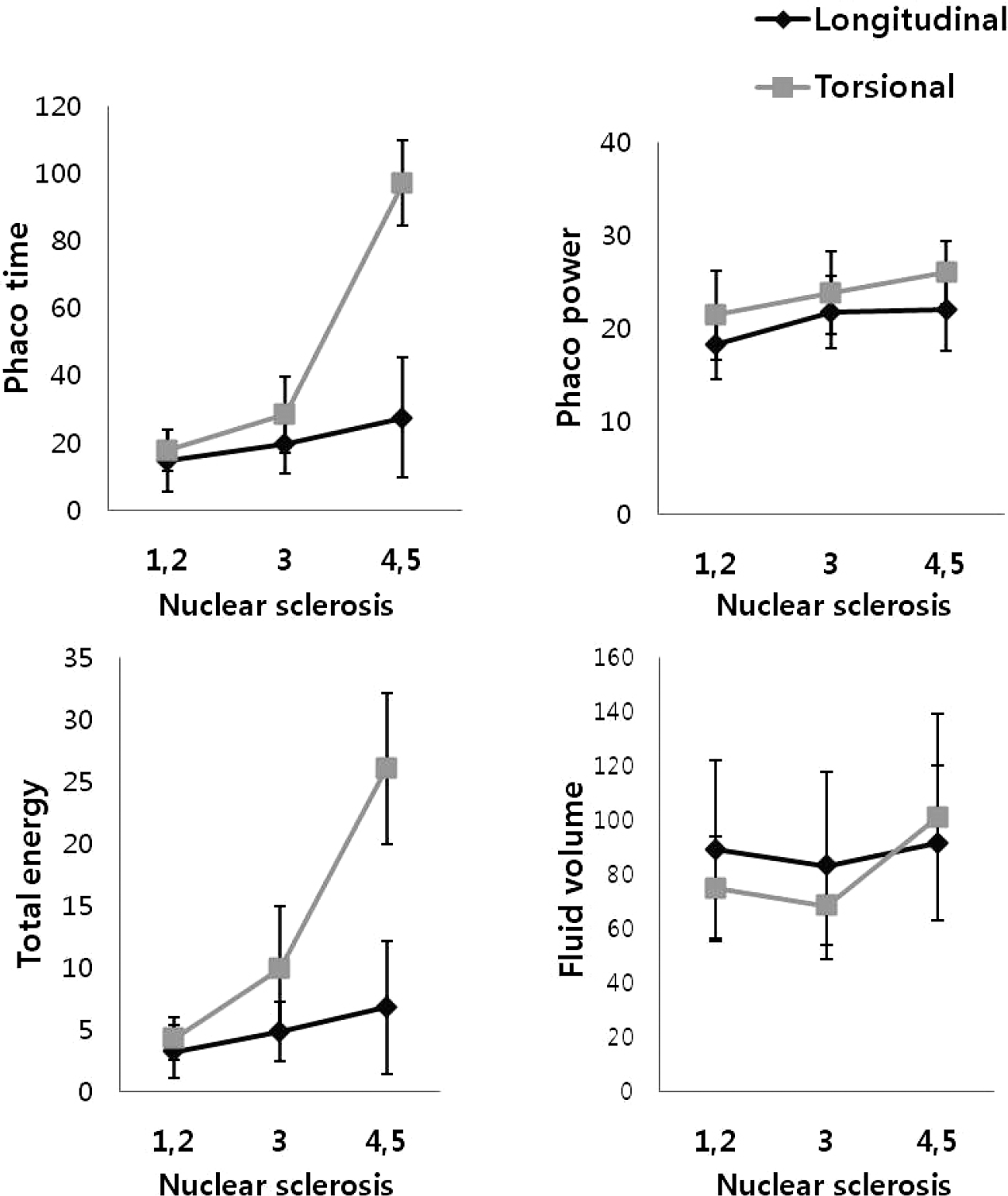
Table 1.
Characteristics of the subjects in the two groups
| Longitudinal (n = 35) | Torsional (n = 39) | p-value∗ | |
|---|---|---|---|
| Sex (n) | |||
| Male:female | 16:19 | 17:22 | 0.391 |
| Eye (n) | |||
| Right:left | 14:21 | 23:16 | 0.091 |
| Age (years) | 70.06 ± 11.99 | 67.21 ± 14.07 | 0.361 |
| NS grade (n, %) | |||
| Grade 1-2 Grade 3 | 11 (31.43)13 (37.14) | 10 (25.64) 18 (46.15) | 0.381 |
| Grade 4-5 | 11 (31.43) | 11 (28.21) | |
Table 2.
Comparison of BCVA, CCT, ECD and endothelial cell loss between the two groups
| Longitudinal (n = 35) | Torsional (n = 39) | p-value∗ | |
|---|---|---|---|
| BCVA (log MAR) | |||
| Preoperative | 0.73 ± 0.54 | 0.92 ± 0.63 | 0.222 |
| Postoperative | |||
| 1 month | 0.15 ± 0.24 | 0.21 ± 0.35 | 0.885 |
| 3 months | 0.15 ± 0.28 | 0.17 ± 0.28 | 0.819 |
| CCT (μ m) | |||
| Preoperative | 536.28 ± 38.86 | 528.65 ± 34.73 | 0.430 |
| Postoperative | |||
| 1 day | 565.89 ± 53.25 | 566.41 ± 62.48 | 0.906 |
| 1 week | 544.46 ± 45.12 | 542.74 ± 53.59 | 0.992 |
| 1 month | 534.43 ± 42.85 | 536.41 ± 46.81 | 0.962 |
| 3 months | 536.33 ± 44.09 | 533.60 ± 45.23 | 0.949 |
| ECD (cell/mm2) | |||
| Preoperative | 2,551 ± 329 | 2,432 ± 485 | 0.346 |
| Postoperative | |||
| 1 week | 2,197± 336 | 2,028 ± 621 | 0.355 |
| 1 month | 2,259 ± 372 | 1,902 ± 799 | 0.035 |
| 3 months | 2,190 ± 515 | 1,878 ± 817 | 0.075 |
| ECL (%) | |||
| Postoperative | |||
| 1 week | 12.96 ± 12.37 | 14.25 ± 19.09 | 0.729 |
| 1 month | 12.48 ± 11.14 | 16.33 ± 21.05 | 0.410 |
| 3 months | 13.17 ± 11.13 | 17.40 ± 20.96 | 0.326 |
Table 3.
Operative parameters in the two groups
| Longitudinal (n = 35) | Torsional (n = 39) | p-value§ | |
|---|---|---|---|
| Ultrasound time (second) | |||
| Mild cataract∗ | 14.90 ± 9.17 | 17.88 ± 6.22 | 0.445 |
| Moderate cataract† | 19.55 ± 8.78 | 28.38 ± 11.21 | 0.005 |
| Hard cataract‡ | 27.50 ± 17.77 | 97.08 ± 12.67 | 0.028 |
| Average phaco power (%) | |||
| Mild cataract∗ | 18.20 ± 3.74 | 21.38 ± 4.81 | 0.097 |
| Moderate cataract† | 21.73 ± 3.82 | 23.76 ± 4.46 | 0.209 |
| Hard cataract‡ | 22.00 ± 4.50 | 26.00 ± 3.38 | 0.027 |
| Total phaco energy | |||
| Mild cataract∗ | 3.20 ± 2.10 | 4.25 ± 1.75 | 0.274 |
| Moderate cataract† | 4.82 ± 2.44 | 9.90 ± 5.02 | 0.178 |
| Hard cataract‡ | 6.80 ± 5.37 | 26.08 ± 9.09 | 0.008 |
| Fluid volume used (mL) | |||
| Mild cataract∗ | 89.13 ± 32.86 | 74.83 ± 19.34 | 0.364 |
| Moderate cataract† | 83.25 ± 34.52 | 68.44 ± 14.24 | 0.458 |
| Hard cataract‡ | 91.75 ± 28.43 | 101.17 ± 38.31 | 0.561 |
Table 4.
Postoperative corneal edema in the two groups
| POD | Corneal edema grade | ||||||
|---|---|---|---|---|---|---|---|
| None∗ | Trace† | Mild‡ | Moderate§ | Severe∏ | p-value# | ||
| 1 day | Longitudinal | 20 | 9 | 4 | 1 | 0 | 0.532 |
| Torsional | 18 | 9 | 7 | 3 | 0 | ||
| 1 week | Longitudinal | 33 | 1 | 0 | 0 | 0 | 0.066 |
| Torsional | 33 | 3 | 1 | 0 | 0 | ||
| 1 month | Longitudinal | 34 | 0 | 0 | 0 | 0 | 1.000 |
| Torsional | 37 | 0 | 0 | 0 | 0 | ||
| 3 months | Longitudinal | 34 | 0 | 0 | 0 | 0 | 1.000 |
| Torsional | 37 | 0 | 0 | 0 | 0 | ||
Table 5.
Postoperative changes of BCVA, CCT, ECD, ECL and corneal edema in cases with hard cataract
| Hard cataract | |||
|---|---|---|---|
| Longitudinal (n = 11) | Torsional (n = 11) | p-value∗ | |
| BCVA (log MAR) | |||
| Preoperative | 1.07 ± 0.61 | 1.34 ± 0.61 | 0.313 |
| Postoperative | |||
| 1 month | 0.29 ± 0.34 | 0.26 ± 0.40 | 0.902 |
| 3 months | 0.28 ± 0.41 | 0.30 ± 0.52 | 0.840 |
| CCT (μ m) | |||
| Preoperative | 538.75 ± 28.54 | 529.11 ± 20.81 | 0.435 |
| Postoperative | |||
| 1 day | 589.14 ± 36.72 | 608.57 ± 79.67 | 0.569 |
| 1 week | 568.00 ± 37.26 | 547.75 ± 22.26 | 0.353 |
| 1 month | 539.20 ± 41.37 | 532.75 ± 17.61 | 0.781 |
| 3 months ECD (cell/mm2) | 538.67 ± 37.28 | 533.04 ± 37.32 | 0.661 |
| Preoperative | 2,490 ± 250 | 2,430± 430 | 0.694 |
| Postoperative | |||
| 1 week | 2,158 ± 300 | 1,496 ± 717 | 0.075 |
| 1 month | 2,153 ± 441 | 1,296 ± 564 | 0.006 |
| 3 months | 2,107 ± 304 | 1,321 ± 828 | 0.033 |
| ECL (%) | |||
| Postoperative | |||
| 1 week | 13.56 ± 11.59 | 36.33 ± 30.92 | 0.136 |
| 1 month | 15.00 ± 12.64 | 45.86 ± 20.29 | 0.003 |
| 3 months | 16.33 ± 15.50 | 38.71 ± 26.49 | 0.042 |
| Corneal edema | |||
| Postoperative | |||
| 1 day | 1.18 ± 0.87 | 1.73 ± 1.01 | 0.190 |
| 1 week | 0.00 ± 0.00 | 0.56 ± 0.73 | 0.020 |
| 1 month | 0.00 ± 0.00 | 0.00 ± 0.00 | 1.000 |
| 3 months | 0.00 ± 0.00 | 0.00 ± 0.00 | 1.000 |
Table 6.
Comparison of surgically induced astigmatism
| POD | Total (n = 74) | Hard cataract (n = 22) | ||||||
|---|---|---|---|---|---|---|---|---|
| Longitudinal | Torsional | p-value∗ | Longitudinal | Torsional | p-value† | |||
| 1 day | 0.80 ± 0.51 | 0.97 ± 0.61 | 0.456 | 0.90 ± 0.75 | 1.06 ± 0.67 | 0.403 | ||
| 1 week | 0.76 ± 0.50 | 0.89 ± 0.64 | 0.383 | 0.81 ± 0.70 | 0.94 ± 0.74 | 0.324 | ||
| 1 month | 0.53 ± 0.44 | 0.60 ± 0.50 | 0.447 | 0.61 ± 0.51 | 0.68 ± 0.53 | 0.536 | ||
| 3 months | 0.51 ± 0.47 | 0.56 ± 0.44 | 0.549 | 0.57 ± 0.67 | 0.64 ± 0.61 | 0.281 | ||
Table 7.
Associations of endothelial cell loss (%) at post-operative 1 month with predisposing factors by multiple linear regression analyses (adjusted R2 =0.535)
Table 8.
Comparison of parameters between the two groups in 15 patients with bilateral cataract surgery
| Longitudinal | Torsional | p-value∗ | |
|---|---|---|---|
| Operative parameter | |||
| Ultrasound time (second) | 19.43 ± 17.38 | 38.21 ± 28.51 | 0.045 |
| Average phaco power (%) | 20.00 ± 4.11 | 25.14 ± 4.54 | 0.003 |
| Total phaco energy | 4.86 ± 4.89 | 14.36 ± 15.65 | 0.026 |
| Fluid volume used (mL) | 85.63 ± 24.75 | 88.38 ± 34.81 | 0.918 |
| BCVA (log MAR) | |||
| Preoperative | 0.63 ± 0.49 | 0.65 ± 0.51 | 0.611 |
| Postoperative | |||
| 1 month | 0.15 ± 0.28 | 0.16 ± 0.32 | 0.769 |
| 3 months | 0.14 ± 0.32 | 0.17 ± 0.32 | 0.860 |
| CCT (μ m) | |||
| Preoperative | 533.94 ± 30.19 | 527.94 ± 40.06 | 0.247 |
| Postoperative | |||
| 1 day | 558.00 ± 36.40 | 554.44 ± 59.15 | 0.721 |
| 1 week | 534.62 ± 34.25 | 523.92 ± 47.89 | 0.244 |
| 1 month | 539.60 ± 45.41 | 526.10 ± 56.91 | 0.207 |
| 3 months | 537.45 ± 42.26 | 526.15 ± 50.32 | 0.268 |
| ECL (%) | |||
| Postoperative | |||
| 1 day | 4.71 ± 4.60 | 7.71 ± 10.28 | 0.266 |
| 1 week | 11.73 ± 10.42 | 15.73 ± 21.52 | 0.505 |
| 1 month | 11.77 ± 8.70 | 18.31 ± 22.02 | 0.228 |
| 3 months | 14.25 ± 8.24 | 17.71 ± 20.21 | 0.351 |
| Corneal edema | |||
| Postoperative | |||
| 1 day | 0.63 ± 0.86 | 0.75 ± 0.88 | 0.580 |
| 1 week | 0.00 ± 0.00 | 0.07 ± 0.25 | 0.334 |
| 1 month | 0.00 ± 0.00 | 0.00 ± 0.00 | 1.000 |
| 3 months | 0.00 ± 0.00 | 0.00 ± 0.00 | 1.000 |
 XML Download