

 PDF
PDF ePub
ePub Citation
 Print
Print


Abstract
Purpose:
To evaluate the long-term clinical effect and safety of YK-KC lens® (LucidKorea Ltd., Seoul, Korea) for keratoconus. Methods: In this study we investigated 152 keratoconic eyes fitted with YK-KC lens® and followed up for at least 5 years. We as-sessed retrospectively self-reported patient comfort, best corrected visual acuity, corneal topographic indices before and after contact lens fitting and complications during contact lens wearing.
Results:
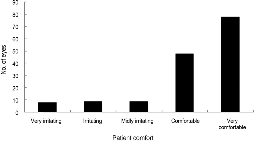
The study included 57 male and 40 female patients with a mean age of 28.6 ± 8.5 years. The mean follow-up was 8.0 ± 2.9 years. Regarding lens comfort, 126 eyes (82.9%) showed self-reported comfort for YK-KC lenses®. The mean best cor-rected visual acuity (log MAR) improved from 0.49 ± 0.42 before lens fitting to 0.19 ± 0.27 after lens wearing, which was statisti-cally significant ( p < 0.001). Based on the keratometric values, after contact lens fitting both Simulated keratometry (Sim K) max and Sim K min tended to be steeper, but these differences were not statistically significant ( p = 0.66 and 0.11, respectively). There were no statistically significant differences between the values before and after fitting with respect to the astigmatic pow-ers ( p = 0.22). Complications observed included punctate or coalesced epithelial corneal staining in 24 eyes (16%), however, persistent full-thickness epithelial defect was not observed.
Go to : 
References
1. Rabinowitz YS. Keratoconus. Surv Ophthalmol. 1998; 42:297–319.

2. Krachmer JH, Feder RS, Belin MW. Keratoconus and related non-inflammatory corneal thinning disorders. Surv Ophthalmol. 1984; 28:293–322.
3. Wollensak G, Spoerl E, Seiler T. Riboflavin/ultraviolet-a-induced collagen crosslinking for the treatment of keratoconus. Am J Ophthalmol. 2003; 135:620–7.
4. Choi SW, Choae WS, Her J. Intrastromal corneal ring segments (Keraring[R]) implantation for the correction of keratoconus. J Korean Ophthalmol Soc. 2011; 52:277–84.
5. Kim MK, Lee JH. Long-term outcome of graft rejection after pene-trating keratoplasty. J Korean Ophthalmol Soc. 1997; 38:1553–60.
6. Kim KH, Ahn K, Chung ES, Chung TY. Comparison of deep ante-rior lamellar keratoplasty and penetrating keratoplasty for keratoconus. J Korean Ophthalmol Soc. 2008; 49:222–9.
7. Kwak NH, Kim MS, Kim JH. Clinical evaluation of keratoconus. J Korean Ophthalmol Soc. 1989; 30:351–6.
8. Kang YS, Park YK, Lee JS. . The effect of the YK lens in keratoconus. Ophthalmic Physiol Opt. 2010; 30:267–73.
9. Yang KM, Kim MK, Lee JL, Kim CS. Effect of the spheric mul-ti-curve lens on changes of corneal topography and endothelial cell in keratoconus. J Korean Ophthalmol Soc. 2004; 45:1427–37.
10. Lee JL, Kim MK. The analysis of management of keratoconus us-ing contact Lens in Koreans. J Korean Ophthalmol Soc. 2004; 45:725–31.
11. Wagner H, Barr JT, Zadnik K. Collaborative Longitudinal Evaluation of Keratoconus (CLEK) Study: methods and findings to date. Cont Lens Anterior Eye. 2007; 30:223–32.
12. Piñero DP, Alió JL, Alesón A. . Corneal volume, pachymetry, and correlation of anterior and posterior corneal shape in sub-clinical and different stages of clinical keratoconus. J Cataract Refract Surg. 2010; 36:814–25.
13. Edrington TB, Szczotka LB, Begley CG. . Repeatability and agreement of two corneal-curvature assessments in keratoconus: keratometry and the first definite apical clearance lens (FDACL) CLEK Study Group. Collaborative Longitudinal Evaluation of Keratoconus. Cornea. 1998; 17:267–77.
14. Buxton JN, Keates RH, Hoefle FB. The contact lens correction of keratoconus. Dabezies O, editor. Contact lenses: The CLAO guide to basic science and clinical practice: update 1. 1st ed. Orlando: Grune & Stratton;1986. p. 1–55.
15. Choi JA, Kim MS. Progression of keratoconus by longitudinal as-sessment with corneal topography. Invest Ophthalmol Vis Sci. 2012; 53:927–35.
16. Edrington TB, Gundel RE, Libassi DP. . Variables affecting rigid contact lens comfort in the collaborative longitudinal evalua-tion of keratoconus (CLEK) study. Optom Vis Sci. 2004; 81:182–8.
17. Barr JT, Gordon MO, Zadnik K. . Photodocumentation of cor-neal scarring Collaborative Longitudinal Evaluation of Keratoconus Study Group. J Refract Surg. 1996; 12:492–500.
18. Caroline PJ, McGuire JR, Doughman DJ. Preliminary report on a new contact lens design for keratoconus. Contact Intraocul Lens. 1978; 4:69–73.
19. Gupta R, Sinha R, Singh P. . Rose-K versus Soper contact lens in keratoconus: a randomized comparative trial. Middle East Afr J Ophthalmol. 2014; 21:50–5.
20. Betts AM, Mitchell GL, Zadnik K. Visual performance and com-fort with the Rose K lens for keratoconus. Optom Vis Sci. 2002; 79:493–501.
21. Patrick JC, Craig WN, Mark PA. The latest lens design for keratoconus. Spectrum. 1997; 8:36–41.
22. Romero-Jiménez M, Santodomingo-Rubido J, González-Méijome JM. An assessment of the optimal lens fit rate in keratoconus sub-jects using three-point-touch and apical touch fitting approaches with the rose K2 lens. Eye Contact Lens. 2013; 39:269–72.
23. McMonnies CW. The biomechanics of keratoconus and rigid con-tact lenses. Eye Contact Lens. 2005; 31:80–92.
24. Wilson SE, Kim WJ. Keratocyte apoptosis: implications on cor-neal wound healing, tissue organization and disease. Invest Ophthalmol Vis Sci. 1998; 39:220–6.
25. Shin DB, Han NS, Kim MK. . Effect of the aspheric RGP lens on corneal topography and endothelial cell in keratoconus. J Korean Ophthalmol Soc. 2004; 45:396–404.
26. Barnett M, Mannis MJ. Contact lenses in the management of keratoconus. Cornea. 2011; 30:1510–6.
27. Smolek MK, Klyce SD. Is keratoconus a true ectasia? An evalua-tion of corneal surface area. Arch Ophthalmol. 2000; 118:1179–86.
28. Jain AK, Sukhija J. Rose-K contact lens for keratoconus. Indian J Ophthalmol. 2007; 55:121–5.
Go to :
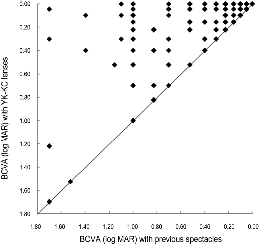 | Figure 2.Change in best-corrected visual activity (BCVA) be-tween spectacles and YK-KC lenses®. Points lying above the solid 45° line correspond to eyes with improved vision with YK-KC lenses®. |
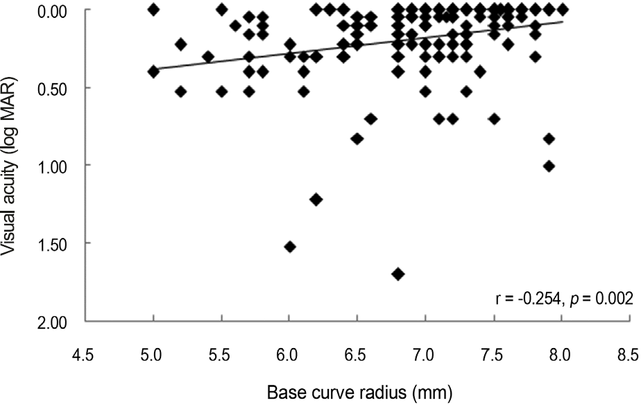 | Figure 3.Distribution of visual acuity when wearing YK-KC lens®, plotted against the base curve radius of the lens. Better visual acuity was achieved when contact lenses with flatter base curve radii were fitted. |
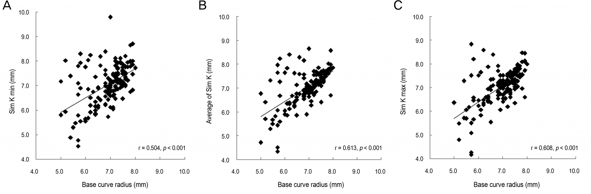 | Figure 4.Correlations between topographic indices and contact lens base curve radius (BCR). 'r' means Pearson's correlation co-efficient between Sim K min and BCR (A), between average of Sim K and BCR (B), between Sim K max and BCR (C), and a rela-tionship is considered to be statistically significant if p < 0.05. Sim K = simulated keratometry; min = minimum. |
 XML Download