Abstract
Purpose
We used computed tomography (CT) scans to describe normal Korean lacrimal gland volume and lacrimal gland size and then examined their correlations with patient age.
Methods
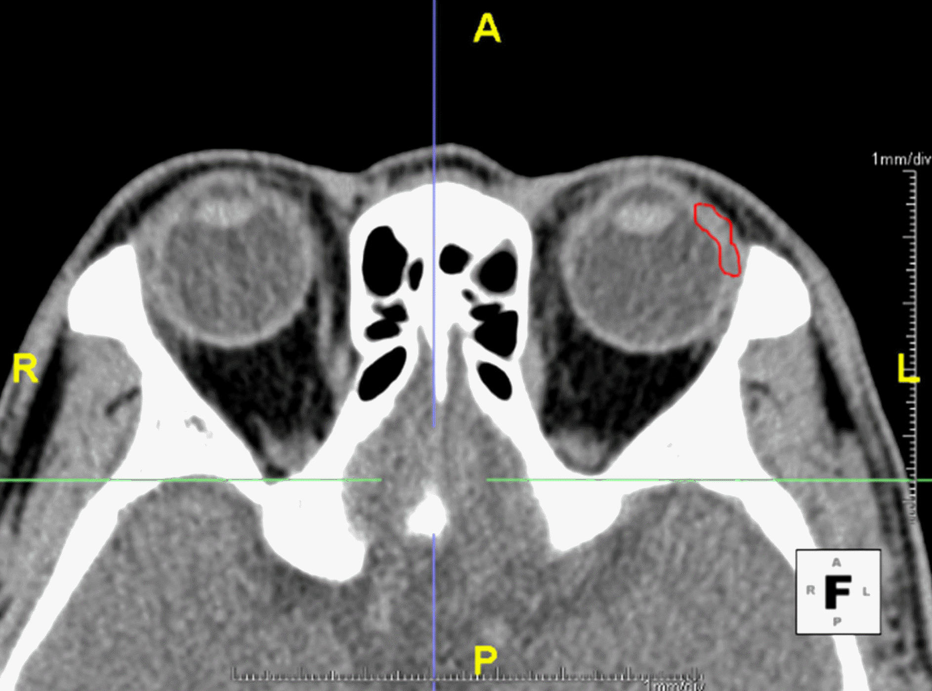
CT scans were obtained in 213 orbits of 111 patients who underwent CT from January to August of 2013. Aquarius iNtuition (TeraRecon, Foster City, CA, USA) software was used to outline the lacrimal gland in consecutive axial slices and to calculate the volume.
Results
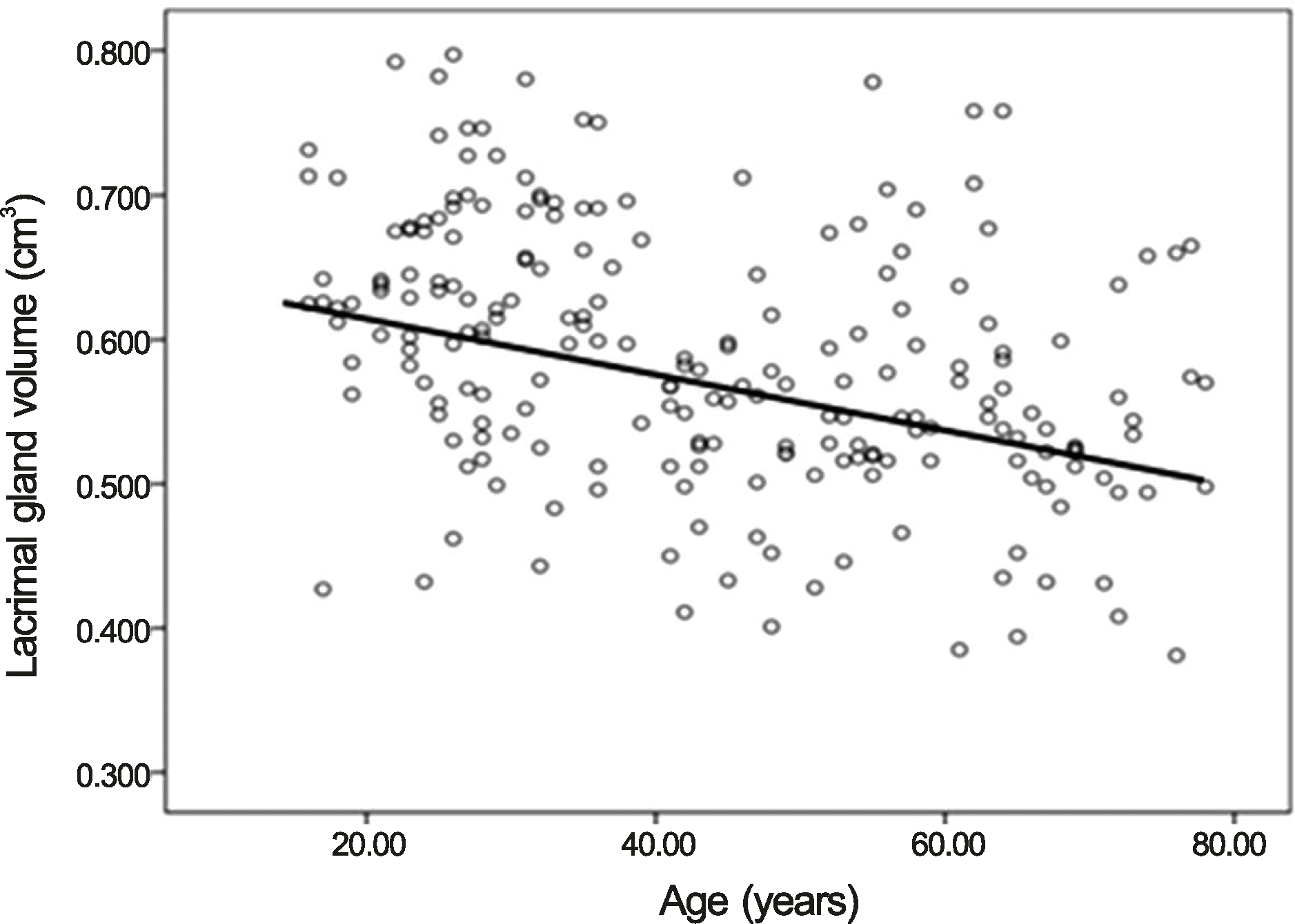
The mean volume of the lacrimal gland was 0.589 cm3 in right orbits (SD = 0.090), 0.583 cm3 in left orbits (SD = 0.289), 0.596 cm3 in males (SD = 0.083), and 0.575 cm3 in females (SD = 0.094). There was no significant difference in mean lacrimal gland volume according to laterality (p = 0.614) or sex (p = 0.102) (2-sample t-tests). We investigated mean lacrimal gland volume in 3 age groups. Mean lacrimal gland volume was 0.630 cm3 (SD = 0.080) for the 20 to 40 year old group, 0.553 cm3 (SD = 0.734) for the 41 to 60 year old group, and 0.544 cm3 (SD = 0.885) for the older than 60 years old group. There was an inverse relationship between gland volume and age (Pearson r = −0.384, p = 0.00).
References
1. Chandler JW, Gillette TE. Immunologic defense mechanisms of the ocular surface. Ophthalmology. 1983; 90:585–91.

2. Obata H. Anatomy and histopathology of the human lacrimal gland. Cornea. 2006; 25(10 Suppl 1):S82–9.

3. Harris MA, Realini T, Hogg JP, Sivak-Callcott JA. CT dimensions of the lacrimal gland in Graves orbitopathy. Ophthal Plast Reconstr Surg. 2012; 28:69–72.

4. Wall J. Extrathyroidal manifestations of Graves' disease. J Clin Endocrinol Metab. 1995; 80:3427–9.

5. Nugent RA, Belkin RI, Neigel JM. . Graves orbitopathy: correlation of CT and clinical findings. Radiology. 1990; 177:675–82.

6. Tamboli DA, Harris MA, Hogg JP. . Computed tomography dimensions of the lacrimal gland in normal Caucasian orbits. Ophthal Plast Reconstr Surg. 2011; 27:453–6.

7. Ueno H, Ariji E, Izumi M. . MR imaging of the lacrimal gland. Age-related and gender-dependent changes in size and structure. Acta Radiol. 1996; 37:714–9.
8. Bingham CM, Castro A, Realini T. . Calculated CT volumes of lacrimal glands in normal Caucasian orbits. Ophthal Plast Reconstr Surg. 2013; 29:157–9.

9. Bingham CM, Harris MA, Realini T. . Calculated computed tomography volumes of lacrimal glands and comparison to clinical findings in patients with thyroid eye disease. Ophthal Plast Reconstr Surg. 2014; 30:116–8.

10. Landis JR, Koch GG. The measurement of observer agreement for categorical data. Biometrics. 1977; 33:159–74.

11. Avetisov SE, Kharlap SI, Markosian AG. . [Ultrasound spatial clinical analysis of the orbital part of the lacrimal gland in health]. Vestn Oftalmol. 2006; 122:14–6.
12. Obata H, Yamamoto S, Horiuchi H, Machinami R. Histopathologic study of human lacrimal gland. Statistical analysis with special reference to aging. Ophthalmology. 1995; 102:678–86.
13. Prager A. [Macroscopic and microscopic investigations on senile atrophy of the lacrimal gland (preliminary report)]. Bibl Ophthalmol. 1966; 69:146–58.
14. Lee JS, Lee H, Kim JW. . Computed tomographic dimensions of the lacrimal gland in healthy orbits. J Craniofac Surg. 2013; 24:712–5.

15. Damato BE, Allan D, Murray SB, Lee WR. Senile atrophy of the human lacrimal gland: the contribution of chronic inflammatory disease. Br J Ophthalmol. 1984; 68:674–80.

16. Roen JL, Stasior OG, Jakobiec FA. Aging changes in the human lacrimal gland: role of the ducts. CLAO J. 1985; 11:237–42.
Figure 1.
Axial CT scan with the outlined entire lacrimal gland using Aquaris intuition software was presented. The palpebral and orbital portion of lacrimal gland were outlined using free hand technique in all consecutive axial slices. A = anterior aspect; P = posterior aspect; R = right; L = left.

Figure 2.
Scatter plot and correlation between age (years) and average lacrimal gland volume (cm3) in normal Korean. There was an inverse relationship between gland volume and age in normal Korean. Pearson r = -0.384 (p = 0.00).

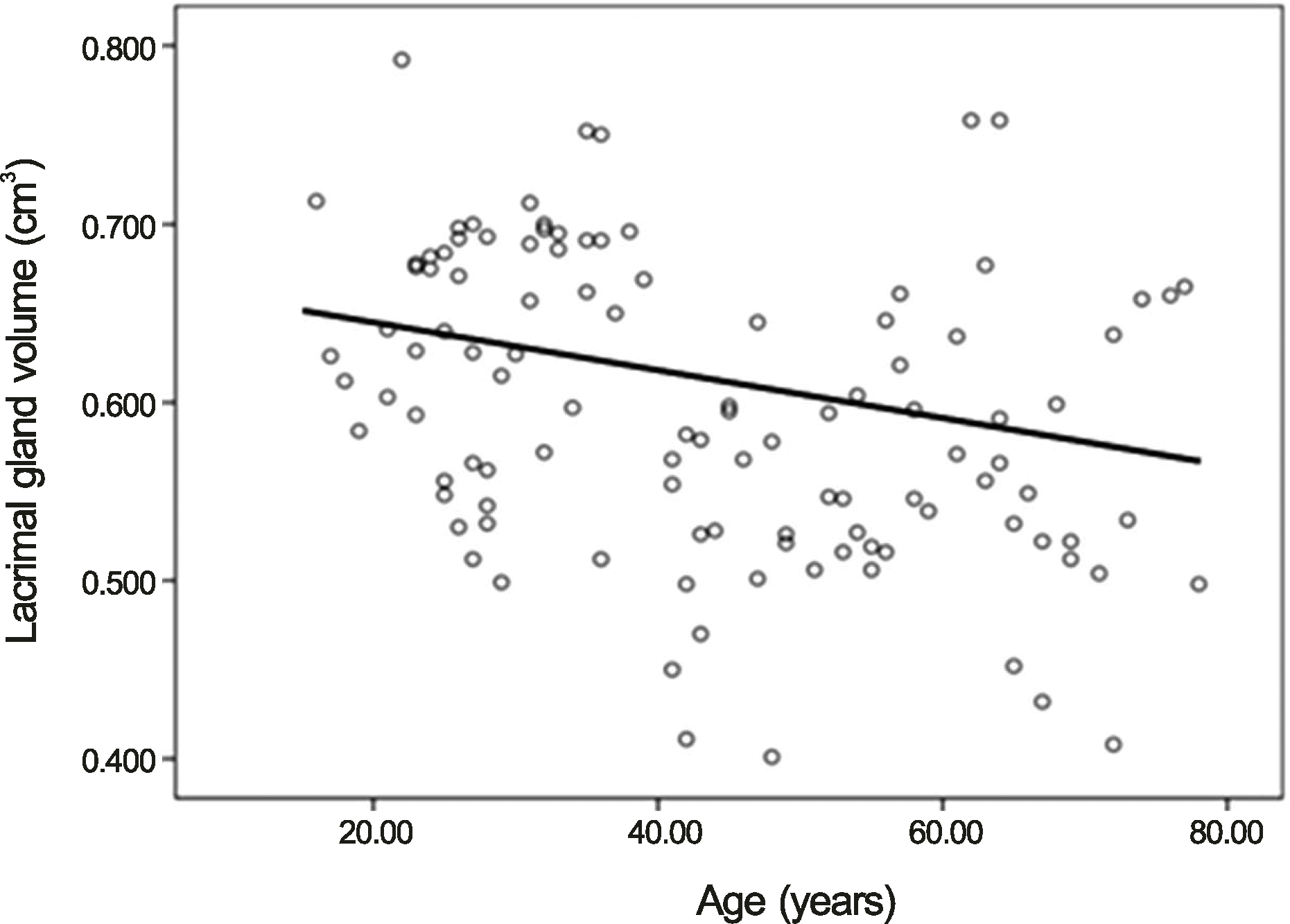
Figure 3.
Scatter plot and correlation between age (years) and average lacrimal gland volume (cm3) in normal Korean male. There was an inverse relationship between gland volume and age in normal Korean male. Pearson r = -0.348 (p = 0.00).

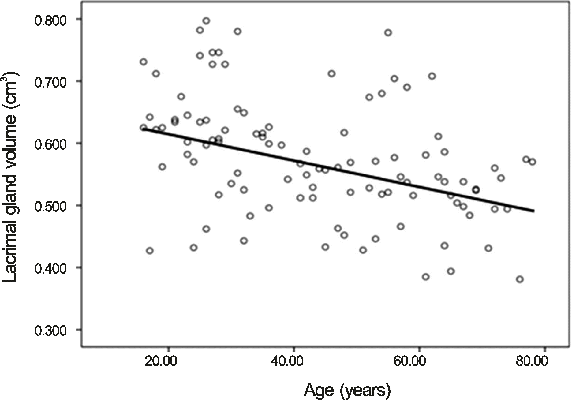
Figure 4.
Scatter plot and correlation between age (years) and average lacrimal gland volume (cm3) in normal Korean female. There was an inverse relationship between gland volume and age in normal Korean female. Pearson r = -0.422 (p = 0.00).


 PDF
PDF ePub
ePub Citation
 Print
Print


 XML Download