Abstract
Purpose
To evaluate the efficacy of 0.05% cyclosporine A on tear film parameters and corneal aberration after cataract surgery.
Methods
Patients who underwent cataract surgery were divided into 2 groups. Patients in Group I (23 eyes) were treated with cyclosporine A from 1 week before surgery to 3 months after surgery. Patients in Group II (24 eyes) underwent surgery without cyclosporine treatment. Tear film break-up time (BUT), Schirmer's test I, Oxford scheme, Ocular surface disease index (OSDI), and corneal aberrations were evaluated before surgery and at 1 and 3 months after surgery.
Results
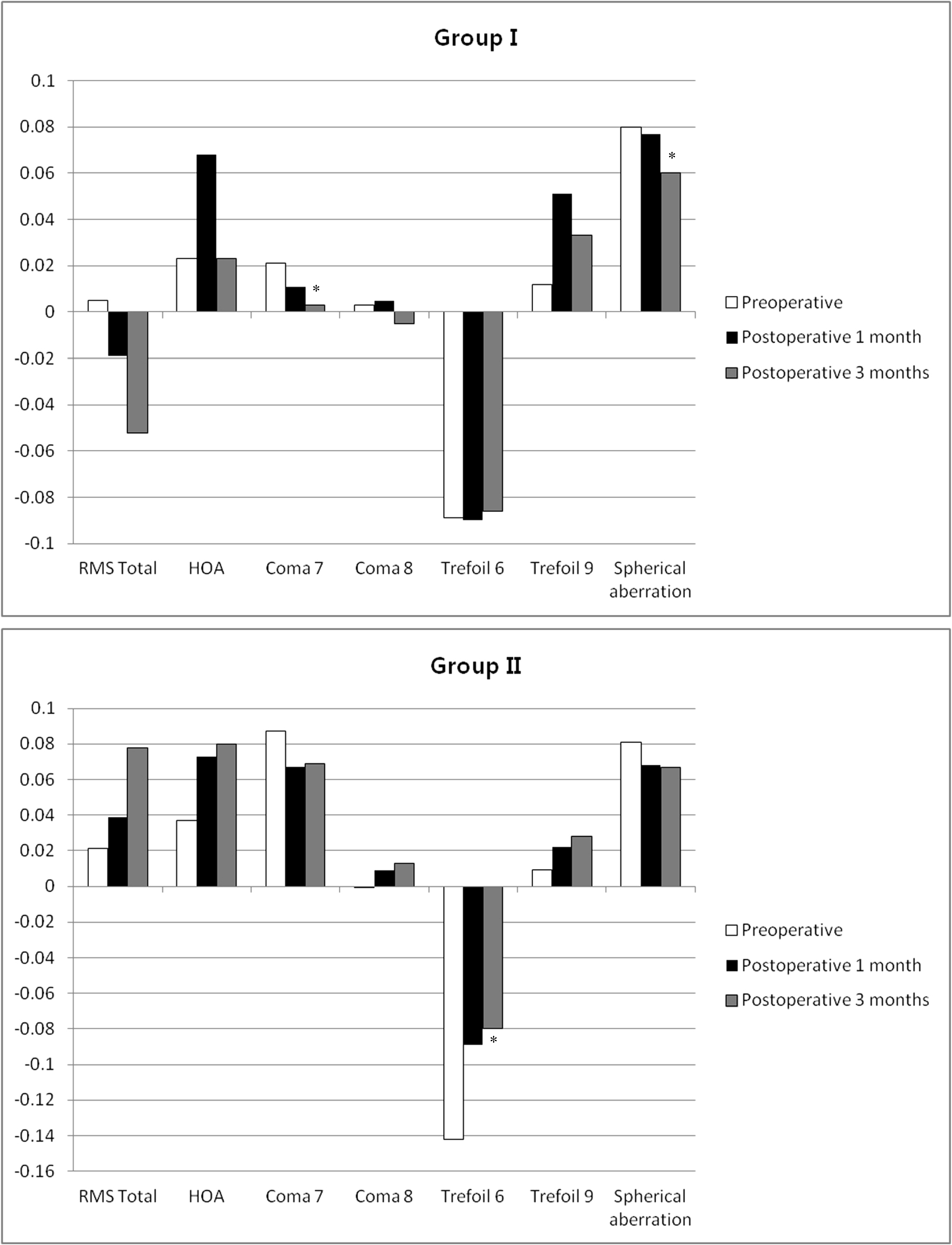
In Group I, BUT was significantly improved at 3 months (p = 0.026) after surgery compared with the preoperative value. OSDI decreased significantly at 1 (p = 0.033) and 3 months (p = 0.003) after surgery compared with the preoperative value. However, there were no significant differences between preoperative and postoperative values of BUT and OSDI in Group II. Schirmer's test results and the Oxford scheme were not significantly changed in either group. Preoperative root mean square (RMS) total values were not different between the 2 groups, but was different at postoperative 3 months (p = 0.015). Group I had a significantly lower value for total RMS than Group II. In Group I, Coma 7 (Z3-1) (p = 0.018) and spherical aberration (Z40) (p = 0.031) were significantly decreased after surgery. In Group II, Trefoil 6 (Z3-3) (p = 0.033) was significantly increased after surgery.
Go to : 
References
1. Apostol S, Filip M, Dragne C, Filip A. Dry eye syndrome. Etiological and therapeutic aspects. Oftalmología. 2003; 59:28–31.
2. Sheppard JD. Guidelines for the treatment of chronic dry eye disease. Manag Care. 2003; 12:20–5.
3. Li XM, Hu L, Hu J, Wang W. Investigation of dry eye disease and analysis of the pathogenic factors in patients after cataract surgery. Cornea. 2007; 26:S16–20.

4. Albietz JM, Lenton LM. Management of the ocular surface and tear film before, during, and after laser in situ keratomileusis. J Refract Surg. 2004; 20:62–71.

5. Begley CG, Caffery B, Nichols K, et al. Results of a dry eye questionnaire from optometric practices in North America. Adv Exp Med Biol. 2002; 506:1009–16.

6. Khanal S, Tomlinson A, Esakowitz L, et al. Changes in corneal sensitivity and tear physiology after phacoemulsification. Ophthalmic Physiol Opt. 2008; 28:127–34.

7. Oh T, Jung Y, Chang D, et al. Changes in the tear film and ocular surface after cataract surgery. Jpn J Ophthalmol. 2012; 56:113–8.

8. Albarran C, Pons AM, Lorente A, et al. Influence of the tear film on optical quality ofthe eye. Cont Lens Anterior Eye. 1997; 20:129–35.
9. Denoyer A, Rabut G, Baudouin C. Tear film aberration dynamics and vision-related quality of life in patients with dry eye disease. Ophthalmology. 2012; 119:1811–8.

10. Choi SH, Shin YI. Changes in higher order aberration according to tear-film instability analyzed by continuous measurement using wavefront. J Korean Ophthalmol Soc. 2012; 53:1076–80.

11. Pflugfelder SC, Tseng SC, Sanabria O, et al. Evaluation of subjective assessments and objective diagnostic tests for diagnosing tear-film disorders known to cause ocular irritation. Cornea. 1998; 17:38–56.

12. Byun YS, Jeon EJ, Chung SK. Clinical effect of cyclosporine 0.05% eye drops in dry eye syndrome patients. J Korean Ophthalmol Soc. 2008; 49:1583–8.

13. Hom MM. Use of cyclosporine 0.05% ophthalmic emulsion for contact lens-intolerant patients. Eye Contact Lens. 2006; 32:109–11.

14. Byun YS, Rho CR, Cho K, et al. Cyclosporine 0.05% ophthalmic emulsion for dry eye in Korea: a prospective, multicenter, open-label, surveillance study. Korean J Ophthalmol. 2011; 25:369–74.

15. Donnenfeld ED, Solomon R, Roberts CW, et al. Cyclosporine 0.05% to improve visual outcomes after multifocal intraocular lens implantation. J Cataract Refract Surg. 2010; 36:1095–100.

16. Chung YW, Oh TH, Chung SK. The effect of topical cyclosporine 0.05% on dry eye after cataract surgery. Korean J Ophthalmol. 2013; 27:167–71.

17. O'Brien PD, Collum LM. Dry eye: diagnosis and current treatment strategies. Curr Allergy Asthma Rep. 2004; 4:314–9.
19. Moon SJ, Lee DJ, Lee KH. Induced astigmatism and high-order aberrations after 1.8-mm, 2.2-mm and 3.0-mm coaxial phacoemulsi- fication incisions. J Korean Ophthalmol Soc. 2011; 52:407–13.
Go to : 
 | Figure 1.Changes of corneal aberrations in each group during follow up. In Group I, Coma 7 (Z3-1), spherical aberration (Z40) were significantly decreased after surgery. In Group II, Trefoil 6 (Z3-3) was significantly increased after surgery. Asterisks mean p < 0.05 compared with preoperative values. RMS = root mean square; HOA = higher order aberrations. |
Table 1.
Patients demographics and variables of tear film in the 2 groups
| Group I (n = 23) | Group II (n = 24) | p-value* | |
|---|---|---|---|
| Age (years) | 67.7 ± 10.6 | 64.8 ± 9.6 | 0.141 |
| Male (%) | 20.5 | 15.9 | 0.683 |
| BUT (sec) | |||
| Preoperative | 5.9 ± 1.0 | 5.9 ± 1.1 | 0.778 |
| Postoperative 1 month | 6.2 ± 1.5 | 5.9 ± 1.3 | 0.527 |
| Postoperative 3 months | 7.0 ± 1.7† | 6.3 ± 1.8 | 0.178 |
| Schirmer test (mm/5 min) | |||
| Preoperative | 10.6 ± 2.4 | 12.0 ± 2.6 | 0.082 |
| Postoperative 1 month | 11.1 ± 2.4 | 11.2 ± 3.4 | 0.804 |
| Postoperative 3 months | 10.6 ± 1.5 | 10.9 ± 3.2 | 0.501 |
| OSDI (total score) | |||
| Preoperative | 20.0 ± 27.8 | 10.1 ± 24.3 | 0.051 |
| Postoperative 1 month | 8.9 ± 14.8† | 6.1 ± 9.3 | 0.783 |
| Postoperative 3 months | 2.4 ± 5.6† | 7.7 ± 10.6 | 0.092 |
| Oxford Scheme | |||
| Preoperative | 1.0 ± 0.6 | 0.8 ± 0.8 | 0.208 |
| Postoperative 1 month | 0.9 ± 0.8 | 0.7 ± 0.7 | 0.351 |
| Postoperative 3 months | 0.8 ± 0.6 | 0.6 ± 0.7 | 0.176 |
Table 2.
Differences of corneal aberrations between groups at each follow-up point
| Group I (n = 23) | Group II (n = 24) | p-value | |
|---|---|---|---|
| RMS Total | |||
| Preoperative | 0.005 ± 0.213 | 0.021 ± 0.214 | 0.741 |
| Postoperative 1 month | -0.019 ± 0.181 | 0.039 ± 0.182 | 0.050 |
| Postoperative 3 months | -0.052 ± 0.127 | 0.078 ± 0.173 | 0.015* |
| HOA | |||
| Preoperative | 0.023 ± 0.166 | 0.037 ± 0.032 | 0.774 |
| Postoperative 1 month | 0.068 ± 0.221 | 0.073 ± 0.159 | 0.966 |
| Postoperative 3 months | 0.023 ± 0.227 | 0.080 ± 0.136 | 0.911 |
| Coma 7, Z3-1 | |||
| Preoperative | 0.021 ± 0.111 | 0.087 ± 0.148 | 0.259 |
| Postoperative 1 month | 0.011 ± 0.177 | 0.067 ± 0.129 | 0.750 |
| Postoperative 3 months | 0.003 ± 0.092 | 0.069 ± 0.117 | 0.103 |
| Coma 8, Z31 | |||
| Preoperative | 0.003 ± 0.070 | -0.001 ± 0.091 | 0.856 |
| Postoperative 1 month | 0.005 ± 0.089 | 0.009 ± 0.088 | 0.496 |
| Postoperative 3 months | -0.005 ± 0.053 | 0.013 ± 0.057 | 0.491 |
| Trefoil 6, Z3-3 | |||
| Preoperative | -0.089 ± 0.086 | -0.142 ± 0.196 | 0.694 |
| Postoperative 1 month | -0.090 ± 0.111 | -0.089 ± 0.166 | 0.503 |
| Postoperative 3 months | -0.086 ± 0.200 | -0.080 ± 0.156 | 0.855 |
| Trefoil 9, Z3+3 | |||
| Preoperative | 0.012 ± 0.080 | 0.009 ± 0.092 | 0.371 |
| Postoperative 1 month | 0.051 ± 0.097 | 0.022 ± 0.089 | 0.099 |
| Postoperative 3 months | 0.033 ± 0.060 | 0.028 ± 0.081 | 0.509 |
| Spherical aberration, Z40 | |||
| Preoperative | 0.080 ± 0.024 | 0.081 ± 0.046 | 0.580 |
| Postoperative 1 month | 0.077 ± 0.042 | 0.068 ± 0.031 | 0.209 |
| Postoperative 3 months | 0.060 ± 0.028 | 0.067 ± 0.027 | 0.643 |


 PDF
PDF ePub
ePub Citation
 Print
Print


 XML Download