Abstract
Purpose
To report a case of peripheral ulcerative keratitis and scleritis in a patient with pustular psoriasis.
Case summary
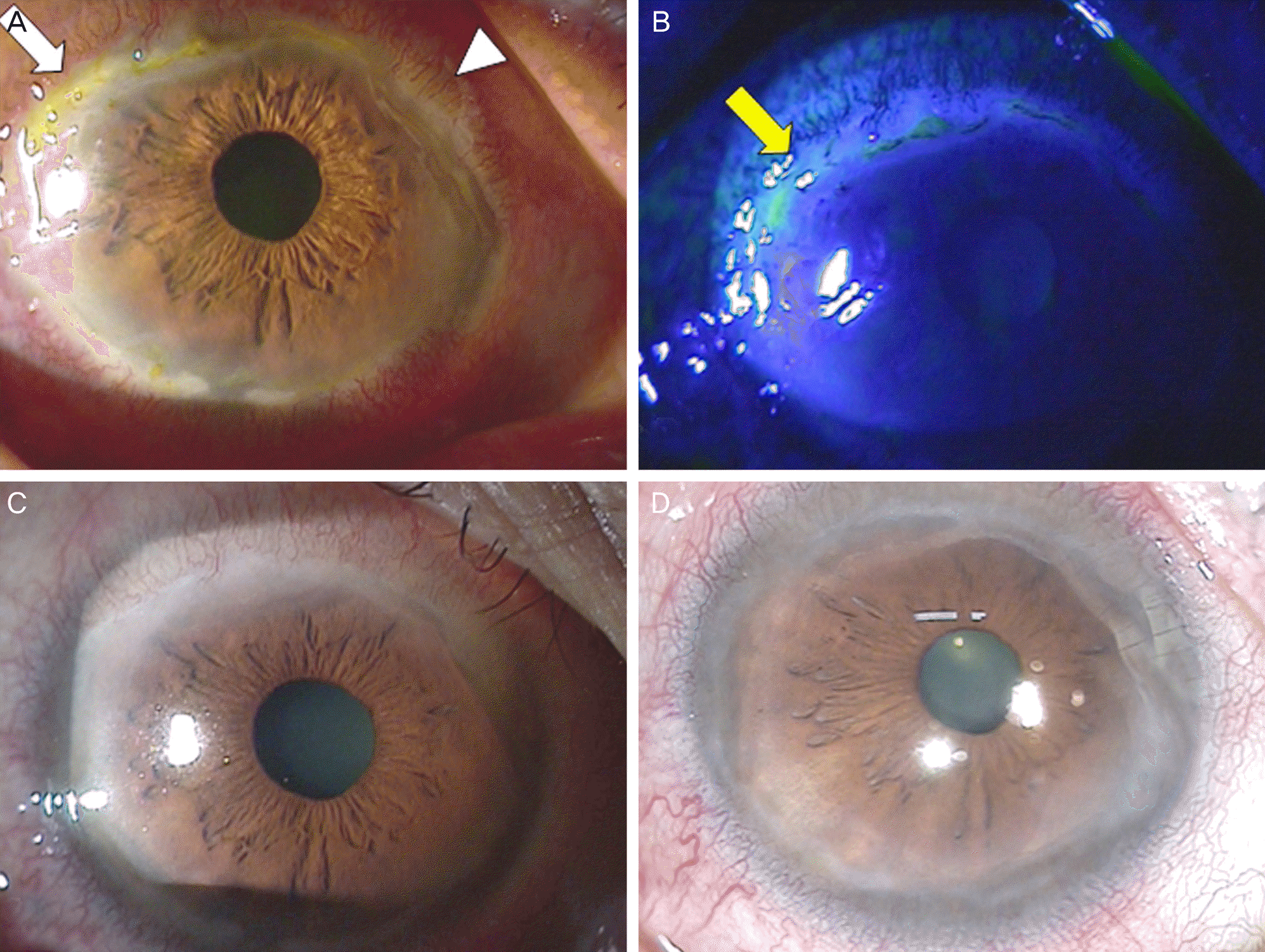
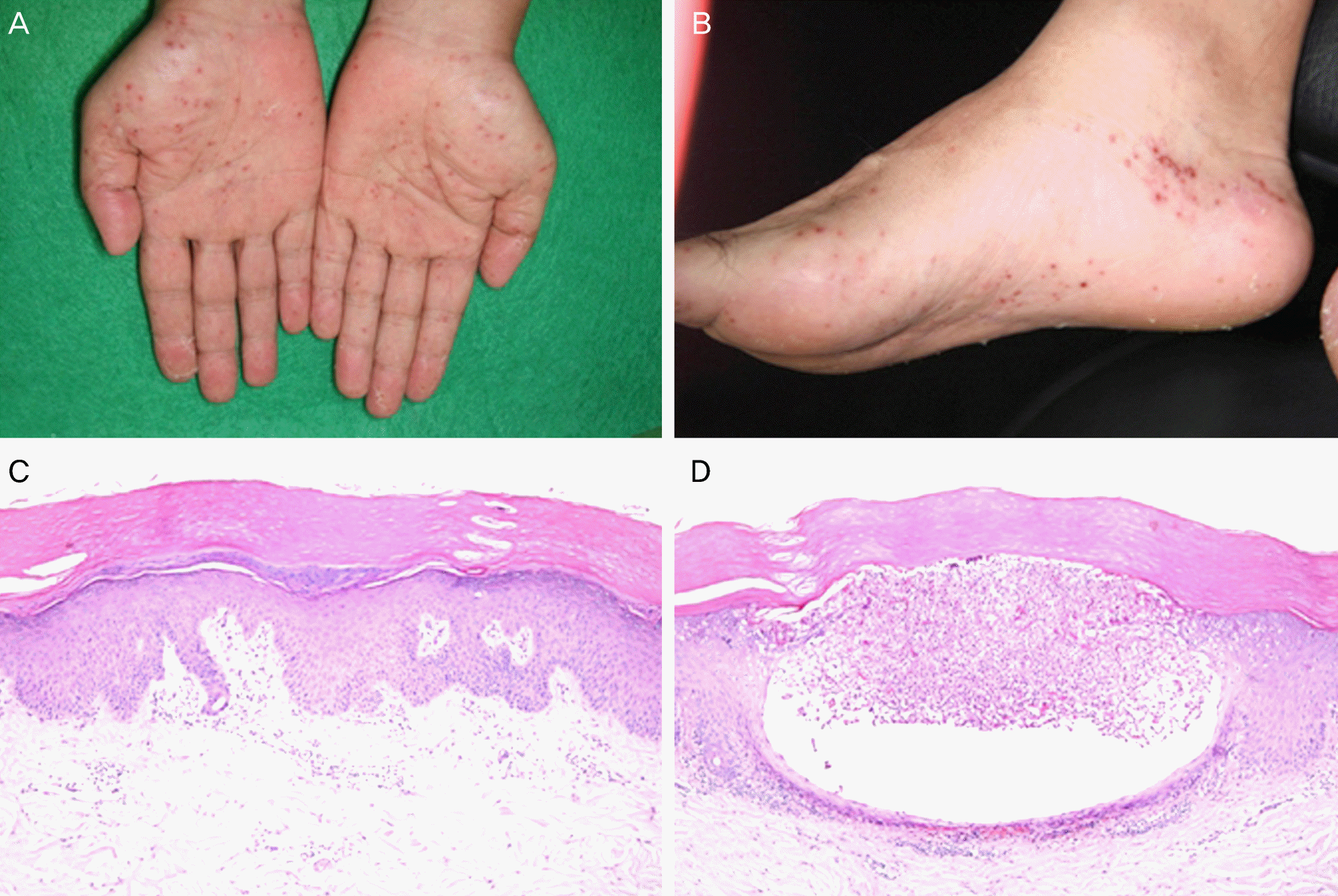
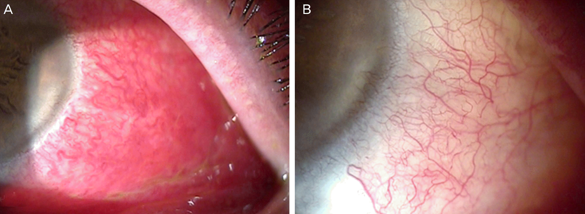
A 62-year-old male presented with skin lesions on the hands and feet and pain in the right eye, which started a few days prior. Corrected visual acuity was 0.5 in the right eye and 0.7 in the left eye at initial visit. Corneal edema, erosion, ulcer and peripheral corneal infiltration of the right eye were observed. However, anterior chamber reaction was not observed. Histological analysis of hand skin lesions indicated pustular psoriasis. The patient was initially treated with topical antibiotics and a combined therapy of oral and topical steroids for ocular symptoms. As a result, the right eye showed slight improvement and the oral steroid was discontinued. One month after the initial visit, scleritis appeared on the left eye and topical and oral steroids were restarted for both eyes. Two months after the initial visit, ocular symptoms were improved significantly and corrected visual acuity was 1.0 in both eyes. The mild peripheral corneal opacity remained in the right eye, but the previous inflammations in both eyes were improved.
References
1. Kilic B, Dogan U, Parlak AH, et al. Ocular findings in patients with psoriasis. Int J Dermatol. 2013; 52:554–9.

2. Moadel K, Perry HD, Donnenfeld ED, et al. Psoriatic corneal abscess. Am J Ophthalmol. 1995; 119:800–1.

3. Chandran NS, Greaves M, Gao F, et al. Psoriasis and the eye: prev-alence of eye disease in Singaporean Asian patients with psoriasis. J Dermatol. 2007; 34:805–10.

4. Hamideh F, Prete PE. Ophthalmologic manifestations of rheumatic diseases. Semin Arthritis Rheum. 2001; 30:217–41.

6. Erbagci I, Erbagci Z, Gungor K, Bekir N. Ocular anterior segment pathologies and tear film changes in patients with psoriasis vulgaris. Acta Med Okayama. 2003; 57:299–303.
7. Varma S, Woboso AF, Lane C, Holt PJ. The peripheral corneal melting syndrome and psoriasis: coincidence or association. Br J Dermatol. 1999; 141:344–6.

8. STUART JA. Ocular psoriasis. Am J Ophthalmol. 1963; 55:615–7.
9. KALDECK R. Ocular psoriasis; clinical review of eleven cases and some comments on treatment. AMA Arch Derm Syphilol. 1953; 68:44–9.
10. Kobayashi T, Naka W, Harada T, Nishikawa T. Association of the acral type of pustular psoriasis, Sjögren's syndrome, systemic lu-pus erythematosus, and Hashimoto's thyroiditis. J Dermatol. 1995; 22:125–8.

11. Gulliver W. Long-term prognosis in patients with psoriasis. Br J Dermatol. 2008; 159(Suppl 2):2–9.

12. Wee SW, Kim JC. Two clinical manifestations of anterior segment associated with systemic lupus erythematosus. J Korean Ophthalmol Soc. 2012; 53:1035–40.

13. Fernandes M, Vemuganti GK, Rao GN. Bilateral periocular psor-iasis: an initial manifestation of acute generalized pustular psoriasis with coexistent Sjögren's syndrome. Clin Experiment Ophthalmol. 2007; 35:763–6.

14. Wirbelauer C. Management of the red eye for the primary care physician. Am J Med. 2006; 119:302–6.

15. Huynh N, Cervantes-Castaneda RA, Bhat P, et al. Biologic re-sponse modifier therapy for psoriatic ocular inflammatory disease. Ocul Immunol Inflamm. 2008; 16:89–93.

16. Sampaio-Barros PD, Conde RA, Bonfiglioli R, et al. Characterization and outcome of uveitis in 350 patients with spondyloarthropathies. Rheumatol Int. 2006; 26:1143–6.

17. Hatchome N, Tagami H. Hypopyon-iridocyclitis as a complication of pustular psoriasis. J Am Acad Dermatol. 1985; 13:828–9.

18. Catsarou-Catsari A, Katsambas A, Theodoropoulos P, Stratigos J. Ophthalmological manifestations in patients with psoriasis. Acta Derm Venereol. 1984; 64:557–9.
Figure 1.
Slit lamp photographs of the right eye. (A, B) The whitish infiltration with ulceration (white arrow), epithelial defects (yellow arrow) and superficial vascularization (white arrow head) at the upper peripheral cornea and perilimbal inflammation in the sclera were noted at initial visit. (C) Two weeks after treatment, the peripheral corneal lesions were improved. (D) Two months after treatment, the lesions were healed with mild peripheral corneal haze.

Figure 2.
Photographs of the patient and histology. (A) Asymptomatic erythematous variable sized pustules and papules were visible in the palm of the hand. (B) Variable sized erythematous pustules were noted in the plantar of the foot. (C, D) The histologic exami-nation of the biopsy tissue revealed an epidermal hyperplasia, hyperkeratosis, parakeratosis, dilated vessels at the tip of the dermal papillae, and perivascular infiltrate of lymphocytes and neutrophils (C) and a large collection of neutrophils with spongiosis in the upper spinous layer and granular layer (D) (Hematoxylin & Eosin stain, x 100).


 PDF
PDF ePub
ePub Citation
 Print
Print


 XML Download