

 PDF
PDF ePub
ePub Citation
 Print
Print


Abstract
Purpose
To investigate the clinical and radiologic manifestations of idiopathic optic perineuritis (OPN), and to evaluate the out-comes of steroid treatment for OPN.
Methods
We reviewed the medical records and radiologic findings of 10 patients (13 eyes) who were diagnosed with OPN and treated with steroid.
Results
The mean age was 56.5 ± 9.3 years (range, 35-77 years) and the sex ratio was equal. The main complaint was de-crease in visual acuity combined with ocular pain during extraocular eye movement in 9 patients. The median visual acuity at the first visit was 0.2 (HM-0.8) and the relative afferent papillary defect was observed in 12 eyes. Additionally, combined orbital dis-eases included posterior scleritis in 1 eye and myositis in 1 eye. Orbit magnetic resonance imaging (MRI) scans demonstrated intraorbital optic nerve sheath enhancement in all patients, occasionally with orbital fat involvement. All patients demonstrated improved visual acuity after high-dose oral steroid therapy (6 patients) or intravenous (IV) pulse steroid therapy (4 patients). Relapse occurred in 4 patients during steroid tapering.
Conclusions
The population in this study was composed predominantly of patients with OPN in their 50's. The primary symptom of OPN was visual acuity decrease combined with ocular pain during extraocular eye movement. Radiologically, orbit MRI scans demonstrated intraorbital optic nerve sheath enhancement. The patients in this study demonstrated good responses to steroid treatment, but clinicians must be aware of the high recurrence rate during steroid tapering in this condition. A combination of clin-ical and radiologic findings was helpful to diagnose OPN.
Go to : 
References
1. Kennerdell JS, Dresner SC. The nonspecific orbital inflammatory syndromes. Surv Ophthalmol. 1984; 29:93–103.

2. Toshniwal P. Optic perineuritis with secondary syphilis. J Clin Neuro-ophthalmol. 1987; 7:6–10.
3. Nassani S, Cocito L, Arcuri T, Favale E. Orbital pseudotumor as a presenting sign of temporal arteritis. Clin Exp Rheumatol. 1995; 13:367–9.
4. Tien RD, Hesselink JR, Szumowski J. MR fat suppression combined with Gd-DTPA enhancement in optic neuritis and perineuritis. J Comput Assist Tomogr. 1991; 15:223–7.
5. Sridharan S, Biswas J. Ocular tuberculosis: an update. Expert Review of Ophthalmology. 2007; 2:845–60.
6. Straatsma BR. Ocular manifestations of Wegener's granulomatosis. Am J Ophthalmol. 1957; 44:789–99.
7. Delaney P. Neurologic manifestations in sarcoidosis. Review of the liferature, with a report of 23 cases. Ann Int Med. 1977; 87:336–45.
8. Purvin V, Kawasaki A, Jacobson DM. Optic perineuritis: clinical and radiographic features. Arch Ophthalmol. 2001; 119:1299–306.
9. Edmunds W, Lawford JB. Examination of optic nerve from cases of amblyopia in diabetes. Trans OphthalmolSoc UK. 1883; 3:160–2.
10. Rush JA, Kennerdell JS, Donin JF. Acute periscleritis: a variant of idiopathic orbital inflammation. 1982; 4:221–30.
11. Dutton JJ, Anderson RL. Idiopathic inflammatory perioptic neu-ritis simulating optic nerve sheath meningioma. Am J Ophthalmol. 1985; 100:424–30.
12. Fay AM, Kane SA, Kazim M, et al. Magnetic resonance imaging of optic perineuritis. J Neuro-ophthalmol. 1997; 17:247–9.
13. Hickman SJ, Miszkiel KA, Plant GT, Miller DH. The optic nerve sheath on MRI in acute optic neuritis. Neuroradiology. 2005; 47:51–5.
14. Perkin GD, Rose FC. Optic neuritis and its differential diagnosis. New York, NY: Oxford University Press Inc.;1979. p. 22.
15. Lee YJ, Chang BL. Clinical manifestations of optic neuritis. J Korean Ophthalmol Soc. 1997; 38:1969–74.
16. Ahn BC, Kim HS, Ahn HS. Clinical profile of the optic neuritis in Korea. J Korean Ophthalmol Soc. 1997; 38:1827–33.
17. Yang DW, Lee SH. Clinical characteristics of optic neuritis according to the presence of abnormal MRI lesions. J Korean Ophthalmol Soc. 2004; 45:273–80.
18. Kang HM, Kim HY. Clinical manifestations of idiopathic optic perineuritis in Korea. J Korean Ophthalmol Soc. 2012; 53:1016–22.
19. Yuen SJ, Rubin PA. Idiopathic orbital inflammation: distribution, clinical features, and treatment outcome. Arch Ophthalmol. 2003; 121:491–9.
20. Gordon LK. Orbital inflammatory disease: a diagnostic and ther-apeutic challenge. Eye (Lond). 2006; 20:1196–206.
Go to :
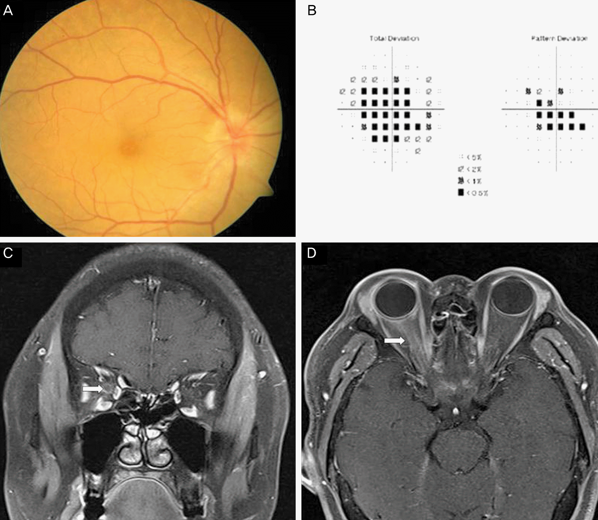 | Figure 1.Right optic disc swelling was observed in the fundus photography (A). Right visual field was seen in Humphery visu-al field test (B). Fat suppression magnetic resonance imaging scan of the orbit with contrast enhancement in axial and coronal section showed that enhancement of the right optic nerve sheath and streakiness of the surrounding orbital fat (white arrow), and posterior sclera thickening in right eye (C, D). |
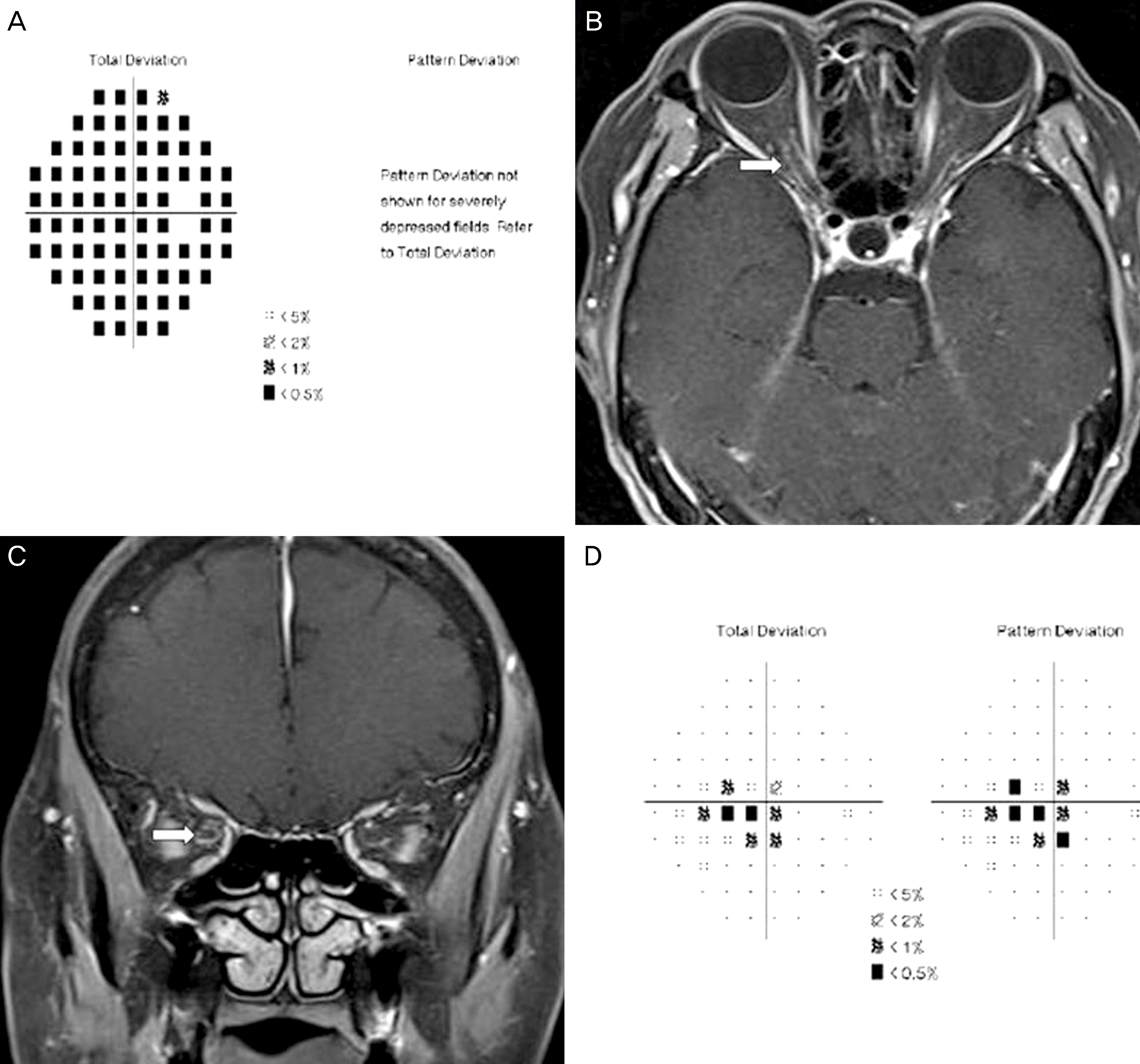 | Figure 2.Right visual field test was seen diffuse visual field defect at initial visit (A). Fat suppression magnetic resonance imaging scan of the orbit with contrast enhancement demonstrated that tram track sign around optic nerve sheath in axial view and donut appearance in optic nerve sheath in coronal view (white arrow) (B, C). Right visual field test revealed rapid improve-ment of visual field defect after steroid treatment, post-treatment day at 5 (D). |
Table 1.
Clinical characteristics of idiopathic optic perineuritis patients
 XML Download