Abstract
Purpose
This study was designed to compare the outcomes in idiopathic epiretinal membrane (ERM) surgery according to sol-vents of indocyanine green (ICG) for internal limiting membrane (ILM) peeling.
Methods
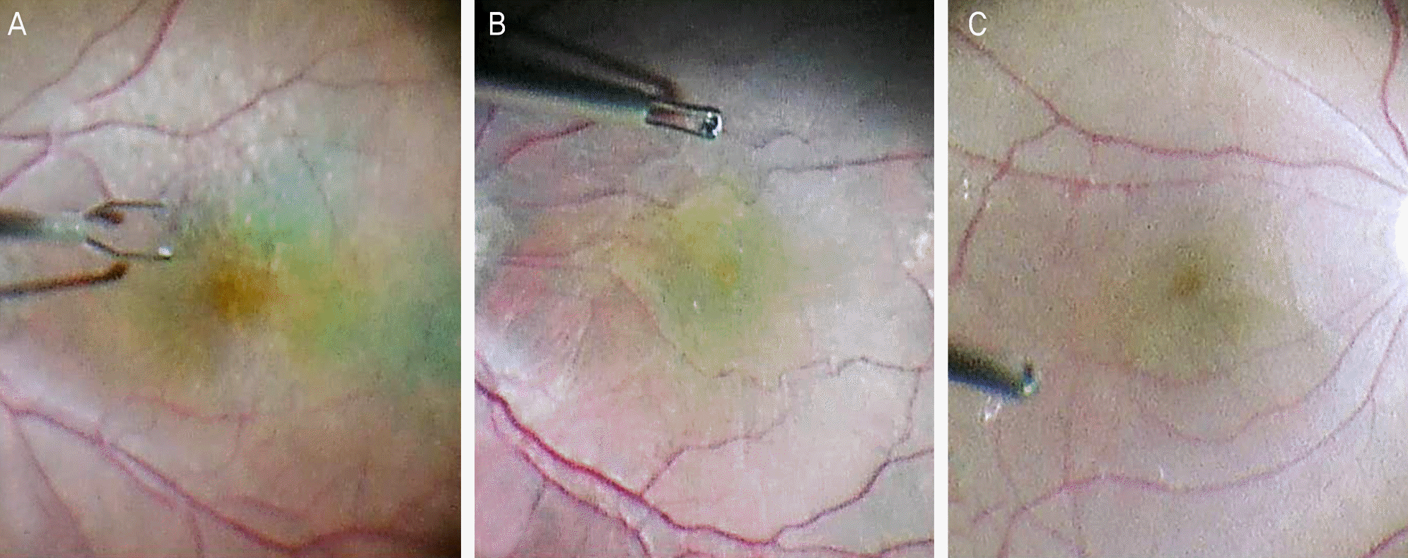
The medical records of 27 patients (27 eyes) with idiopathic ERM who had undergone pars plana vitrectomy with ICG staining for ILM peeling were retrospectively reviewed. The patients were divided into two groups according to solvents of 0.25% ICG solutions. Solvents used were balanced salt solution (BSS) in group I (15 eyes) and 5% glucose in group II (12 eyes). The severity of ERM, the duration of symptoms, the preoperative and postoperative best corrected visual acuity (BCVA) values, the visibility of the stained ILM (Good, Fair, Poor), and the postoperative complications were compared in the two groups.
Results
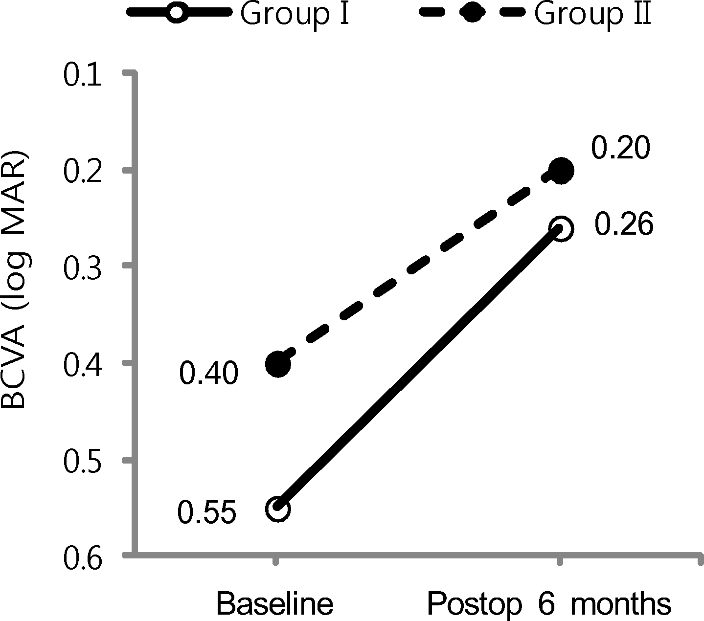
There was no statistically significant difference in the severity of ERM, the duration of symptoms and the preoperative BCVA in the two groups. The postoperative BCVA was significantly improved in both groups, and the difference was not statisti-cally significant (p = 0.675). There was a significantly smaller number of eyes with poor ILM staining in group II than in group I (p = 0.014). No complications such as recurrence of ERM, atrophy of the retinal pigment epithelium (RPE) or retinal detachment were observed in the two groups.
Go to : 
References
1. Machemer R. The surgical removal of epiretinal macular mem-branes (macular puckers). Klin Monbl Augenheilkd. 1978; 173:36–42.
3. Margherio RR, Cox MS Jr, Trese MT, et al. Removal of epimacular membranes. Ophthalmology. 1985; 92:1075–83.

4. McDonald HR, Verre WP, Aaberg TM. Surgical management of idiopathic epiretinal membranes. Ophthalmology. 1986; 93:978–83.

5. Pesin SR, Olk RJ, Grand MG, et al. Vitrectomy for premacular fibroplasia. Prognostic factors, long-term follow-up, and time course of visual improvement. Ophthalmology. 1991; 98:1109–14.
6. Donati G, Kapetanios AD, Pournaras CJ. Complications of surgery for epiretinal membranes. Graefes Arch Clin Exp Ophthalmol. 1998; 236:739–46.

7. Benhamou N, Massin P, Spolaore R, et al. Surgical management of epiretinal membrane in young patients. Am J Ophthalmol. 2002; 133:358–64.
8. Massin P, Paques M, Masri H, et al. Visual outcome of surgery for epiretinal membranes with macular pseudoholes. Ophthalmology. 1999; 106:580–5.

9. Zarbin MA, Michels RG, Green WR. Epiretinal membrane con-tracture associated with macular prolapse. Am J Ophthalmol. 1990; 110:610–8.

10. Clarkson JG, Green WR, Massof D. A histopathologic review of 168 cases of preretinal membrane. Am J Ophthalmol. 1977; 84:1–17.

11. Michels RG. A clinical and histopathologic study of epiretinal membranes affecting the macula and removed by vitreous surgery. Trans Am Ophthalmol Soc. 1982; 80:580–656.
12. Smiddy WE, Green WR, Michels RG, de la Cruz Z. Ultrastructural studies of vitreomacular traction syndrome. Am J Ophthalmol. 1989; 107:177–85.

13. Fine BS. Limiting membranes of the sensory retina and pigment epithelium. An electron microscopic study. Arch Ophthalmol. 1961; 66:847–60.
14. Burk SE, Da Mata AP, Snyder ME, et al. Indocyanine green-assisted peeling of the retinal internal limiting membrane. Ophthalmology. 2000; 107:2010–4.
15. Park DW, Dugel PU, Garda J, et al. Macular pucker removal with and without internal limiting membrane peeling: pilot study. Ophthalmology. 2003; 110:62–4.

16. Kwok AK, Lai TY, Yuen KS. Epiretinal membrane surgery with or without internal limiting membrane peeling. Clinical and Experimental Ophthalmology. 2005; 33:379–85.

17. Bovey EH, Uffer S, Achache F. Surgery for epimacular membrane: impact of retinal internal limiting membrane removal on functional outcome. Retina. 2004; 24:728–35.
18. Konstantinidis L, Uffer S, Bovey EH. Ultrastructural changes of the internal limiting membrane removed during indocyanine green assisted peeling versus conventional surgery for idiopathic mac-ular epiretinal membrane. Retina. 2009; 29:380–6.

19. Kim TW, Song SJ, Chung H, Yu HG. Internal limiting membrane peeling in surgical treatment of macular epiretinal membrane. J Korean Ophthalmol Soc. 2005; 46:989–94.
20. Kim YC, Kim KS. The effect of internal limiting membrane peeling in treatment of idiopathic epiretinal membrane. J Korean Ophthalmol Soc. 2007; 48:1067–72.

21. Gandorfer A, Messmer EM, Ulbig MW, Kampik A. Indocyanine green selectively stains the internal limiting membrane. Am J Ophthalmol. 2001; 131:387–8.

22. Da Mata AP, Burk SE, Riemann CD, et al. Indocyanine green-as-sisted peeling of the retinal internal limiting membrane during vi-trectomy surgery for macular hole repair. Ophthalmology. 2001; 108:1187–92.

23. Kwok AK, Lai TY, Li WW, et al. Indocyanine green-assisted in-ternal limiting membrane removal in epiretinal membrane surgery: a clinical and histologic study. Am J Ophthalmol. 2004; 138:194–9.

24. von Jagow B, Hoing A, Gandorfer A, et al. Functional outcome of indocyanine green-assisted macular surgery: 7-year follow-up. Retina. 2009; 29:1249–56.
25. Lanzetta P, Polito A, Del Borrello M, et al. Idiopathic macular hole surgery with low-concentration infracyanine green-assisted peeling ofthe internal limiting membrane. Am J Ophthalmol. 2006; 142:771–6.
26. Lai MM, Williams GA. Anatomical and visual outcomes of idio-pathic macular hole surgery with internal limiting membrane re-moval using low-concentration indocyanine green. Retina. 2007; 27:477–82.

27. Kwok AK, Lai TY, Yew DT, Li WW. Internal limiting membrane staining with various concentrations of indocyanine green dye un-der air in macular surgeries. Am J Ophthalmol. 2003; 136:223–30.

28. Kanda S, Uemura A, Yamashita T, et al. Visual field defects after intravitreous administration of indocyanine green in macular hole surgery. Arch Ophthalmol. 2004; 122:1447–51.
29. Gandorfer A, Haritoglou C, Gass CA, et al. Indocyanine green-as-sisted peeling of the internal limiting membrane may cause retinal damage. Am J Ophthalmol. 2001; 132:431–3.

30. Hillenkamp J, Saikia P, Gora F, et al. Macular function and mor-phology after peeling of idiopathic epiretinal membrane with and without the assistance of indocyanine green. Br J Ophthalmol. 2005; 89:437–43.

31. Sippy BD, Engelbrecht NE, Hubbard GB, et al. Indocyanine green effect on cultured human retinal pigment epithelial cells: implication for macular hole surgery. Am J Ophthalmol. 2001; 132:433–5.

32. Enaida H, Sakamoto T, Hisatomi T, et al. Morphological and functional damage of the retina caused by intravitreous indocyanine green in rat eyes. Graefes Arch Clin Exp Ophthalmol. 2002; 240:209–13.

33. Haritoglou C, Gandorfer A, Gass CA, et al. The effect of in-docyanine-green on functional outcome of macular pucker surgery. Am J Ophthalmol. 2003; 135:328–37.

34. Gandorfer A, Haritoglou C, Gandorfer A, Kampik A. Retinal dam-age from indocyanine green in experimental macular surgery. Invest Ophthalmol Vis Sci. 2003; 44:316–23.

35. Maia M, Haller JA, Pieramici DJ, et al. Retinal pigment epithelial abnormalities after internal limiting membrane peeling guided by indocyanine green staining. Retina. 2004; 24:157–60.

36. Stalmans P, Van Aken EH, Veckeneer M, et al. Toxic effect of in-docyanine green on retinal pigment epithelium related to osmotic effects of the solvent. Am J Ophthalmol. 2002; 134:282–5.

37. Stalmans P, Himpens B. Confocal imaging of Ca2+ signaling in cultured rat retinal pigment epithelial cells during mechanical and pharmacologic stimulation. Invest Ophthalmol Vis Sci. 1997; 38:176–87.
38. Haritoglou C, Gandorfer A, Schaumberger M, et al. Light-absorbing properties and osmolarity of indocyanine-green depending on concentration and solvent medium. Invest Ophthalmol Vis Sci. 2003; 44:2722–9.

39. Ho JD, Chen HC, Chen SN, Tsai RJ. Reduction of indocyanine green-associated photosensitizing toxicity in retinal pigment epi-thelium by sodium elimination. Arch Ophthalmol. 2004; 122:871–8.
Go to : 
 | Figure 1.Surgeon's view. Visual quality of the stained internal limiting membrane (ILM). (A) Good stain. (B) Fair stain. (C) Poor stain. |
 | Figure 2.Changes in best corrected visual acuity (BCVA) be-tween preoperation (Preop) and postoperation (Postop). |
Table 1.
Comparison of patients’ demographics between two groups
| Group I* | Group II† | Total | p-value | |
|---|---|---|---|---|
| No. of eyes | 15 | 12 | 27 | |
| Age (years) | 63.7 ± 6.0 | 66.0 ± 6.8 | 64.7 ± 6.3 | 0.464‡ |
| Sex (M:F) | 9:6 | 4:8 | 13:14 | 0.168§ |
| Periods of Sx (months) | 16.4 ± 23.7 | 11.2 ± 14.0 | 13.9 ± 19.4 | 0.496‡ |
| Baseline BCVA (log MAR) | 0.55 ± 0.26 | 0.40 ± 0.22 | 0.49 ± 0.25 | 0.172‡ |
| Lens state (phakic:pseudophakic) | 15:0 | 10:2 | 25:2 | 0.188§ |
| Phacovitrectomy:Vitrectomy | 14:1 | 10:2 | 24:3 | 0.569§ |
| ERM stage (%) | 1.000§ | |||
| Stage 1 | 7 (46.7) | 5 (41.7) | 12 (44.4) | |
| Stage 2 | 7 (46.7) | 6 (50.0) | 13 (48.1) | |
| Stage 3 | 1 (6.7) | 1 (8.3) | 2 (7.4) |
Table 2.
Visualization of internal limiting membrane and number of indocyanine green (ICG) injection with different solvents
|
No. of eyes (%) |
p-value | |||
|---|---|---|---|---|
| Group I* | Group II† | Total | ||
| Visualization of ILM (%) | 0.014‡ | |||
| Poor | 7 (46.7) | 1 (8.3) | 8 (29.6) | |
| Fair | 6 (40.0) | 3 (25.0) | 9 (33.3) | |
| Good | 2 (6.7) | 8 (66.7) | 10 (37.0) | |
| No. of ICG injections | 1.2 ± 0.4 | 1.1 ± 0.3 | 1.2 ± 0.4 | 0.405§ |
Table 3.
Comparison of the anatomical and functional success after surgery between two groups
|
No. of eyes (%) |
p-value‡ | ||
|---|---|---|---|
| Group I* | Group II† | ||
| Anatomical success | |||
| Complete ILM removal | 15 (100.0) | 12 (100.0) | 1.000 |
| Remained retinal wrinkling | 10 (66.7) | 4 (33.3) | 0.085 |
| Foveal contour | 5 (33.3) | 9 (75.0) | 0.031 |
| Functional success§ | 10 (66.7) | 8 (66.7) | 1.000 |


 PDF
PDF ePub
ePub Citation
 Print
Print


 XML Download