Abstract
Purpose
To evaluate the target refraction of patients with binocular myopia and monocular cataract after intraocular lens (IOL) implantation.
Methods
This study comprised 199 patients with binocular myopia (axial length >25 mm) and monocular cataract after IOL im-plantation for the removal of the monocular cataract. The research was conducted using a questionnaire method and by performing statistical analysis of the refractive outcomes.
Results
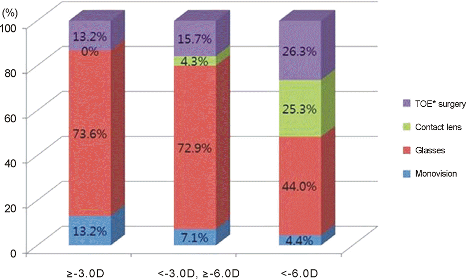
The patients are grouped into 3 domains (<-3 D group, −3∼-6 D group, >-6 D group). There were no statistically sig-nificant differences among the 3 groups; whereas the satisfaction of the present corrected state was statistically low in the >-6 D group (p < 0.05). The satisfaction of the corrected state was statistically high in the group of postoperative anisometropia under 3 D (p < 0.05). There was no statistical difference between the groups on the satisfaction of target diopter (-2∼-3 D or emme-tropia). In terms of correction method, glasses were worn in 119 patients (60%), contact lenses were worn in 26 patients (13%), and monovision was used in 14 patients (7%) were used respectively. Forty patients (20%) with implanted IOL in both eyes did not use any of the correction methods above. Except for the contact lens group, the general satisfaction and the satisfaction of the present corrected state were statistically lower than the other group (p < 0.05). There was no statistically significant differ-ence among the 3 groups in the percentage of cataract surgery in the fellow eye.
Go to : 
References
1. Jung SK, Lee JH, Kakizaki H, Jee D. Prevalence of myopia and its association with body stature and educational level in 19-year-old male conscripts in seoul, South Korea. Invest Ophthalmol Vis Sci. 2012; 53:5579–83.

2. Mangione CM, Lee PP, Pitts J, et al. Psychometric properties of the National Eye Institute Visual Function Questionnaire (NEI-VFQ). NEI-VFQ Field Test Investigators. Arch Ophthalmol. 1998; 116:1496–504.
3. Mangione CM, Lee PP, Gutierrez PR, et al. Development of the 25-item National Eye Institute Visual Function Questionnaire. Arch Ophthalmol. 2001; 119:1050–8.
4. Heo JW, Yoon HS, Shin JP, et al. A validation and reliability study of the Korean Version of National Eye Institute Visual Function Questionnaire 25. J Korean Ophthalmol Soc. 2010; 51:1354–67.

5. Stenström S. Untersuchungen über die variation und kovariation der optischen elemente des menschlichen Auges. Acta Ophthalmol. 1946; 26:101–3.
6. Johannsdottir KR, Stelmach LB. Monovision: a review of the sci-entific literature. Optom Vis Sci. 2001; 78:646–51.
8. Ito M, Shimizu K, Amano R, Handa T. Assessment of visual per-formance in pseudophakic monovision. J Cataract Refract Surg. 2009; 35:710–4.

9. Ito M, Shimizu K, Iida Y, Amano R. Five-year clinical study of patients with pseudophakic monovision. J Cataract Refract Surg. 2012; 38:1440–5.

11. Finkelman YM, Nq JQ, Barrett GD. Patient satisfaction and visual function after pseudophakic monovision. J Cataract Refract Surg. 2009; 35:998–1002.

12. Xiao J, Jiang C, Zhang M. Pseudophakic monovision is an important surgical approach to being spectacle-free. Indian J Ophthalmol. 2011; 59:481–5.

13. Marques FF, Sato RM, Chiacchio BB, et al. Evaluation of visual performance and patient satisfaction with pseudophakic mono-vision technique. Arq Bras Oftalmol. 2009; 72:164–8.

Go to : 
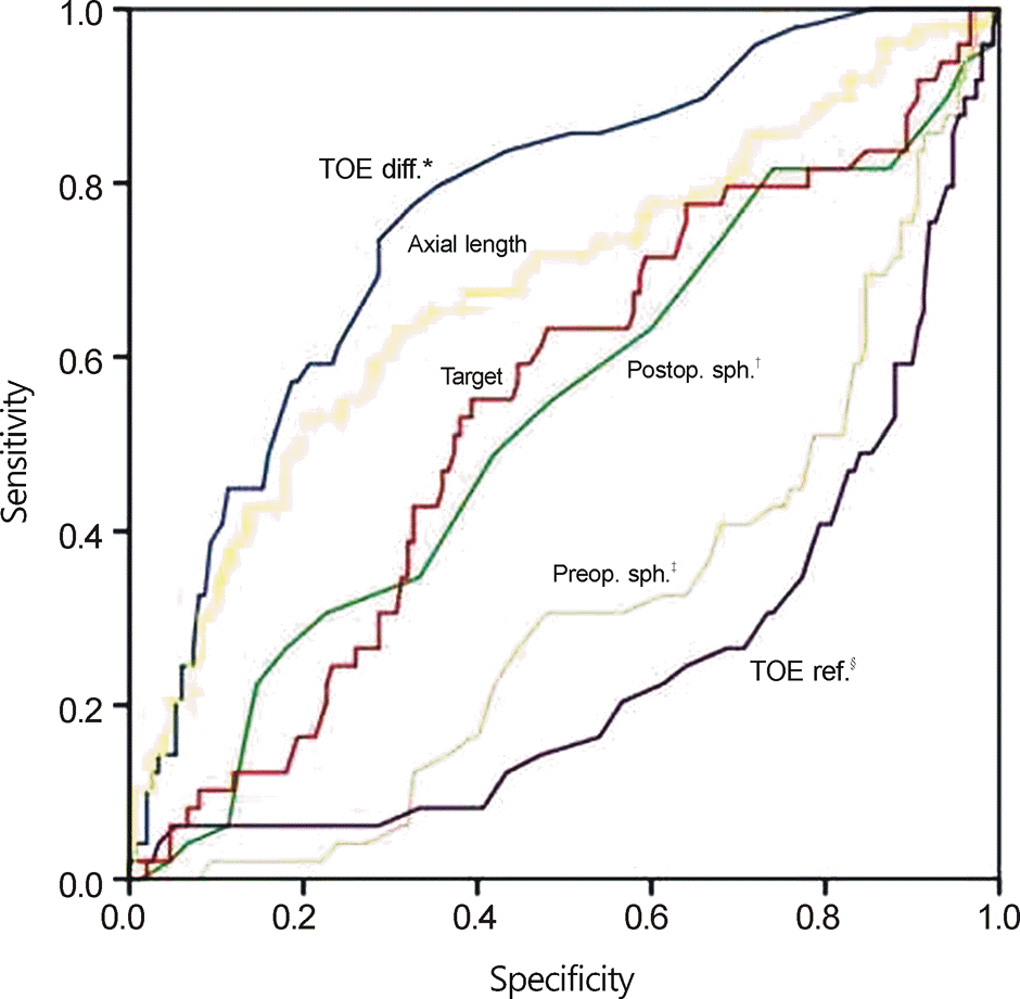
 | Figure 1.Receiver operating characteristic (ROC) curve. *The refractive difference with the other eye; †Postoperative spher-ical equivalent; ‡Preoperative spherical equivalent; §The re-fractive amount of the other eye. |
Table 1.
Demographics of the study group
Table 2.
The amount of preoperative refraction
Table 3.
The satisfaction score about the amount of preoperative refraction
Table 4.
The satisfaction score about the selection of target refraction
Table 5.
The satisfaction score about the amount of preoperative refraction and the target refraction
Table 6.
The number of the patients grouped by the correction methods of anisometropia
Table 7.
The mean ages of the patients grouped by the correction methods of anisometropia
| ≤-3.0 D | -3.0∼-6.0 D | >-6.0 D | Total | |
|---|---|---|---|---|
| Monovision | 64.60 ± 11.67 | 56.40 ± 8.85 | 56.25 ± 7.41 | 59.29 ± 9.78 |
| Glasses | 59.00 ± 10.74 | 57.80 ± 11.08 | 59.13 ± 9.44 | 58.53 ± 10.41 |
| Contact lens | - | 45.33 ± 4.16* | 49.65 ± 10.92† | 49.15 ± 10.41 |
| TOE surgery | 65.6 ± 12.24 | 56.00 ± 13.96 | 60.17 ± 12.59 | 59.70 ± 12.55 |
Table 8.
Comparisons of the preoperative and postoperative refraction differences on both eyes, the target refraction about the surgery, the correction state according to the correction methods of anisometropia
Table 9.
The satisfaction score about the surgery and the correction state according to the correction methods of anisometropia


 PDF
PDF ePub
ePub Citation
 Print
Print


 XML Download